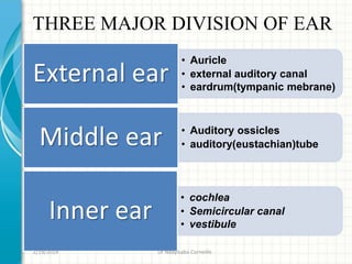
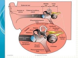
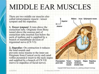
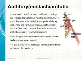
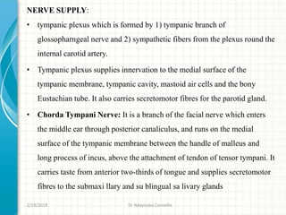
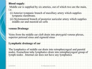
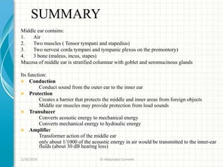
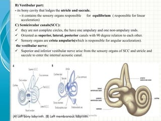
The document discusses the anatomy of the ear, which is divided into three main parts:
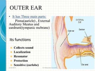
1. The outer ear collects sound and directs it to the middle ear. The middle ear contains small bones that transmit sound to the inner ear.
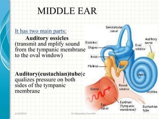
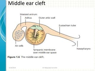
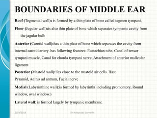
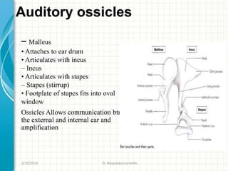
2. The middle ear contains three small bones called the ossicles that amplify sound waves. It also contains two muscles and connects to the inner ear via the cochlea.
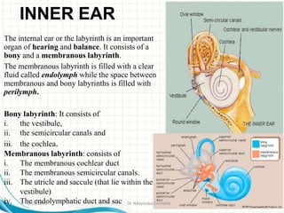
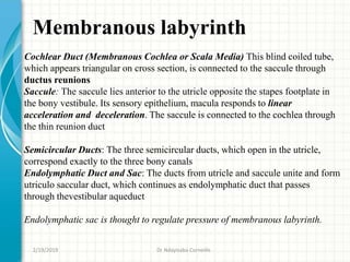
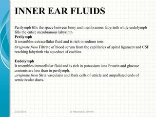
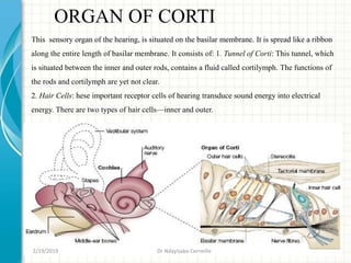
3. The inner ear contains the cochlea for hearing and balance organs like the semicircular canals and vestibule. The cochlea converts sound waves into nerve signals that are transmitted to the brain.
The ear can be divided into three main sections:
1. The outer ear collects sound waves and directs them to the eardrum.
2. The middle ear contains three small bones that amplify vibrations and transmit them to the inner ear. It also contains two muscles and two nerves.
3. The inner ear contains the cochlea for hearing and semicircular canals for balance. Within the cochlea is the organ of Corti which contains hair cells that transduce sound vibrations into nerve signals.
The document provides an overview of the anatomy of the ear, which is divided into three main parts:
1. The external ear includes the pinna, external auditory canal, and tympanic membrane.
2. The middle ear contains the ossicles (malleus, incus, stapes), muscles, lining, and connections to structures like the Eustachian tube and mastoid air cells.
3. The internal ear, or labyrinth, houses the bony and membranous structures involved in balance and hearing, including the vestibule, semicircular canals, and cochlea.
The ear is divided into three parts - external, middle, and internal. The external ear consists of the pinna and external auditory meatus. The pinna has elevations like the helix and depressions like the concha. The middle ear contains the auditory ossicles and transmits sound from the tympanic membrane to the inner ear. The internal ear contains the membranous labyrinth within the bony labyrinth, and is responsible for hearing and balance. It includes the cochlea, saccule, utricle and semicircular canals.
The document summarizes the anatomy and function of the ear. It describes the three parts of the ear - external, middle, and inner ear. The external ear includes the pinna and ear canal. The middle ear contains the auditory ossicles and muscles. The inner ear has the bony labyrinth and membranous labyrinth, including the cochlea for hearing and vestibule for balance. Common ear disorders like otitis media and types of hearing loss are also summarized.
The document discusses the anatomy of the ear, including:
- The ear is divided into external, middle, and inner sections. The external ear collects sound waves and the middle ear transmits vibrations via ossicles. The inner ear contains receptors for hearing and balance.
- The external ear includes the pinna, external auditory meatus, and tympanic membrane. The middle ear contains the ossicles, muscles, and nerves. The inner ear contains the cochlear and vestibular nerves which carry signals for hearing and balance.
- Key structures in the middle ear include the ossicles (malleus, incus, stapes), which transmit vibrations, and the muscles (tensor
The document summarizes the anatomy and physiology of the ear. It is divided into three main parts: the external ear, middle ear, and inner ear. The external ear collects sound waves and directs them through the external auditory canal to the tympanic membrane. The middle ear contains three small bones (ossicles) that transmit sound vibrations from the tympanic membrane to the inner ear. The inner ear, or labyrinth, contains the cochlea for hearing and semicircular canals for balance. It converts sound vibrations into neural signals that are sent to the brain.
Provides a detailed description of the gross anatomy of the ear for undergraduate medical students; i.e. parts of the ear, structures found, their blood supply, their innervation, developmental origins & their functions. It also includes examples of common disorders associated with those parts.
This document discusses the anatomy and functions of the ear. It describes the external, middle, and inner ear in detail. The external ear includes the pinna, external auditory canal, and eardrum. The middle ear contains three small bones called ossicles that transmit sound from the eardrum to the inner ear. The inner ear includes the cochlea for hearing and semicircular canals for balance. The eustachian tube connects the middle ear to the throat and helps equalize pressure. The ear has important functions of hearing sound waves and maintaining balance.
The document summarizes the anatomy and function of the ear. It describes the three main divisions of the ear - the outer, middle and inner ear. The outer ear collects sound waves. The middle ear contains the ossicles that transmit vibrations from the eardrum to the inner ear. The inner ear converts vibrations to neural signals via hair cells in the cochlea and balances functions in the semicircular canals. The ear detects sound and maintains equilibrium through these specialized structures that work together to transmit physical vibrations to the brain for processing.
The Ear, Anatomy, Physiology, Clinical diseases, and pathology, hearing testsHamzehKYacoub
Ear is composed of three parts: External ear, middle ear, and the Inner ear.
Hearing tests (Rinne's and Weber's tests).
Most important hearing and ear diseases are included.
The common ENT disorders includes Ear Disorder: Ear Infections,Hearing Disorders and Deafness, Meniere's Disease. acute otitis media, tonsillopharyngitis, sore throat, adenoid disorders, epistaxis, nasal congestion and rhinorrhea, sinusitis, and ear foreign bodies and nasal foreign bodies etc.
The middle ear is an air-filled cavity located between the inner ear and outer ear. It contains three small bones called ossicles that transmit sound vibrations from the eardrum to the inner ear. The middle ear is divided into several compartments and has six boundaries: roof, floor, medial, lateral, anterior and posterior walls. It contains the ossicles (malleus, incus, stapes), two small muscles (tensor tympani and stapedius), and several nerves including the facial nerve. The mastoid air cells are air spaces in the mastoid bone behind the middle ear that communicate with the middle ear and petrous apex. The middle ear and mastoid air cells are important anatomical structures involved
The ear has three main parts - the external, middle, and inner ear. The external ear consists of the auricle and external acoustic meatus. The middle ear contains the auditory ossicles and connects to the inner ear and nasopharynx. The inner ear houses the vestibulocochlear organ including the cochlea for hearing and semicircular canals and vestibule for balance. Sound waves cause the tympanic membrane and ossicles to vibrate, transmitting the signal through fluid-filled ducts to hair cells which convert it to neural impulses in the cochlear and vestibular nerves.
This document provides an overview of anatomy and disorders of the ear, nose, and throat. It begins with a review of the anatomy and physiology of the external, middle, and inner ear. It then discusses various disorders that can affect each part of the ear, such as cerumen impaction, foreign bodies, and external otitis in the external ear. Assessment techniques including history, physical exam, and diagnostic tests are also covered. The document provides nursing management strategies for some common ear, nose, and throat conditions.
Human ear, organ of hearing and equilibrium that detects and analyzes sound by transduction (or the conversion of sound waves into electrochemical impulses) and maintains the sense of balance (equilibrium).
The document describes the anatomy and physiology of the ear, nose, and tongue. It begins by defining the three main parts of the ear - external, middle, and inner ear. It then provides details on the structures within each part, including the pinna, external auditory canal, tympanic membrane, auditory ossicles, cochlea, vestibule and semicircular canals. It further discusses the physiology of sound conduction and disorders like otitis media. Regarding the nose, it outlines the nasal cavity, septum, turbinates and their roles in breathing, air conditioning and smell. Common nasal disorders are also listed. Finally, it details the structures of the tongue such as the pap
The document summarizes the structure and function of the human ear. It describes the ear as having three main parts: the outer, middle, and inner ear. The outer ear collects sound waves and directs them through the auditory canal to the middle ear, where the vibrations are transmitted through three small bones to the inner ear. In the inner ear, fluid waves stimulate hair cells to generate nerve impulses that travel to the brain for hearing and balance. The ear detects sound properties like pitch from frequency and volume from amplitude to enable hearing perception.
The middle ear is a six-walled air-filled cavity located in the petrous part of the temporal bone between the inner and outer ear. It has three divisions - the epitympanum above the eardrum, the mesotympanum opposite the eardrum, and the hypotympanum below the eardrum. The middle ear contains the three ossicles (malleus, incus, stapes) that transmit sound vibrations from the eardrum to the inner ear. It is lined by epithelium and contains the tensor tympani and stapedius muscles that help dampen loud sounds. Blood supply comes from branches of the maxillary and posterior auricular arteries.
Anatomy of external and middle ear by dr. faisal rahmanFaisalRahman153
This includes anatomy of external and middle ear with their clinical co relations. Embryology is also discussed here. Pinna, External auditory canal, Tympanic membrane, Middle ear Cleft, Mastoid and Auditory tube topics are included.
The ear is divided into three parts - external, middle, and inner ear. The external ear includes the auricle and external auditory canal. The middle ear, or tympanic cavity, contains the three ossicles (malleus, incus, stapes) and two small muscles (tensor tympani and stapedius). It is separated from the inner ear by the oval and round windows. The complex anatomy of the middle ear, including its walls, spaces, folds, and vascular and neural supply, allow it to efficiently transmit sound vibrations from the outer to inner ear.
The document provides information about the anatomy and functions of the human ear. It describes the three main parts of the ear - outer, middle and inner ear. It details the structures within each part, including the pinna, ear canal, eardrum, ossicles and cochlea. The functions of hearing and balance are explained. Finally, common ear problems like infections, injuries and conditions affecting the ear structures are listed.
Anatomy of ear for undergraduate students ( MBBS ) .
Basic anatomy of ear with its clinical importance
nerve supply of ear
blood supply of ear
middle ear ossicles
malleus incus stapes
muscles of middle ear
mastoid air cells
pinna tympanic membrane
lymphatics of ear
walls of middle ear
organ of corti
middle ear cleft
facial recess
sinus tympani
chorda tympani
embryology of ear
The document summarizes the anatomy of the middle ear. It describes the structures found within the middle ear cavity including the ossicles (malleus, incus, stapes), muscles (tensor tympani, stapedius), nerves (facial, glossopharyngeal) and blood supply. It also details the six walls that make up the middle ear cavity - roof, floor, medial, lateral, anterior and posterior walls - and their anatomical features and relationships.
The surgical importance of the ear lies in addressing various conditions affecting hearing, balance, and overall ear health. Otolaryngologists (ear, nose, and throat specialists) often perform surgical procedures to treat a range of ear-related issues. Understanding the surgical importance of the ear is essential for otolaryngologists and surgeons specializing in ear, nose, and throat (ENT) procedures. These surgeries aim to treat various ear conditions, improve hearing, and enhance overall ear health.
The document summarizes the anatomy and physiology of the human ear. It is divided into three main sections - the external, middle, and inner ear. The external ear includes the pinna, external auditory canal, and eardrum. The middle ear contains the three smallest bones in the body known as the auditory ossicles which transmit sound from the eardrum to the inner ear. The inner ear is made up of the semicircular canals for balance and the cochlea, which converts sound waves into nerve signals for hearing.
Local Advanced Lung Cancer: Artificial Intelligence, Synergetics, Complex Sys...Oleg Kshivets
Overall life span (LS) was 1671.7±1721.6 days and cumulative 5YS reached 62.4%, 10 years – 50.4%, 20 years – 44.6%. 94 LCP lived more than 5 years without cancer (LS=2958.6±1723.6 days), 22 – more than 10 years (LS=5571±1841.8 days). 67 LCP died because of LC (LS=471.9±344 days). AT significantly improved 5YS (68% vs. 53.7%) (P=0.028 by log-rank test). Cox modeling displayed that 5YS of LCP significantly depended on: N0-N12, T3-4, blood cell circuit, cell ratio factors (ratio between cancer cells-CC and blood cells subpopulations), LC cell dynamics, recalcification time, heparin tolerance, prothrombin index, protein, AT, procedure type (P=0.000-0.031). Neural networks, genetic algorithm selection and bootstrap simulation revealed relationships between 5YS and N0-12 (rank=1), thrombocytes/CC (rank=2), segmented neutrophils/CC (3), eosinophils/CC (4), erythrocytes/CC (5), healthy cells/CC (6), lymphocytes/CC (7), stick neutrophils/CC (8), leucocytes/CC (9), monocytes/CC (10). Correct prediction of 5YS was 100% by neural networks computing (error=0.000; area under ROC curve=1.0).
These lecture slides, by Dr Sidra Arshad, offer a quick overview of the physiological basis of a normal electrocardiogram.
Learning objectives:
1. Define an electrocardiogram (ECG) and electrocardiography
2. Describe how dipoles generated by the heart produce the waveforms of the ECG
3. Describe the components of a normal electrocardiogram of a typical bipolar lead (limb II)
4. Differentiate between intervals and segments
5. Enlist some common indications for obtaining an ECG
6. Describe the flow of current around the heart during the cardiac cycle
7. Discuss the placement and polarity of the leads of electrocardiograph
8. Describe the normal electrocardiograms recorded from the limb leads and explain the physiological basis of the different records that are obtained
9. Define mean electrical vector (axis) of the heart and give the normal range
10. Define the mean QRS vector
11. Describe the axes of leads (hexagonal reference system)
12. Comprehend the vectorial analysis of the normal ECG
13. Determine the mean electrical axis of the ventricular QRS and appreciate the mean axis deviation
14. Explain the concepts of current of injury, J point, and their significance
Study Resources:
1. Chapter 11, Guyton and Hall Textbook of Medical Physiology, 14th edition
2. Chapter 9, Human Physiology - From Cells to Systems, Lauralee Sherwood, 9th edition
3. Chapter 29, Ganong’s Review of Medical Physiology, 26th edition
4. Electrocardiogram, StatPearls - https://www.ncbi.nlm.nih.gov/books/NBK549803/
5. ECG in Medical Practice by ABM Abdullah, 4th edition
6. Chapter 3, Cardiology Explained, https://www.ncbi.nlm.nih.gov/books/NBK2214/
7. ECG Basics, http://www.nataliescasebook.com/tag/e-c-g-basics
TEST BANK For Community Health Nursing A Canadian Perspective, 5th Edition by...Donc Test
TEST BANK For Community Health Nursing A Canadian Perspective, 5th Edition by Stamler, Verified Chapters 1 - 33, Complete Newest Version Community Health Nursing A Canadian Perspective, 5th Edition by Stamler, Verified Chapters 1 - 33, Complete Newest Version Community Health Nursing A Canadian Perspective, 5th Edition by Stamler Community Health Nursing A Canadian Perspective, 5th Edition TEST BANK by Stamler Test Bank For Community Health Nursing A Canadian Perspective, 5th Edition Pdf Chapters Download Test Bank For Community Health Nursing A Canadian Perspective, 5th Edition Pdf Download Stuvia Test Bank For Community Health Nursing A Canadian Perspective, 5th Edition Study Guide Test Bank For Community Health Nursing A Canadian Perspective, 5th Edition Ebook Download Stuvia Test Bank For Community Health Nursing A Canadian Perspective, 5th Edition Questions and Answers Quizlet Test Bank For Community Health Nursing A Canadian Perspective, 5th Edition Studocu Test Bank For Community Health Nursing A Canadian Perspective, 5th Edition Quizlet Test Bank For Community Health Nursing A Canadian Perspective, 5th Edition Stuvia Community Health Nursing A Canadian Perspective, 5th Edition Pdf Chapters Download Community Health Nursing A Canadian Perspective, 5th Edition Pdf Download Course Hero Community Health Nursing A Canadian Perspective, 5th Edition Answers Quizlet Community Health Nursing A Canadian Perspective, 5th Edition Ebook Download Course hero Community Health Nursing A Canadian Perspective, 5th Edition Questions and Answers Community Health Nursing A Canadian Perspective, 5th Edition Studocu Community Health Nursing A Canadian Perspective, 5th Edition Quizlet Community Health Nursing A Canadian Perspective, 5th Edition Stuvia Community Health Nursing A Canadian Perspective, 5th Edition Test Bank Pdf Chapters Download Community Health Nursing A Canadian Perspective, 5th Edition Test Bank Pdf Download Stuvia Community Health Nursing A Canadian Perspective, 5th Edition Test Bank Study Guide Questions and Answers Community Health Nursing A Canadian Perspective, 5th Edition Test Bank Ebook Download Stuvia Community Health Nursing A Canadian Perspective, 5th Edition Test Bank Questions Quizlet Community Health Nursing A Canadian Perspective, 5th Edition Test Bank Studocu Community Health Nursing A Canadian Perspective, 5th Edition Test Bank Quizlet Community Health Nursing A Canadian Perspective, 5th Edition Test Bank Stuvia
Osteoporosis - Definition , Evaluation and Management .pdfJim Jacob Roy
Osteoporosis is an increasing cause of morbidity among the elderly.
In this document , a brief outline of osteoporosis is given , including the risk factors of osteoporosis fractures , the indications for testing bone mineral density and the management of osteoporosis
Muktapishti is a traditional Ayurvedic preparation made from Shoditha Mukta (Purified Pearl), is believed to help regulate thyroid function and reduce symptoms of hyperthyroidism due to its cooling and balancing properties. Clinical evidence on its efficacy remains limited, necessitating further research to validate its therapeutic benefits.
Does Over-Masturbation Contribute to Chronic Prostatitis.pptxwalterHu5
In some case, your chronic prostatitis may be related to over-masturbation. Generally, natural medicine Diuretic and Anti-inflammatory Pill can help mee get a cure.
Integrating Ayurveda into Parkinson’s Management: A Holistic ApproachAyurveda ForAll
Explore the benefits of combining Ayurveda with conventional Parkinson's treatments. Learn how a holistic approach can manage symptoms, enhance well-being, and balance body energies. Discover the steps to safely integrate Ayurvedic practices into your Parkinson’s care plan, including expert guidance on diet, herbal remedies, and lifestyle modifications.
4. OUTER EAR
Its functions
Collects sound
Localization
Resonator
Protection
Sensitive (earlobe)
• It has Three main parts:
Pinna(auricle) , External
Auditory Meatus and
eardrum(tympanic mebrane)
2/19/2019 Dr Ndayisaba Corneille
5. Pinna(auricle)
The visible portion that is commonly referred to
as "the ear" ,It consists of cartilege and skin
Helps localize sound sources and directs them
towards the external auditory meatus and on to
the tympanic membrane
• Lymphatics;drain into parotid group,upper deep
cervical and mastoid lymphnodes
• Veins;dain into into external jugular,common
facial vein
• Arteries;posterior auricular branch of external
carotid artery,ant.auricular branch of superficial
temporal and a branch of occipital artery
2/19/2019 Dr Ndayisaba Corneille
6. Auriculotemporal nerve (CN V3): It is a
branch of mandibular division of trigeminal
nerve and supplies anterosuperior part of lateral
surface of pinna including tragusand crus of
helix.
CN VII (facial nerve): It innervates the skin of
lateral concha and antihelix, lobule and
mastoid.
CN X (vagus nerve): Its auricular branch
(Arnold’s nerve) supplies to concha and post
auricular skin.
Greater auricular nerve (C2,3): This nerve of
cervical plexus supplies most of the medial
surface of auricle and posterior part of lateral
surface and the postauricular region.
Lesser occipital nerve (C2): This nerve of
cervical plexus supplies upper part of medial
surface of auricle and postauricular region.
Nerve supply
2/19/2019 Dr Ndayisaba Corneille
7. Is a curved tube about 2.5cm that lies in the temporal bone and leads from the
auricle to the eardrum (tympanic memb)
Near to the exterior of its opening there is a few hairs and specialized
sebaceous(oil)glands called ceruminous glands that secrete cerumen(earwax)
The combination of hairs and cerumen help prevent dust and foreign objects
from entering the ear and cleaning mechanism of ear
NERVE SUPLY
(i) Anterior wall and roof: auriculotemporal (V3) supplies anterosuperior wall
of external auditory canal
(ii) Posterior wall and floor: auricular branch of vagus nerve(CNX) supplies to
inferoposterior external auditory canal.
Posterior wall of the auditory canal also receives sensory fibres of cranial N VII
(facial nerve) through auricular branch of vagus.
External Auditory Meatus
2/19/2019 Dr Ndayisaba Corneille
8. Eardrum(tympanic membrane)
It is a thin,semitransparent partiton between the external auditory canal and
middle ear.
It has two parts :(a) Pars Tensa:
It forms most of tympanic membrane..
Its periphery is thickened to form a fibro cartilaginous ring called the
annulus tympanicus which fits in the tympanic sulcus.
The central part is tented inwards at the level of the tip of malleus and is
called the umbo.
(b) Pars Flaccida (Shrapnel's Membrane)
• This is situated above the lateral process of malleus between the notch of
Rivinus and the anterior and posterior malleolar folds.
It has three layers:
(i) Outer epithelial layer, which is continuous with the skin lining the
meatus. no hairs and glands
(ii) Middle fibrous layer, which encloses the handle of malleus and has three
types of fibres-the radial, circular and the parabolic.
(iii) Inner mucosal layer, which is continuous with the mucosa of the middle
ear
2/19/2019 Dr Ndayisaba Corneille
9. 1. Malleus 2,6
2. Anterior mallear fold
3. Post mallear fold
4. Pars flaccida
5. projection of long process of
incus.
6. Pars tensa
7. Annular ligament.
Tympanic membrane
NERVE SUPPLY
(i) Anterior half of lateral surface:
auriculotemporal(V 3)
(ii) Posterior half of lateral surface: auricular
branch of vagus nerve (CN X) (Arnold’s nerve)
(iii) Medial surface: Tympanic branch of CN
IX glossopharyngeal nerve (Jacobson's nerve).
2/19/2019 Dr Ndayisaba Corneille
10. MIDDLE EAR
It has two main parts:
Auditory ossicles
(transmit and mplify sound
from the tympanic membrane
to the oval window)
Auditory(eustachian)tube(e
qualizes pressure on both
sides of the tympanic
membrane
2/19/2019 Dr Ndayisaba Corneille
11. MIDDLE EAR
The middle ear together with the Eustachian tube,
aditus,antrum and mastoid air cells is called the
middle ear cleft.
its lined with mucous membrane and filled with
air.
Aditus and Antrum: Aditus is an opening through
which the attic communicates with the antrum.
The Mastoid and its Air Cell System: The
mastoid consists of bone cortex with a
"honeycomb“ of air cells underneath.
Depending on development of air cell, three types
of mastoid have been described: Well-pneumatised
or cellular; Diploetic; Sclerotic or acellular
It is divided into:
I. Mesotympanum(lying opposite to parsa
tensa).
II. Epitmpanum or attic( lying above parsa
tensa but medial to shrapnell’s membrane
and the bony lateral attic wall)
III. Hypotympanum( lying below the level of
parca tensa).
2/19/2019 Dr Ndayisaba Corneille
13. BOUNDARIES OF MIDDLE EAR
Roof (Tegmental wall): is formed by a thin plate of bone called tegmen tympani.
Floor (Jugular wall):is also thin plate of bone which separates tympanic cavity from
the jagular bulb
Anterior (Carotid wall):has a thin plate of bone which separates the cavity from
internal carotid artery. has following features: Eustachian tube, Canal of tensor
tympani muscle, Canal for chorda tympani nerve, Attachment of anterior malleolar
ligament
Posterior (Mastoid wall):lies close to the mastoid air cells. Has:
Pyramid, Aditus ad antrum, Facial nerve
Medial (Labyrinthine wall):is formed by labyrinth( including promontory, Round
window, oval window.)
Lateral wall: is formed largely by tympanic membrane.
2/19/2019 Dr Ndayisaba Corneille
14. Auditory ossicles
– Malleus
• Attaches to ear drum
• Articulates with incus
– Incus
• Articulates with stapes
– Stapes (stirrup)
• Footplate of stapes fits into oval
window
Ossicles Allows communication btn
the external and internal ear and
amplification
2/19/2019 Dr Ndayisaba Corneille
15. MIDDLE EAR MUSCLES
There are two middle ear muscles also
called intratympanic muscle : tensor
tympani and the stapedius.
1. Tensor tympani: It runs above the
eustachian tube. Originate from Bony
tunnel above the osseous part of
eustachian tube.inserted Just below the
neck of malleus and is supplied by a
branch of mandibular division of
trigeminal nerve (CN V3)
2. Stapedius: On contraction it reduces
the loud sounds and
prevents noise trauma to the inner ear.
Originate from Conical cavity and canal
within pyramid.it insert to the neck stapes
and supplied by a branch of CN VII
(nerve to stapedius of facial nerve).
2/19/2019 Dr Ndayisaba Corneille
16. Auditory(eustachian)tube
• It consists of both of both bone and hyaline cartilage
and connects the middle ear with the nasopharynx.it is
normally closed at its medial(pharyngeal)end;during
swallowing and yawning,it opens,then atmospheric
pressure from throat enters or leaves the middle ear
until int.pressure is =to external pressure.
• When the pressures are balanced,the eardrum vibrates
freely as soundwaves strike it
• It is also a route where pathogens can travel from throat
and nose to the middle ear
2/19/2019 Dr Ndayisaba Corneille
17. NERVE SUPPLY:
• tympanic plexus which is formed by 1) tympanic branch of
glossopharngeal nerve and 2) sympathetic fibers from the plexus round the
internal carotid artery.
• Tympanic plexus supplies innervation to the medial surface of the
tympanic membrane, tympanic cavity, mastoid air cells and the bony
Eustachian tube. It also carries secretomotor fibres for the parotid gland.
• Chorda Tympani Nerve: It is a branch of the facial nerve which enters
the middle ear through posterior canaliculus, and runs on the medial
surface of the tympanic membrane between the handle of malleus and
long process of incus, above the attachment of tendon of tensor tympani. It
carries taste from anterior two-thirds of tongue and supplies secretomotor
fibres to the submaxi llary and su blingual sa livary glands
2/19/2019 Dr Ndayisaba Corneille
18. Blood supply:
Middle ear is supplied by six arteries, out of which two are the main,
i.e.
(i) Anterior tympanic branch of maxillary artery which supplies
tympanic membrane.
(ii) Stylomastoid branch of posterior auricular artery which supplies
middle ear and mastoid air cells
venous Drainage
Veins from the middle ear cleft drain into pterygoid venous plexus,
superior petrosal sinus and sigmoid sinus
Lymphatic drainage of ear
The lymphatics of middle ear drain into retropharyngeal and parotid
nodes. Eustachian tube lymphatics drain into retropharyngeal group of
lymph nodes . Internal ear does not have any lymphatics
2/19/2019 Dr Ndayisaba Corneille
19. SUMMARY
Middle ear contains:
1. Air
2. Two muscles ( Tensor tympani and stapedius)
3. Two nerves( corda tympani and tympanic plexus on the promontory)
4. 3 bone (maleus, incus, stapes)
Mucosa of middle ear is stratified columnar with goblet and seromucinous glands
Its function:
Conduction
– Conduct sound from the outer ear to the inner ear
Protection
– Creates a barrier that protects the middle and inner areas from foreign objects
– Middle ear muscles may provide protection from loud sounds
Transducer
– Converts acoustic energy to mechanical energy
– Converts mechanical energy to hydraulic energy
Amplifier
– Transformer action of the middle ear
– only about 1/1000 of the acoustic energy in air would be transmitted to the inner-ear
fluids (about 30 dB hearing loss)
2/19/2019 Dr Ndayisaba Corneille
20. INNER EAR
The internal ear or the labyrinth is an important
organ of hearing and balance. It consists of a
bony and a membranous labyrinth.
The membranous labyrinth is filled with a clear
fluid called endolymph while the space between
membranous and bony labyrinths is filled with
perilymph.
Bony labyrinth: It consists of
i. the vestibule,
ii. the semicircular canals and
iii. the cochlea.
Membranous labyrinth: consists of
i. The membranous cochlear duct
ii. The membranous semicircular canals.
iii. The utricle and saccule (that lie within the
vestibule)
iv. The endolymphatic duct and sac
2/19/2019 Dr Ndayisaba Corneille
21. A. Cochlear part
it is fluid filled organ.
it is bony coiled up on axis like
a snail’s shell (central pyramid called
modiolus)
Its basal turn forms the promontory.
It has three compartment
a. Scala vestibule
b. scala tympani
Above 2 are filled with perilymph and
communicate with each other at apex of cochlea through Helicotrema
c. scala media(cochlear duct): its blind coiled tube and it appears triangular on cross-
section and its three wall are formed by:
i. basilar membrane which supports the organ of corti( inner, outer hair cells and
tectorial mem)
ii. The Reissner’s membrane which separate it from scala vestibuli,
iii. The stria vasculars which contains vascular epithelium and is concerned with
secretion of endolymph.
2/19/2019 Dr Ndayisaba Corneille
23. B) Vestibular part:
- its bony cavity that lodges the utricle and saccule.
- it contains the sensory organs responsible for equilibrium ( responsible for linear
acceleration)
C) Semicircular canals(SCC):
they are not complete circles, the have one ampulary and one non-ampulary ends.
Oriented as superior, lateral, posterior canals with 90 degree relation to each other.
Sensory organs are crista ampularis(which is responsible for angular acceleration).
the vestibular nerve:
Superior and inferior vestibular nerve arise from the sensory organs of SCC and utricle and
saccule to enter the internal acoustic canal.
2/19/2019 Dr Ndayisaba Corneille
24. Membranous labyrinth
Cochlear Duct (Membranous Cochlea or Scala Media) This blind coiled tube,
which appears triangular on cross section, is connected to the saccule through
ductus reunions
Saccule: The saccule lies anterior to the utricle opposite the stapes footplate in
the bony vestibule. Its sensory epithelium, macula responds to linear
acceleration and deceleration. The saccule is connected to the cochlea through
the thin reunion duct
Semicircular Ducts: The three semicircular ducts, which open in the utricle,
correspond exactly to the three bony canals
Endolymphatic Duct and Sac: The ducts from utricle and saccule unite and form
utriculo saccular duct, which continues as endolymphatic duct that passes
through thevestibular aqueduct
Endolymphatic sac is thought to regulate pressure of membranous labyrinth.
2/19/2019 Dr Ndayisaba Corneille
25. INNER EAR FLUIDS
Perilymph fills the space between bony and membranous labyrinth while endolymph
fills the entire membranous labyrinth
Perilymph
It resembles extracellular fluid and is rich in sodium ions
Originate from Filtrate of blood serum from the capillaries of spiral ligament and CSF
reaching labyrinth via aqueduct of cochlea.
Endolymph
It resembles intracellular fluid and is rich in potassium ions Protein and glucose
contents are less than in perilymph.
„originate from Stria vascularis and Dark cells of utricle and ampullated ends of
semicircular ducts.
2/19/2019 Dr Ndayisaba Corneille
26. This sensory organ of the hearing, is situated on the basilar membrane. It is spread like a ribbon
along the entire length of basilar membrane. It consists of: 1. Tunnel of Corti: This tunnel, which
is situated between the inner and outer rods, contains a fluid called cortilymph. The functions of
the rods and cortilymph are yet not clear.
2. Hair Cells: hese important receptor cells of hearing transduce sound energy into electrical
energy. There are two types of hair cells—inner and outer.
ORGAN OF CORTI
2/19/2019 Dr Ndayisaba Corneille
27. REFERENCE
1,Harold Ludman and Patrick J Bradley 2007 Ear, Nose and Throat
Fifth Edition
2,Mohan Bansal 2013 Diseases of Ear, Nose and Throat First edition
published by jaypee brothers medical ;New Delhi in India
3, Ghada M W F. Ear Anatomy. Glob J Otolaryngol 2017
2/19/2019 Dr Ndayisaba Corneille