SlideShare a Scribd company logo
1 of 12
Download to read offline
Anatomy of EAR
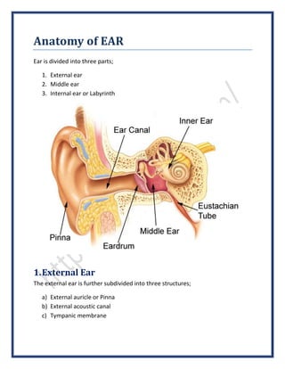
Ear is divided into three parts;
1. External ear
2. Middle ear
3. Internal ear or Labyrinth
1.External Ear
The external ear is further subdivided into three structures;
a) External auricle or Pinna
b) External acoustic canal
c) Tympanic membrane
a)External Auricle Or Pinna
The auricle or pinna forms the outer part of
the external auditory canal and consists of a
single piece of elastic cartilage covered with
skin. Various elevations and depressions are
present on the lateral aspect of the pinna. The
deepest depression of the external ear is
called Concha.
The entire pinna (except its lobule & outer
part of external acoustic canal) is made up of a
cartilaginous framework which is a single piece
of yellow elastic cartilage covered with skin. The skin is loosely attached to
perichondrium on the medial aspect (cranial) of pinna while it is in close
adherence to perichondrium on its lateral surface.
Clinical: - The furuncles formed in ear are the most painful than any other site as the
skin is closely adherent to perichondrium & little space is left. When a furuncle is
formed in ear, high pressure is built which causes sensation of intense pain.
Elevated outer margin of auricle or pinna is called Helix. Another smaller elevation
opposite to it is called Anti-Helix. A large conical structure in the base of ear in
front anti-helix is called Tragus. Behind the tragus & at the termination of anti-
helix, a smaller conical structure is called Anti-Tragus. Above the tragus, the
termination of elevated margin i-e helix is downward & backward which is called
Curs of Helix. Curs of helix covers a space called Cymba Conchae, an important
landmark used in mastoid surgery to reach mastoid antrum.
The area between tragus and the crus of helix is called Incisura Terminalis, which
lacks cartilage. An incision through the incisura terminalis does not cut through
the cartilage and is done usually for endaural approach in surgery of external
auditory canal or mastoid.
b)External Acoustic Canal/External Auditory Canal
The external auditory canal extends from the bottom of the concha to the
tympanic membrane. It is about 24 mm in length along its posterior aspect. It is
not a straight tube, its lateral part is directed upwards, backwards, forwards and
medially while its inner part is directed downwards, forwards and medially.
Therefore, for the examination of the tympanic membrane the pinna is pulled
upwards, backwards and laterally so as to form the External Auditory Canal a
straight canal.
The external auditory canal is divided into two parts;
a) Outer part/cartilaginous part
b) Inner part/bony part
Outer or lateral one third is cartilaginous which forms almost 8 mm of the
external auditory canal. The cartilage of the outer part is continuous with the
cartilage of the pinna. The skin covering the cartilaginous part is covered with
thick skin and contains ceruminous and pilosebaceous glands which secretes ear
wax. Hair are present only in the outer cartilaginous part, therefore furuncles are
seen only in the outer cartilaginous part.
Inner or medial two third is bony in nature and forms 16 mm of the total auditory
canal. Thin skin is present over the bony part. This part lacks the hair as well as
ceruminous glands.
Relations Of External Acoustic Canal
The relations of the external auditory canal are as followings,
a) Anteriorly: - Temporomandibular joint (TMJ)
b) Posteriorly: - Mastoid air cells and facial nerve
c) Superiorly: - Middle cranial fossa
d) Inferiorly: - Parotid gland
c)Tympanic Membrane
Tympanic membrane separates the external ear from the middle ear and it is set
in oblique position therefore, its posterosuperior part is more lateral than the
anteroinferior part. Tympanic membrane is almost 10 mm in length, 9 mm wide
and 0.1 mm in thickness.
Tympanic membrane can be divided into two parts;
a) Pars Tensa
b) Pars Flaccida
Pars Tensa forms the major portion of the tympanic membrane. Its peripheral
part is thickened to form a fibrocartilaginous ring called annulus tympanicus. The
central part of the pars tensa is tented inwards at the level of tip of the malleus
and is called Umbo.
Pars Flaccida forms the minor portion of the tympanic membrane and it is
situated above the lateral process of the malleus.
Tympanic membrane consists of three layers;
a) Outer epithelial layer, which is continuous with the skin of the external
auditory canal.
b) Middle fibrous layer, which consists of three types of fibers-the radial,
circular and parabolic fibers.
c) Inner mucosal layer, which is continuous with the mucosa of the middle ear
cavity.
Nerve Supply of the External Ear
i. Pinna
a) Greater auricular nerve (C2, C3) supplies the most of the medial surface of
the pinna and posterior part of lateral surface.
b) Lesser occipital nerve (C2) supplies the upper part of the medial surface.
c) Auriculotemporal nerve (V3) supplies the tragus and helix.
d) Auricular branch of the Vagus nerve (CN X) supplies the concha.
e) Facial nerve supplies the concha as well as retroauricular groove.
ii. External Auditory Canal
a) Auriculotemporal nerve (V3) supplies the anterior wall and roof of external
auditory canal.
b) Auricular branch of Vagus nerve (CN X) supplies the posterior wall and floor
of the external auditory canal.
c) The sensory fibers of the CN VII through auricular branch of vagus nerve
supply the posterior wall of the external auditory canal.
iii. Tympanic Membrane
a) Auriculotemporal nerve (V3) supplies the anterior aspect of the lateral
surface of the tympanic membrane
b) Auricular branch of the Vagus Nerve (CN X) supplies the posterior half of
the lateral surface of the tympanic membrane
c) Tympanic branch of the glossopharyngeal nerve (CN IX) supplies the medial
aspect of the tympanic membrane
2.Middle Ear
The middle ear cavity along with mastoid antrum, Eustachian tube, auditus and
mastoid air cells is called Middle Ear Cleft.
The middle ear is divided into;
 Mestotympanum
 Epitympanum
 Hypotympanum
The middle ear is like a six-sided box containing a roof, a floor, medial, lateral,
anterior and posterior walls.
a) The roof is formed by tegmen tympanic.
b) The floor is formed by the thin plate of bone.
c) The anterior wall is formed by the thin plate of bone which separates the
internal carotid artery from the middle ear cavity. The anterior wall
contains the opening of the Eustachian tube.
d) The posterior wall lies close to the mastoid air cells. Facial nerve lies in the
posterior wall of the middle ear.
e) The lateral wall is formed by the tympanic membrane.
f) The medial wall is formed by labyrinth. The medial wall presents a bulging
surface called promontory which is due to the basal coil of the cochlea.
i. Mastoid Antrum
Mastoid antrum is a large, air containing space which is connected to the middle
ear via auditus. Its roof is formed by tegmen antri, which is the continuation of
the tegmen tympani and it separates the mastoid antrum from the middle cranial
fossa. The lateral wall of the mastoid antrum is formed by thin plate of bone.
ii. Auditus
Auditus is an opening through which the mastoid antrum communicates with
middle ear cavity. The facial nerve passes just below the auditus.
iii. Mastoid Air Cells
The mastoid consists of bone with a “honeycomb” appearance of the mastoid.
The mastoid cells are well developed and pneumatized and their intervening
septa are thin. Depending upon the location the mastoid air cells are divided into
following types,
a) Zygomatic cells
b) Perisinus cells
c) Perilabyrinth cells
d) Tegmen cells
e) Peritubal cells
f) Retrofacial cells
g) Squamous cells
h) Tip cells
i) Marginal cells
Ossicles of Middle Ear
The Ossicles conduct sound waves from the tympanic membrane to the oval
window and then into the inner ear. There are three Ossicles present in the
middle ear cavity,
a) Malleus
b) Incus
c) Stapes
The malleus has a head, neck, handle, a lateral process and an anterior process.
The handle of malleus is embedded in the fibrous layer of the tympanic
membrane. The lateral process of the malleus makes the knob like projection of
the ear-drum.
The incus has a body and a short process as well as a long process. Both the body
and short process of the incus lie in the attic of middle ear cavity. The long
process is attached to the stapes.
The stapes has head, neck, anterior & posterior crura and footplate. The footplate
of the stapes is attached to the oval window of the labyrinth by annular ligament
Muscles of the Middle Ear
There are two intra-tympanic muscles (muscles of the middle ear);
a) Tensor tympani
b) Stapedius
Tensor tympani muscle is attached to the malleus and creates tension in the
tympanic membrane. Stapedius is attached to the neck of the stapes and prevent
the inner air from noise pollution by dampening very loud sounds.
Lining of Middle Ear
The mucous membrane of the middle ear is continuous with the mucous
membrane of the nasopharynx. The middle ear cavity is lined with ciliated
columnar epithelium in the anterior wall while the epithelium changes to cuboidal
epithelium in the posterior wall of middle ear cavity.
Blood Supply of the Middle Ear
Ear is supplied by six arteries, two are the major arteries while four are minor
arteries.
a) Anterior tympanic branch of the maxillary artery supplies the tympanic
membrane
b) Stylomastoid branch of auricular artery supplies the middle ear and
mastoid air cells
c) Tympanic branch of the internal carotid artery
d) Superior branch of middle meningeal artery
e) Branch from the artery of the pterygoid canal
f) Petrosal branch of middle meningeal artery
3.Internal Ear
The internal ear also called labyrinth is an important organ for hearing and
maintaining balance of the body. The internal ear consists of bony labyrinth as
well as Membranous Labyrinth. The membranous labyrinth is filled with fluid
called Endolymph.
Bony Labyrinth
The bony labyrinth contains three parts;
a) Vestibule
b) Semicircular canals
c) Cochlea
The vestibule is the main chamber of the labyrinth. The oval window lies in the
lateral wall of the vestibule. In the posterosuperior part of the vestibule are the
openings of the semicircular canals which are five in number.
The semicircular canals are three in number, the lateral, superior and posterior.
Each canal has an ampulla at its end which opens into vestibule. There are five
openings of the three semicircular canals in the posterosuperior part of the
vestibule.
The bony cochlea makes 2.5 to 2.75 turns around the central pyramid of the bone
called modiolus. The base of the modiolus is directed towards the internal
acoustic meatus. The bony cochlea contains three compartments;
i. Scala vestibuli
ii. Scala tympani
iii. Scala media
The scala vestibuli and scala tympani are filled with perilymph and communicates
with each other with an opening called Helicotrema.
Membranous Labyrinth
The membranous labyrinth consists of cochlear ducts, utericle and saccule, three
semicircular ducts and endolymphatic duct.
The cochlear duct also called Scala Media or Membranous Cochlea is a coiled
tube. On cross-section it appears triangular in appearance. Its three walls are
formed by
a) The Basilar Membrane which provides support to the Organ of Corti.
b) The Reissner’s Membrane which separates the scala media from scala
vestibuli
c) The Stria Vascularis which contains the vascular epithelium containing the
secretory epithelium for secretion of the endolymph
The utericle is present in the posterior part of the bony vestibule and receives the
five openings of the three semicircular ducts. Utericle is also connected to the
saccule through utericulosaccular duct. The sensory epithelium of the utericle is
called macula concerned with acceleration and deceleration.
The saccule is also present in the bony vestibule. Its sensory epithelium is also
called macula. It is also concerned with the acceleration and deceleration.
Endolymphatic duct is formed by the union of two ducts, one from each utericle
and saccule.
Blood Supply of Labyrinth
The entire labyrinth receives blood supply through labyrinthine artery, which is a
branch of anterior-inferior cerebellar artery. Sometimes labyrinthine artery also
originates from the basilar artery.
The venous drainage is through three veins;
a) Internal auditory vein
b) Vein of cochlear duct
c) Vein of vestibular duct
Lymphatic Drainage Of Ear
The lymph from concha, tragus, fossa triangularis and external auditory canal is
drained into preauricular and parotid lymph nodes. The lymph from lymph and
antitragus is drained into inferior auricular lymph nodes. The lymph from helix
and antihelix is drained into posterior auricular and deep jugular lymph nodes
Lymph from the middle ear is drained into the parotid and retropharyngeal lymph
nodes. The lymph from Eustachian tube is drained into retropharyngeal lymph
nodes.
There are no more lymphatics and lymph nodes in the inner ear.
Feedback us @ http://mukhdoom.com for more data & info.
Like us @ https://www.facebook.com/StairsOfSuccess

More Related Content

What's hot

The pneumatic system of the temporal bone
The pneumatic system of the temporal boneThe pneumatic system of the temporal bone
The pneumatic system of the temporal boneSurbhi narayan
 
Physiology of hearing
Physiology of hearingPhysiology of hearing
Physiology of hearingajusarma
 
Anatomy of ear
Anatomy of ear Anatomy of ear
Anatomy of ear amit jha
 
Anatomy & Physiology Of Vestibular System
Anatomy & Physiology Of Vestibular SystemAnatomy & Physiology Of Vestibular System
Anatomy & Physiology Of Vestibular SystemPrasanna Datta
 
Congenital anomalies of inner ear
Congenital anomalies of inner earCongenital anomalies of inner ear
Congenital anomalies of inner earChanmiki Sayoo
 
Anatomy of ear
Anatomy of earAnatomy of ear
Anatomy of ear11032013
 
Spaces of middle ear and their surgical importance
Spaces of middle ear  and their surgical importanceSpaces of middle ear  and their surgical importance
Spaces of middle ear and their surgical importanceDr Soumya Singh
 
Mucosal folds and ventilation of middle ear
Mucosal folds and ventilation of middle ear  Mucosal folds and ventilation of middle ear
Mucosal folds and ventilation of middle ear AlkaKapil
 
Nasal septum and its diseases
Nasal septum and its diseasesNasal septum and its diseases
Nasal septum and its diseasesVinay Bhat
 
Middle ear anatomy
Middle ear anatomyMiddle ear anatomy
Middle ear anatomyAmro1988
 
Nasal septum & its diseases
Nasal septum & its diseasesNasal septum & its diseases
Nasal septum & its diseasesNavas Shareef
 
Nasal Cavity and Sphenoid Sinus Anatomy
Nasal Cavity and Sphenoid Sinus AnatomyNasal Cavity and Sphenoid Sinus Anatomy
Nasal Cavity and Sphenoid Sinus AnatomyFarrukh Javeed
 
Anatomy of inner ear
Anatomy of inner earAnatomy of inner ear
Anatomy of inner earRazal M
 
11 surgery for otosclerosis.ppt copy
11 surgery for otosclerosis.ppt   copy11 surgery for otosclerosis.ppt   copy
11 surgery for otosclerosis.ppt copysocial service
 
Inner ear anatomy & physiology of hearing
Inner ear anatomy & physiology of hearingInner ear anatomy & physiology of hearing
Inner ear anatomy & physiology of hearingDR PRASANN RASANIA
 
Endoscopic anatomy of ethmoid bone
Endoscopic anatomy of ethmoid boneEndoscopic anatomy of ethmoid bone
Endoscopic anatomy of ethmoid boneSaaketh Boddupalli
 

What's hot (20)

The pneumatic system of the temporal bone
The pneumatic system of the temporal boneThe pneumatic system of the temporal bone
The pneumatic system of the temporal bone
 
Physiology of hearing
Physiology of hearingPhysiology of hearing
Physiology of hearing
 
Anatomy of ear
Anatomy of ear Anatomy of ear
Anatomy of ear
 
Anatomy & Physiology Of Vestibular System
Anatomy & Physiology Of Vestibular SystemAnatomy & Physiology Of Vestibular System
Anatomy & Physiology Of Vestibular System
 
Congenital anomalies of inner ear
Congenital anomalies of inner earCongenital anomalies of inner ear
Congenital anomalies of inner ear
 
Anatomy of ear
Anatomy of earAnatomy of ear
Anatomy of ear
 
Inner ear
Inner earInner ear
Inner ear
 
Development of ear ppt
Development of ear pptDevelopment of ear ppt
Development of ear ppt
 
Spaces of middle ear and their surgical importance
Spaces of middle ear  and their surgical importanceSpaces of middle ear  and their surgical importance
Spaces of middle ear and their surgical importance
 
Mucosal folds and ventilation of middle ear
Mucosal folds and ventilation of middle ear  Mucosal folds and ventilation of middle ear
Mucosal folds and ventilation of middle ear
 
Nasal septum and its diseases
Nasal septum and its diseasesNasal septum and its diseases
Nasal septum and its diseases
 
Tympanic membrane
Tympanic membraneTympanic membrane
Tympanic membrane
 
Middle ear anatomy
Middle ear anatomyMiddle ear anatomy
Middle ear anatomy
 
Nasal septum & its diseases
Nasal septum & its diseasesNasal septum & its diseases
Nasal septum & its diseases
 
Phonosurgery
PhonosurgeryPhonosurgery
Phonosurgery
 
Nasal Cavity and Sphenoid Sinus Anatomy
Nasal Cavity and Sphenoid Sinus AnatomyNasal Cavity and Sphenoid Sinus Anatomy
Nasal Cavity and Sphenoid Sinus Anatomy
 
Anatomy of inner ear
Anatomy of inner earAnatomy of inner ear
Anatomy of inner ear
 
11 surgery for otosclerosis.ppt copy
11 surgery for otosclerosis.ppt   copy11 surgery for otosclerosis.ppt   copy
11 surgery for otosclerosis.ppt copy
 
Inner ear anatomy & physiology of hearing
Inner ear anatomy & physiology of hearingInner ear anatomy & physiology of hearing
Inner ear anatomy & physiology of hearing
 
Endoscopic anatomy of ethmoid bone
Endoscopic anatomy of ethmoid boneEndoscopic anatomy of ethmoid bone
Endoscopic anatomy of ethmoid bone
 

Viewers also liked

Anatomy and physiology of ear by maghan das
Anatomy and physiology of ear by maghan dasAnatomy and physiology of ear by maghan das
Anatomy and physiology of ear by maghan dasMaghan Das
 
Middle ear by arifa
Middle      ear by arifaMiddle      ear by arifa
Middle ear by arifaarifahussain
 
Inner ear anatomy
Inner ear anatomy Inner ear anatomy
Inner ear anatomy yatish88
 
The mechanism of hearing
The mechanism of hearingThe mechanism of hearing
The mechanism of hearingAjay Manickam
 
The Human Ear and Sound
The Human Ear and SoundThe Human Ear and Sound
The Human Ear and Soundbarbburckart
 
Anatomy of ear
Anatomy of earAnatomy of ear
Anatomy of earENTDOST
 
Ct scan temporal bone Dr Zeeshan Ahmad
Ct scan temporal bone Dr Zeeshan AhmadCt scan temporal bone Dr Zeeshan Ahmad
Ct scan temporal bone Dr Zeeshan AhmadDr Zeeshan Ahmad
 
Ct temporal bone
Ct temporal bone Ct temporal bone
Ct temporal bone Yasha Gupta
 
Anatomy of the ear
Anatomy of the earAnatomy of the ear
Anatomy of the earNancyDecker
 
IMAGING OF TEMPORAL BONE
IMAGING OF TEMPORAL BONEIMAGING OF TEMPORAL BONE
IMAGING OF TEMPORAL BONESameer Peer
 
Gross anatomy of the ‘ear’
Gross anatomy of the ‘ear’Gross anatomy of the ‘ear’
Gross anatomy of the ‘ear’Lucidante1
 
English Project 7 peaches
English Project 7 peachesEnglish Project 7 peaches
English Project 7 peachesMansour Odeibat
 
How to Excel In interviews - master 4
How to Excel In interviews - master 4How to Excel In interviews - master 4
How to Excel In interviews - master 4Joseph Fallarino
 
Firstpresentation
FirstpresentationFirstpresentation
Firstpresentationbgreen97
 

Viewers also liked (20)

anatomy of ear
anatomy of earanatomy of ear
anatomy of ear
 
Anatomy and physiology of ear by maghan das
Anatomy and physiology of ear by maghan dasAnatomy and physiology of ear by maghan das
Anatomy and physiology of ear by maghan das
 
Anatomy of ear
Anatomy of earAnatomy of ear
Anatomy of ear
 
Middle ear by arifa
Middle      ear by arifaMiddle      ear by arifa
Middle ear by arifa
 
Inner ear anatomy
Inner ear anatomy Inner ear anatomy
Inner ear anatomy
 
The mechanism of hearing
The mechanism of hearingThe mechanism of hearing
The mechanism of hearing
 
The Human Ear and Sound
The Human Ear and SoundThe Human Ear and Sound
The Human Ear and Sound
 
Anatomy of ear
Anatomy of earAnatomy of ear
Anatomy of ear
 
Ct scan temporal bone Dr Zeeshan Ahmad
Ct scan temporal bone Dr Zeeshan AhmadCt scan temporal bone Dr Zeeshan Ahmad
Ct scan temporal bone Dr Zeeshan Ahmad
 
Ct temporal bone
Ct temporal bone Ct temporal bone
Ct temporal bone
 
The human ear
The human earThe human ear
The human ear
 
Anatomy of the ear
Anatomy of the earAnatomy of the ear
Anatomy of the ear
 
Anatomy and physiology of the ear
Anatomy and physiology of the earAnatomy and physiology of the ear
Anatomy and physiology of the ear
 
IMAGING OF TEMPORAL BONE
IMAGING OF TEMPORAL BONEIMAGING OF TEMPORAL BONE
IMAGING OF TEMPORAL BONE
 
Gross anatomy of the ‘ear’
Gross anatomy of the ‘ear’Gross anatomy of the ‘ear’
Gross anatomy of the ‘ear’
 
pedsovet 14-15
pedsovet 14-15pedsovet 14-15
pedsovet 14-15
 
Donne e motori
Donne e motoriDonne e motori
Donne e motori
 
English Project 7 peaches
English Project 7 peachesEnglish Project 7 peaches
English Project 7 peaches
 
How to Excel In interviews - master 4
How to Excel In interviews - master 4How to Excel In interviews - master 4
How to Excel In interviews - master 4
 
Firstpresentation
FirstpresentationFirstpresentation
Firstpresentation
 

Similar to Anatomy of Ear | SurgicoMed.com

Ear anatomy (1)-1.pptx
Ear anatomy (1)-1.pptxEar anatomy (1)-1.pptx
Ear anatomy (1)-1.pptxsafatullah50
 
Anatomy of the ear
Anatomy of the earAnatomy of the ear
Anatomy of the earRafid Rashid
 
Middle ear presentation
Middle ear presentationMiddle ear presentation
Middle ear presentationSomnath Saha
 
EXTERNAL ACOUSTIC MEATUS, MIDDLE EAR
EXTERNAL ACOUSTIC MEATUS, MIDDLE EAREXTERNAL ACOUSTIC MEATUS, MIDDLE EAR
EXTERNAL ACOUSTIC MEATUS, MIDDLE EARDHABHAI
 
middle ear anatomy
middle ear anatomymiddle ear anatomy
middle ear anatomyShafiq38
 
Anatomy of the ear (ecture 2) by dr, noura 2018
Anatomy of the ear (ecture 2) by dr, noura 2018Anatomy of the ear (ecture 2) by dr, noura 2018
Anatomy of the ear (ecture 2) by dr, noura 2018Dr. Noura El Tahawy
 
anatomy and embrology of middle and inner ear
anatomy and embrology of middle and inner earanatomy and embrology of middle and inner ear
anatomy and embrology of middle and inner earaayushi402162
 
earanatomy-190219150351.pdf
earanatomy-190219150351.pdfearanatomy-190219150351.pdf
earanatomy-190219150351.pdfAliyahJohanis1
 
ANATOMY OF EXTERNAL AUDITORY CANAL
ANATOMY OF EXTERNAL AUDITORY CANALANATOMY OF EXTERNAL AUDITORY CANAL
ANATOMY OF EXTERNAL AUDITORY CANALKanu Saha
 
External &;middle ear anatomy
External &;middle ear anatomyExternal &;middle ear anatomy
External &;middle ear anatomyDr Lovely Jain
 
Anatomy of ear for UG.pptx
Anatomy of ear for UG.pptxAnatomy of ear for UG.pptx
Anatomy of ear for UG.pptxAlkaKapil
 
anatomy of ear nn.pptx
anatomy of ear nn.pptxanatomy of ear nn.pptx
anatomy of ear nn.pptxAzeemHamdani
 
Middle ear anatomy
Middle ear anatomyMiddle ear anatomy
Middle ear anatomykbn2914
 

Similar to Anatomy of Ear | SurgicoMed.com (20)

Ear anatomy (1)-1.pptx
Ear anatomy (1)-1.pptxEar anatomy (1)-1.pptx
Ear anatomy (1)-1.pptx
 
Anatomy of the ear
Anatomy of the earAnatomy of the ear
Anatomy of the ear
 
Anatomy of Ear
Anatomy of EarAnatomy of Ear
Anatomy of Ear
 
Middle ear presentation
Middle ear presentationMiddle ear presentation
Middle ear presentation
 
Anatomy of human ear
Anatomy of human earAnatomy of human ear
Anatomy of human ear
 
EXTERNAL ACOUSTIC MEATUS, MIDDLE EAR
EXTERNAL ACOUSTIC MEATUS, MIDDLE EAREXTERNAL ACOUSTIC MEATUS, MIDDLE EAR
EXTERNAL ACOUSTIC MEATUS, MIDDLE EAR
 
ear anatomy.pptx
ear anatomy.pptxear anatomy.pptx
ear anatomy.pptx
 
middle ear anatomy
middle ear anatomymiddle ear anatomy
middle ear anatomy
 
Anatomy of the ear (ecture 2) by dr, noura 2018
Anatomy of the ear (ecture 2) by dr, noura 2018Anatomy of the ear (ecture 2) by dr, noura 2018
Anatomy of the ear (ecture 2) by dr, noura 2018
 
anatomy and embrology of middle and inner ear
anatomy and embrology of middle and inner earanatomy and embrology of middle and inner ear
anatomy and embrology of middle and inner ear
 
Anatomy of temporal bone final
Anatomy of temporal bone finalAnatomy of temporal bone final
Anatomy of temporal bone final
 
earanatomy-190219150351.pdf
earanatomy-190219150351.pdfearanatomy-190219150351.pdf
earanatomy-190219150351.pdf
 
Ear Anatomy
Ear AnatomyEar Anatomy
Ear Anatomy
 
Anatomy of ear and mastoid
Anatomy of ear and mastoidAnatomy of ear and mastoid
Anatomy of ear and mastoid
 
ANATOMY OF EXTERNAL AUDITORY CANAL
ANATOMY OF EXTERNAL AUDITORY CANALANATOMY OF EXTERNAL AUDITORY CANAL
ANATOMY OF EXTERNAL AUDITORY CANAL
 
External &;middle ear anatomy
External &;middle ear anatomyExternal &;middle ear anatomy
External &;middle ear anatomy
 
Anatomy of ear for UG.pptx
Anatomy of ear for UG.pptxAnatomy of ear for UG.pptx
Anatomy of ear for UG.pptx
 
anatomy of ear nn.pptx
anatomy of ear nn.pptxanatomy of ear nn.pptx
anatomy of ear nn.pptx
 
Anatomy Of Ear.pdf
Anatomy Of Ear.pdfAnatomy Of Ear.pdf
Anatomy Of Ear.pdf
 
Middle ear anatomy
Middle ear anatomyMiddle ear anatomy
Middle ear anatomy
 

More from Mukhdoom BaharAli

25 Important Cases In Ear Nose Throat | SurgicoMed.com
25 Important Cases In Ear Nose Throat | SurgicoMed.com25 Important Cases In Ear Nose Throat | SurgicoMed.com
25 Important Cases In Ear Nose Throat | SurgicoMed.comMukhdoom BaharAli
 
Dns (Deviated Nasal Septum) | SurgicoMed.com
Dns (Deviated Nasal Septum) | SurgicoMed.comDns (Deviated Nasal Septum) | SurgicoMed.com
Dns (Deviated Nasal Septum) | SurgicoMed.comMukhdoom BaharAli
 
Anatomy of the Nose | SurgicoMed.com
Anatomy of the Nose | SurgicoMed.comAnatomy of the Nose | SurgicoMed.com
Anatomy of the Nose | SurgicoMed.comMukhdoom BaharAli
 
Housing & its Basic Requirements | SurgicoMed.com
Housing & its Basic Requirements | SurgicoMed.comHousing & its Basic Requirements | SurgicoMed.com
Housing & its Basic Requirements | SurgicoMed.comMukhdoom BaharAli
 
Concept of Disease Control | SurgicoMed.com
Concept of Disease Control | SurgicoMed.comConcept of Disease Control | SurgicoMed.com
Concept of Disease Control | SurgicoMed.comMukhdoom BaharAli
 
Biostatistics (Basic Definitions & Concepts) | SurgicoMed.com
Biostatistics (Basic Definitions & Concepts) | SurgicoMed.comBiostatistics (Basic Definitions & Concepts) | SurgicoMed.com
Biostatistics (Basic Definitions & Concepts) | SurgicoMed.comMukhdoom BaharAli
 
An Introduction To Community Medicine (Basic Definitions) | SurgicoMed.com
An Introduction To Community Medicine (Basic Definitions) | SurgicoMed.comAn Introduction To Community Medicine (Basic Definitions) | SurgicoMed.com
An Introduction To Community Medicine (Basic Definitions) | SurgicoMed.comMukhdoom BaharAli
 
Typhoid Fever | SurgicoMed.com
Typhoid Fever | SurgicoMed.comTyphoid Fever | SurgicoMed.com
Typhoid Fever | SurgicoMed.comMukhdoom BaharAli
 
Tuberculosis | SurgicoMed.com
Tuberculosis | SurgicoMed.comTuberculosis | SurgicoMed.com
Tuberculosis | SurgicoMed.comMukhdoom BaharAli
 
Hydronephrosis | SurgicoMed.com
Hydronephrosis | SurgicoMed.comHydronephrosis | SurgicoMed.com
Hydronephrosis | SurgicoMed.comMukhdoom BaharAli
 

More from Mukhdoom BaharAli (11)

Ent ospe | SurgicoMed.com
Ent ospe | SurgicoMed.comEnt ospe | SurgicoMed.com
Ent ospe | SurgicoMed.com
 
25 Important Cases In Ear Nose Throat | SurgicoMed.com
25 Important Cases In Ear Nose Throat | SurgicoMed.com25 Important Cases In Ear Nose Throat | SurgicoMed.com
25 Important Cases In Ear Nose Throat | SurgicoMed.com
 
Dns (Deviated Nasal Septum) | SurgicoMed.com
Dns (Deviated Nasal Septum) | SurgicoMed.comDns (Deviated Nasal Septum) | SurgicoMed.com
Dns (Deviated Nasal Septum) | SurgicoMed.com
 
Anatomy of the Nose | SurgicoMed.com
Anatomy of the Nose | SurgicoMed.comAnatomy of the Nose | SurgicoMed.com
Anatomy of the Nose | SurgicoMed.com
 
Housing & its Basic Requirements | SurgicoMed.com
Housing & its Basic Requirements | SurgicoMed.comHousing & its Basic Requirements | SurgicoMed.com
Housing & its Basic Requirements | SurgicoMed.com
 
Concept of Disease Control | SurgicoMed.com
Concept of Disease Control | SurgicoMed.comConcept of Disease Control | SurgicoMed.com
Concept of Disease Control | SurgicoMed.com
 
Biostatistics (Basic Definitions & Concepts) | SurgicoMed.com
Biostatistics (Basic Definitions & Concepts) | SurgicoMed.comBiostatistics (Basic Definitions & Concepts) | SurgicoMed.com
Biostatistics (Basic Definitions & Concepts) | SurgicoMed.com
 
An Introduction To Community Medicine (Basic Definitions) | SurgicoMed.com
An Introduction To Community Medicine (Basic Definitions) | SurgicoMed.comAn Introduction To Community Medicine (Basic Definitions) | SurgicoMed.com
An Introduction To Community Medicine (Basic Definitions) | SurgicoMed.com
 
Typhoid Fever | SurgicoMed.com
Typhoid Fever | SurgicoMed.comTyphoid Fever | SurgicoMed.com
Typhoid Fever | SurgicoMed.com
 
Tuberculosis | SurgicoMed.com
Tuberculosis | SurgicoMed.comTuberculosis | SurgicoMed.com
Tuberculosis | SurgicoMed.com
 
Hydronephrosis | SurgicoMed.com
Hydronephrosis | SurgicoMed.comHydronephrosis | SurgicoMed.com
Hydronephrosis | SurgicoMed.com
 

Recently uploaded

How to Make a Pirate ship Primary Education.pptx
How to Make a Pirate ship Primary Education.pptxHow to Make a Pirate ship Primary Education.pptx
How to Make a Pirate ship Primary Education.pptxmanuelaromero2013
 
Enzyme, Pharmaceutical Aids, Miscellaneous Last Part of Chapter no 5th.pdf
Enzyme, Pharmaceutical Aids, Miscellaneous Last Part of Chapter no 5th.pdfEnzyme, Pharmaceutical Aids, Miscellaneous Last Part of Chapter no 5th.pdf
Enzyme, Pharmaceutical Aids, Miscellaneous Last Part of Chapter no 5th.pdfSumit Tiwari
 
call girls in Kamla Market (DELHI) 🔝 >༒9953330565🔝 genuine Escort Service 🔝✔️✔️
call girls in Kamla Market (DELHI) 🔝 >༒9953330565🔝 genuine Escort Service 🔝✔️✔️call girls in Kamla Market (DELHI) 🔝 >༒9953330565🔝 genuine Escort Service 🔝✔️✔️
call girls in Kamla Market (DELHI) 🔝 >༒9953330565🔝 genuine Escort Service 🔝✔️✔️9953056974 Low Rate Call Girls In Saket, Delhi NCR
 
Introduction to AI in Higher Education_draft.pptx
Introduction to AI in Higher Education_draft.pptxIntroduction to AI in Higher Education_draft.pptx
Introduction to AI in Higher Education_draft.pptxpboyjonauth
 
Alper Gobel In Media Res Media Component
Alper Gobel In Media Res Media ComponentAlper Gobel In Media Res Media Component
Alper Gobel In Media Res Media ComponentInMediaRes1
 
How to Configure Email Server in Odoo 17
How to Configure Email Server in Odoo 17How to Configure Email Server in Odoo 17
How to Configure Email Server in Odoo 17Celine George
 
ECONOMIC CONTEXT - LONG FORM TV DRAMA - PPT
ECONOMIC CONTEXT - LONG FORM TV DRAMA - PPTECONOMIC CONTEXT - LONG FORM TV DRAMA - PPT
ECONOMIC CONTEXT - LONG FORM TV DRAMA - PPTiammrhaywood
 
Crayon Activity Handout For the Crayon A
Crayon Activity Handout For the Crayon ACrayon Activity Handout For the Crayon A
Crayon Activity Handout For the Crayon AUnboundStockton
 
Sanyam Choudhary Chemistry practical.pdf
Sanyam Choudhary Chemistry practical.pdfSanyam Choudhary Chemistry practical.pdf
Sanyam Choudhary Chemistry practical.pdfsanyamsingh5019
 
Software Engineering Methodologies (overview)
Software Engineering Methodologies (overview)Software Engineering Methodologies (overview)
Software Engineering Methodologies (overview)eniolaolutunde
 
SOCIAL AND HISTORICAL CONTEXT - LFTVD.pptx
SOCIAL AND HISTORICAL CONTEXT - LFTVD.pptxSOCIAL AND HISTORICAL CONTEXT - LFTVD.pptx
SOCIAL AND HISTORICAL CONTEXT - LFTVD.pptxiammrhaywood
 
Hybridoma Technology ( Production , Purification , and Application )
Hybridoma Technology  ( Production , Purification , and Application  ) Hybridoma Technology  ( Production , Purification , and Application  )
Hybridoma Technology ( Production , Purification , and Application ) Sakshi Ghasle
 
Science lesson Moon for 4th quarter lesson
Science lesson Moon for 4th quarter lessonScience lesson Moon for 4th quarter lesson
Science lesson Moon for 4th quarter lessonJericReyAuditor
 
Organic Name Reactions for the students and aspirants of Chemistry12th.pptx
Organic Name Reactions  for the students and aspirants of Chemistry12th.pptxOrganic Name Reactions  for the students and aspirants of Chemistry12th.pptx
Organic Name Reactions for the students and aspirants of Chemistry12th.pptxVS Mahajan Coaching Centre
 
Final demo Grade 9 for demo Plan dessert.pptx
Final demo Grade 9 for demo Plan dessert.pptxFinal demo Grade 9 for demo Plan dessert.pptx
Final demo Grade 9 for demo Plan dessert.pptxAvyJaneVismanos
 
BASLIQ CURRENT LOOKBOOK LOOKBOOK(1) (1).pdf
BASLIQ CURRENT LOOKBOOK  LOOKBOOK(1) (1).pdfBASLIQ CURRENT LOOKBOOK  LOOKBOOK(1) (1).pdf
BASLIQ CURRENT LOOKBOOK LOOKBOOK(1) (1).pdfSoniaTolstoy
 
POINT- BIOCHEMISTRY SEM 2 ENZYMES UNIT 5.pptx
POINT- BIOCHEMISTRY SEM 2 ENZYMES UNIT 5.pptxPOINT- BIOCHEMISTRY SEM 2 ENZYMES UNIT 5.pptx
POINT- BIOCHEMISTRY SEM 2 ENZYMES UNIT 5.pptxSayali Powar
 

Recently uploaded (20)

How to Make a Pirate ship Primary Education.pptx
How to Make a Pirate ship Primary Education.pptxHow to Make a Pirate ship Primary Education.pptx
How to Make a Pirate ship Primary Education.pptx
 
Enzyme, Pharmaceutical Aids, Miscellaneous Last Part of Chapter no 5th.pdf
Enzyme, Pharmaceutical Aids, Miscellaneous Last Part of Chapter no 5th.pdfEnzyme, Pharmaceutical Aids, Miscellaneous Last Part of Chapter no 5th.pdf
Enzyme, Pharmaceutical Aids, Miscellaneous Last Part of Chapter no 5th.pdf
 
9953330565 Low Rate Call Girls In Rohini Delhi NCR
9953330565 Low Rate Call Girls In Rohini  Delhi NCR9953330565 Low Rate Call Girls In Rohini  Delhi NCR
9953330565 Low Rate Call Girls In Rohini Delhi NCR
 
call girls in Kamla Market (DELHI) 🔝 >༒9953330565🔝 genuine Escort Service 🔝✔️✔️
call girls in Kamla Market (DELHI) 🔝 >༒9953330565🔝 genuine Escort Service 🔝✔️✔️call girls in Kamla Market (DELHI) 🔝 >༒9953330565🔝 genuine Escort Service 🔝✔️✔️
call girls in Kamla Market (DELHI) 🔝 >༒9953330565🔝 genuine Escort Service 🔝✔️✔️
 
Introduction to AI in Higher Education_draft.pptx
Introduction to AI in Higher Education_draft.pptxIntroduction to AI in Higher Education_draft.pptx
Introduction to AI in Higher Education_draft.pptx
 
Alper Gobel In Media Res Media Component
Alper Gobel In Media Res Media ComponentAlper Gobel In Media Res Media Component
Alper Gobel In Media Res Media Component
 
Model Call Girl in Tilak Nagar Delhi reach out to us at 🔝9953056974🔝
Model Call Girl in Tilak Nagar Delhi reach out to us at 🔝9953056974🔝Model Call Girl in Tilak Nagar Delhi reach out to us at 🔝9953056974🔝
Model Call Girl in Tilak Nagar Delhi reach out to us at 🔝9953056974🔝
 
How to Configure Email Server in Odoo 17
How to Configure Email Server in Odoo 17How to Configure Email Server in Odoo 17
How to Configure Email Server in Odoo 17
 
ECONOMIC CONTEXT - LONG FORM TV DRAMA - PPT
ECONOMIC CONTEXT - LONG FORM TV DRAMA - PPTECONOMIC CONTEXT - LONG FORM TV DRAMA - PPT
ECONOMIC CONTEXT - LONG FORM TV DRAMA - PPT
 
Crayon Activity Handout For the Crayon A
Crayon Activity Handout For the Crayon ACrayon Activity Handout For the Crayon A
Crayon Activity Handout For the Crayon A
 
Sanyam Choudhary Chemistry practical.pdf
Sanyam Choudhary Chemistry practical.pdfSanyam Choudhary Chemistry practical.pdf
Sanyam Choudhary Chemistry practical.pdf
 
Software Engineering Methodologies (overview)
Software Engineering Methodologies (overview)Software Engineering Methodologies (overview)
Software Engineering Methodologies (overview)
 
SOCIAL AND HISTORICAL CONTEXT - LFTVD.pptx
SOCIAL AND HISTORICAL CONTEXT - LFTVD.pptxSOCIAL AND HISTORICAL CONTEXT - LFTVD.pptx
SOCIAL AND HISTORICAL CONTEXT - LFTVD.pptx
 
Hybridoma Technology ( Production , Purification , and Application )
Hybridoma Technology  ( Production , Purification , and Application  ) Hybridoma Technology  ( Production , Purification , and Application  )
Hybridoma Technology ( Production , Purification , and Application )
 
Science lesson Moon for 4th quarter lesson
Science lesson Moon for 4th quarter lessonScience lesson Moon for 4th quarter lesson
Science lesson Moon for 4th quarter lesson
 
Organic Name Reactions for the students and aspirants of Chemistry12th.pptx
Organic Name Reactions  for the students and aspirants of Chemistry12th.pptxOrganic Name Reactions  for the students and aspirants of Chemistry12th.pptx
Organic Name Reactions for the students and aspirants of Chemistry12th.pptx
 
Final demo Grade 9 for demo Plan dessert.pptx
Final demo Grade 9 for demo Plan dessert.pptxFinal demo Grade 9 for demo Plan dessert.pptx
Final demo Grade 9 for demo Plan dessert.pptx
 
BASLIQ CURRENT LOOKBOOK LOOKBOOK(1) (1).pdf
BASLIQ CURRENT LOOKBOOK  LOOKBOOK(1) (1).pdfBASLIQ CURRENT LOOKBOOK  LOOKBOOK(1) (1).pdf
BASLIQ CURRENT LOOKBOOK LOOKBOOK(1) (1).pdf
 
TataKelola dan KamSiber Kecerdasan Buatan v022.pdf
TataKelola dan KamSiber Kecerdasan Buatan v022.pdfTataKelola dan KamSiber Kecerdasan Buatan v022.pdf
TataKelola dan KamSiber Kecerdasan Buatan v022.pdf
 
POINT- BIOCHEMISTRY SEM 2 ENZYMES UNIT 5.pptx
POINT- BIOCHEMISTRY SEM 2 ENZYMES UNIT 5.pptxPOINT- BIOCHEMISTRY SEM 2 ENZYMES UNIT 5.pptx
POINT- BIOCHEMISTRY SEM 2 ENZYMES UNIT 5.pptx
 

Anatomy of Ear | SurgicoMed.com

  • 1. Anatomy of EAR Ear is divided into three parts; 1. External ear 2. Middle ear 3. Internal ear or Labyrinth 1.External Ear The external ear is further subdivided into three structures; a) External auricle or Pinna b) External acoustic canal c) Tympanic membrane
  • 2. a)External Auricle Or Pinna The auricle or pinna forms the outer part of the external auditory canal and consists of a single piece of elastic cartilage covered with skin. Various elevations and depressions are present on the lateral aspect of the pinna. The deepest depression of the external ear is called Concha. The entire pinna (except its lobule & outer part of external acoustic canal) is made up of a cartilaginous framework which is a single piece of yellow elastic cartilage covered with skin. The skin is loosely attached to perichondrium on the medial aspect (cranial) of pinna while it is in close adherence to perichondrium on its lateral surface. Clinical: - The furuncles formed in ear are the most painful than any other site as the skin is closely adherent to perichondrium & little space is left. When a furuncle is formed in ear, high pressure is built which causes sensation of intense pain. Elevated outer margin of auricle or pinna is called Helix. Another smaller elevation opposite to it is called Anti-Helix. A large conical structure in the base of ear in front anti-helix is called Tragus. Behind the tragus & at the termination of anti- helix, a smaller conical structure is called Anti-Tragus. Above the tragus, the termination of elevated margin i-e helix is downward & backward which is called Curs of Helix. Curs of helix covers a space called Cymba Conchae, an important landmark used in mastoid surgery to reach mastoid antrum. The area between tragus and the crus of helix is called Incisura Terminalis, which lacks cartilage. An incision through the incisura terminalis does not cut through the cartilage and is done usually for endaural approach in surgery of external auditory canal or mastoid. b)External Acoustic Canal/External Auditory Canal The external auditory canal extends from the bottom of the concha to the tympanic membrane. It is about 24 mm in length along its posterior aspect. It is not a straight tube, its lateral part is directed upwards, backwards, forwards and
  • 3. medially while its inner part is directed downwards, forwards and medially. Therefore, for the examination of the tympanic membrane the pinna is pulled upwards, backwards and laterally so as to form the External Auditory Canal a straight canal. The external auditory canal is divided into two parts; a) Outer part/cartilaginous part b) Inner part/bony part Outer or lateral one third is cartilaginous which forms almost 8 mm of the external auditory canal. The cartilage of the outer part is continuous with the cartilage of the pinna. The skin covering the cartilaginous part is covered with thick skin and contains ceruminous and pilosebaceous glands which secretes ear wax. Hair are present only in the outer cartilaginous part, therefore furuncles are seen only in the outer cartilaginous part. Inner or medial two third is bony in nature and forms 16 mm of the total auditory canal. Thin skin is present over the bony part. This part lacks the hair as well as ceruminous glands.
  • 4. Relations Of External Acoustic Canal The relations of the external auditory canal are as followings, a) Anteriorly: - Temporomandibular joint (TMJ) b) Posteriorly: - Mastoid air cells and facial nerve c) Superiorly: - Middle cranial fossa d) Inferiorly: - Parotid gland c)Tympanic Membrane Tympanic membrane separates the external ear from the middle ear and it is set in oblique position therefore, its posterosuperior part is more lateral than the anteroinferior part. Tympanic membrane is almost 10 mm in length, 9 mm wide and 0.1 mm in thickness. Tympanic membrane can be divided into two parts; a) Pars Tensa b) Pars Flaccida Pars Tensa forms the major portion of the tympanic membrane. Its peripheral part is thickened to form a fibrocartilaginous ring called annulus tympanicus. The central part of the pars tensa is tented inwards at the level of tip of the malleus and is called Umbo.
  • 5. Pars Flaccida forms the minor portion of the tympanic membrane and it is situated above the lateral process of the malleus. Tympanic membrane consists of three layers; a) Outer epithelial layer, which is continuous with the skin of the external auditory canal. b) Middle fibrous layer, which consists of three types of fibers-the radial, circular and parabolic fibers. c) Inner mucosal layer, which is continuous with the mucosa of the middle ear cavity. Nerve Supply of the External Ear i. Pinna a) Greater auricular nerve (C2, C3) supplies the most of the medial surface of the pinna and posterior part of lateral surface. b) Lesser occipital nerve (C2) supplies the upper part of the medial surface. c) Auriculotemporal nerve (V3) supplies the tragus and helix. d) Auricular branch of the Vagus nerve (CN X) supplies the concha. e) Facial nerve supplies the concha as well as retroauricular groove. ii. External Auditory Canal a) Auriculotemporal nerve (V3) supplies the anterior wall and roof of external auditory canal.
  • 6. b) Auricular branch of Vagus nerve (CN X) supplies the posterior wall and floor of the external auditory canal. c) The sensory fibers of the CN VII through auricular branch of vagus nerve supply the posterior wall of the external auditory canal. iii. Tympanic Membrane a) Auriculotemporal nerve (V3) supplies the anterior aspect of the lateral surface of the tympanic membrane b) Auricular branch of the Vagus Nerve (CN X) supplies the posterior half of the lateral surface of the tympanic membrane c) Tympanic branch of the glossopharyngeal nerve (CN IX) supplies the medial aspect of the tympanic membrane 2.Middle Ear The middle ear cavity along with mastoid antrum, Eustachian tube, auditus and mastoid air cells is called Middle Ear Cleft. The middle ear is divided into;  Mestotympanum  Epitympanum  Hypotympanum
  • 7. The middle ear is like a six-sided box containing a roof, a floor, medial, lateral, anterior and posterior walls. a) The roof is formed by tegmen tympanic. b) The floor is formed by the thin plate of bone. c) The anterior wall is formed by the thin plate of bone which separates the internal carotid artery from the middle ear cavity. The anterior wall contains the opening of the Eustachian tube. d) The posterior wall lies close to the mastoid air cells. Facial nerve lies in the posterior wall of the middle ear. e) The lateral wall is formed by the tympanic membrane. f) The medial wall is formed by labyrinth. The medial wall presents a bulging surface called promontory which is due to the basal coil of the cochlea. i. Mastoid Antrum Mastoid antrum is a large, air containing space which is connected to the middle ear via auditus. Its roof is formed by tegmen antri, which is the continuation of the tegmen tympani and it separates the mastoid antrum from the middle cranial fossa. The lateral wall of the mastoid antrum is formed by thin plate of bone. ii. Auditus Auditus is an opening through which the mastoid antrum communicates with middle ear cavity. The facial nerve passes just below the auditus. iii. Mastoid Air Cells The mastoid consists of bone with a “honeycomb” appearance of the mastoid. The mastoid cells are well developed and pneumatized and their intervening septa are thin. Depending upon the location the mastoid air cells are divided into following types, a) Zygomatic cells b) Perisinus cells c) Perilabyrinth cells d) Tegmen cells
  • 8. e) Peritubal cells f) Retrofacial cells g) Squamous cells h) Tip cells i) Marginal cells Ossicles of Middle Ear The Ossicles conduct sound waves from the tympanic membrane to the oval window and then into the inner ear. There are three Ossicles present in the middle ear cavity, a) Malleus b) Incus c) Stapes The malleus has a head, neck, handle, a lateral process and an anterior process. The handle of malleus is embedded in the fibrous layer of the tympanic membrane. The lateral process of the malleus makes the knob like projection of the ear-drum. The incus has a body and a short process as well as a long process. Both the body and short process of the incus lie in the attic of middle ear cavity. The long process is attached to the stapes.
  • 9. The stapes has head, neck, anterior & posterior crura and footplate. The footplate of the stapes is attached to the oval window of the labyrinth by annular ligament Muscles of the Middle Ear There are two intra-tympanic muscles (muscles of the middle ear); a) Tensor tympani b) Stapedius
  • 10. Tensor tympani muscle is attached to the malleus and creates tension in the tympanic membrane. Stapedius is attached to the neck of the stapes and prevent the inner air from noise pollution by dampening very loud sounds. Lining of Middle Ear The mucous membrane of the middle ear is continuous with the mucous membrane of the nasopharynx. The middle ear cavity is lined with ciliated columnar epithelium in the anterior wall while the epithelium changes to cuboidal epithelium in the posterior wall of middle ear cavity. Blood Supply of the Middle Ear Ear is supplied by six arteries, two are the major arteries while four are minor arteries. a) Anterior tympanic branch of the maxillary artery supplies the tympanic membrane b) Stylomastoid branch of auricular artery supplies the middle ear and mastoid air cells c) Tympanic branch of the internal carotid artery d) Superior branch of middle meningeal artery e) Branch from the artery of the pterygoid canal f) Petrosal branch of middle meningeal artery 3.Internal Ear The internal ear also called labyrinth is an important organ for hearing and maintaining balance of the body. The internal ear consists of bony labyrinth as well as Membranous Labyrinth. The membranous labyrinth is filled with fluid called Endolymph. Bony Labyrinth The bony labyrinth contains three parts;
  • 11. a) Vestibule b) Semicircular canals c) Cochlea The vestibule is the main chamber of the labyrinth. The oval window lies in the lateral wall of the vestibule. In the posterosuperior part of the vestibule are the openings of the semicircular canals which are five in number. The semicircular canals are three in number, the lateral, superior and posterior. Each canal has an ampulla at its end which opens into vestibule. There are five openings of the three semicircular canals in the posterosuperior part of the vestibule. The bony cochlea makes 2.5 to 2.75 turns around the central pyramid of the bone called modiolus. The base of the modiolus is directed towards the internal acoustic meatus. The bony cochlea contains three compartments; i. Scala vestibuli ii. Scala tympani iii. Scala media The scala vestibuli and scala tympani are filled with perilymph and communicates with each other with an opening called Helicotrema. Membranous Labyrinth The membranous labyrinth consists of cochlear ducts, utericle and saccule, three semicircular ducts and endolymphatic duct. The cochlear duct also called Scala Media or Membranous Cochlea is a coiled tube. On cross-section it appears triangular in appearance. Its three walls are formed by a) The Basilar Membrane which provides support to the Organ of Corti. b) The Reissner’s Membrane which separates the scala media from scala vestibuli c) The Stria Vascularis which contains the vascular epithelium containing the secretory epithelium for secretion of the endolymph
  • 12. The utericle is present in the posterior part of the bony vestibule and receives the five openings of the three semicircular ducts. Utericle is also connected to the saccule through utericulosaccular duct. The sensory epithelium of the utericle is called macula concerned with acceleration and deceleration. The saccule is also present in the bony vestibule. Its sensory epithelium is also called macula. It is also concerned with the acceleration and deceleration. Endolymphatic duct is formed by the union of two ducts, one from each utericle and saccule. Blood Supply of Labyrinth The entire labyrinth receives blood supply through labyrinthine artery, which is a branch of anterior-inferior cerebellar artery. Sometimes labyrinthine artery also originates from the basilar artery. The venous drainage is through three veins; a) Internal auditory vein b) Vein of cochlear duct c) Vein of vestibular duct Lymphatic Drainage Of Ear The lymph from concha, tragus, fossa triangularis and external auditory canal is drained into preauricular and parotid lymph nodes. The lymph from lymph and antitragus is drained into inferior auricular lymph nodes. The lymph from helix and antihelix is drained into posterior auricular and deep jugular lymph nodes Lymph from the middle ear is drained into the parotid and retropharyngeal lymph nodes. The lymph from Eustachian tube is drained into retropharyngeal lymph nodes. There are no more lymphatics and lymph nodes in the inner ear. Feedback us @ http://mukhdoom.com for more data & info. Like us @ https://www.facebook.com/StairsOfSuccess