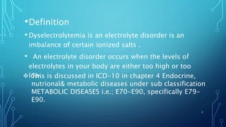
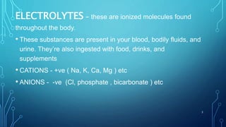
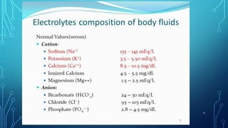
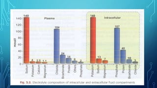
PRESENTER:
Dr. S. Keerthi
Dept. Of Paediatrics,
J.S.P.S Govt Homoeopathic Medical College,
Ramanthapur, Hyderabad.
MODERATOR:
Dr. RAJANI CHANDER, M.D (Hom)
H.O.D, Prof & P.G. Guide,
Dept. Of Paediatrics,
J.S.P.S Govt Homoeopathic Medical College,
Ramanthapur, Hyderabad.
1
•Definition
• Dyselectrolytemia is an electrolyte disorder is an
imbalance of certain ionized salts .
• An electrolyte disorder occurs when the levels of
electrolytes in your body are either too high or too
low. This is discussed in ICD-10 in chapter 4 Endocrine,
nutrional& metabolic diseases under sub classification
METABOLIC DISEASES i.e.; E70-E90, specifically E79-
E90.
2
ELECTROLYTES – these are ionized molecules found
throughout the body.
• These substances are present in your blood, bodily fluids, and
urine. They’re also ingested with food, drinks, and
supplements
• CATIONS - +ve ( Na, K, Ca, Mg ) etc
• ANIONS - -ve (Cl, phosphate , bicarbonate ) etc
3
Normal levels of electrolytes
4
Ecf icf electrolytes and
exchange
5
General functions of electrolytes
 Help to balance pH and acid base balance in body
Facilitate the transport of fluids
Regulating the functions of endocrine , neuromuscular and
excretory systems .
6
•Causes of electrolyte disorders
• Electrolyte disorders are most often caused by a loss
of bodily fluids through prolonged vomiting,
diarrhoea, or sweating.
• They may also develop due to fluid loss related to
burns.
• Certain medications can cause electrolyte disorders as
well.
7
 Types of electrolyte disorder
• Sodium: hypernatremia and hyponatremia
• Potassium: hyperkalaemia and hypokalaemia
• Calcium: hypercalcemia and hypocalcaemia
• Chloride: hyperchloremia and hypochloraemia
Magnesium: hypermagnesemia and hypomagnesemia
• Phosphate: hyperphosphatemia or hypophosphatemia
8
Sodium
• Normal range 135 -145 mEq/l
• Sodium helps to balance fluid levels in body
• Daily sodium requirement is 2 to 3 mEq/kg body weight although
intakes are generally well in excess.
9
10
HYPONATRAEMIA:
Hyponatremia, defined as plasma sodium less than
135mEq/l
Commonly results from excessive loss of sodium
from excessive sweating, vomiting, diarrhea, burns
and the administration of diuretics
Becomes symptomatic when the levels fall below
125mEq/ml or the decline is acute i.e, in < 24 hrs.
Broadly classified as
a)Hypovolemic hyponatremia
b)Normovolemic hyponatremia
c)Hypervolemic hyponatremia
11
 HYPOVOLEMIC HYPONATRAEMIA
A) Renal loss: - Diuretics
- Osmotic diuresis
- Renal salt wasting
- Adrenal insufficiency
- Pseudo-hypo-aldosteronism
B) Extra renal loss: - Diarrhoea, Vomitings,
Sweat
- Fistulas, Drains
- Cerebral salt wasting
syndrome
- Effusions, Ascites
12
 NORMOVOLEMIC HYPONATREMIA
This is caused by conditions that predispose to
SIADH
i.e., A) Inflammation of CNS- Meningitis,
Encephalitis
B) Pulmonary- severe Asthma, Pneumonia
C) Drugs
D) Others- tumours, postoperative
13
 HYPERVOLEMIC HYPONATREMIA:
Caused by - Congestive heart failure
- Cirrhosis of liver
- Nephrotic syndrome
- Acute or Chronic renal failure
14
 CLINICAL SYMPTOMS:
 Milder symptoms include- headache,
nausea, vomitings, lethargy, confusion
 In advanced stages, there may be-
seizures, coma, decorticate posturing,
dilated pupils, anisocoria, papilledema,
cardiac arrhythmias, myocardial ischaemia,
central diabetes insipidus, cerebral
oedema
15
16
17
Hypernatremia
• Hypernatremia is defined as increase in serum
sodium
concentration to levels more than 150 mEq/l
• The major cause of hypernatremia Is loss of body
water, inadequate intake of water, a lack antidiuretic
hormone (ADH), or excessive intake of Sodium (e.g.
solutions with high sodium )
18
• The most objective sign of hypernatremia is
lethargy or mental status changes, which
proceeds to coma
and convulsions. With acute and severe
hypernatremia,
the osmotic shift of water from neurons leads to
shrinkage
of the brain and tearing of the meningeal vessels
and
intracranial hemorrhage; slowly developing
hypematremia
19
POTASSIUM
• Main ICF component
• Normal range 3.5-5.0Meq/dl
• Sources-meat, bones, fruits and potatoes
• Main Hormones for regulation -Aldosterone and
insulin
• Functions:
1)excitability of nerve and muscle tissue
2)contractibility of cardiac, skeletal and smooth
muscles
•
20
Regulation
 Aldosterone :it causes increasing of sodium
absorption and potassium excretion maintaining
balance
2)it also leads to loss in saliva, sweat etc
 Alkalosis : in which hydrogen ions which are
exchanged in place of potassium into cells
 INSULIN :causes potassium uptake by Na+-
k+activity
21
HYPOKALEMIA
• when potassium levels falls below 3.5mE/dL.
• If it is less than 2.5mE/dl causes abdominal
distension and paralytic ileus
Causes/etiology:
 Reduced intake
 Malnutrition
 High renal loss - Diuretics, osmotic diuretics
Tubular defects -renal tubular acidosis
22
 Acid base disturbances - alkalosis
 Endocrinopathies-Cushing syndrome, primary
aldosteronism , thyrotoxicosis
 High extrarenal loss: GIT- Diarrhea, vomiting
,frequent enemas, Profuse sweating
 Decrease in muscle mass myopathies
23
 Symptoms:
 Weakness of skeletal muscles
 Hypotonia
 Hyporeflexia
 Abdominal distension
 Paralytic ileus
 Respiratory distress
 Prolonged loss -polyuria, polydipsia
 Cardiac-arrythmia
24
o ECG CHANGES -depressed ST ,Flat/inversed T Wave
prolonged P-R interval
25
HYPERKALAMIA
Causes
Acidosis
Renal insufficiency
Diseases including aldosterone and insulin functions
 Increased k+ intake
o Packed cell transfuion
o Cell injury
o Packed cell transfusion
26
• Decreased excretion
o Renal failure
o Ut obstruction
o Addison disease
o Angiotensin receptor blockers
27
Symptoms:
 Nausea
 Vomiting’s
 Paresthesia
 Skeletal fatigue
 ECG CHANGES -T wave TALL, prolonged PR interval, flat Pa
28
CALCIUM:
98% 0f calcium is in Skeleton
Functions:
 Blood coagulation
 Cellular communication
 Exocytosis
 Muscle contraction
 Neuromuscular transmission
29
HYPO CALCEMIA:
LESS THAN 8mg/dl or ionized ca+2 4mg/dl
Causes:
 Aplasia of thyroid gland
 Pseudo hypo parathyroidism
 Mutations in calcium sensing receptors
 Vit D deficiency, resistance to Vit D actions
30
 Hypo magnesemia
 Hyper phosphatemia
 Mal absorption
 Renal tubular acidosis
 Acute pancreatitis
 Drugs: corticosteroids or ptenytoin
31
Symptoms:
 CNS irritability
 Poor muscular contractility
 Poor feeding
 Vomiting’s
 Abdominal distension
 Muscle twitching ,cramps
 Tetany and signs of nerve irritability
 Carpopedal spasm and stridor
 Chavokestick sigh (twitching of orbiculo occcular )
32
33
RICKETS
• most
common
presentation
in children
34
35
HYPER CALCEMIA :
12mg/dl causes symptoms
Causes:
 Neonatal hypoparathyroidism
 familiar hypocaloric hyper calcemia
 excess ca+2supplementation
 Ewing sarcoma
 neuroblastoma
 Rhabdo sarcoma
 vit D Or A excess ,phosphate deficiency
 prolonged immobilization
36
 symptoms:
 coma
 lethargy
 confusion
 hyporeflexia
 muscle weakness
 constipation
 nephrolithiasis
 polyuria
 Ectopic -conjunctivitis,
 pancreatitis 37
MAGNESIUM
• It is 3rd moat abundant intracellular cation
• It is mostly bound to proteins
 Functions
• It helps in protein carbohydrate and fat metabolism
• Regulation of parathyroid hormone function
• Functioning of normal cell membrane
 Source
Green leafy vegetables, cereals, nuts and meat.
38
ABSORBTION
• Parathyroid hormone and glucocorticoids
increases its absorption
• Vitamin D and PTH also enhances its absorption
• In kidneys it is absorbed mainly in thick
ascending loop of henle
39
40
• Increased intestinal motility and calcium also
decrease magnesium absorption.
• Vitamin D and parathyroid hormone (PTH) may
enhance
absorption, although this effect is limited. Intestinal
absorption does
increase when intake is decreased
Hypermagnesemia
• When magnesium is greater than 2.5mg/dl
Causes
• Mg containing antacids
• In neonates whose mother was given magnesium
sulphate to prevent eclamsia
41
Symptoms
• Vomiting
• feeding difficulty
• Lethargy
• Weakness and dizziness
42
43
HYPOMAGNESEMIA
• Hypomagnesemia with secondary hypocalcaemia,
a rare autosomal recessive disorder, is caused by
decreased intestinal absorption of magnesium and
renal magnesium wasting.
• Poor intake
• Insulin administration
• Pancreatitis
• Intrauterine growth retardation
• Infants of diabetic mothers
44
• GASTROINTESTINAL DISORDERS
• Diarrhea
Nasogastric suction or emesis
Inflammatory bowel disease
Small bowel resection or bypass
Pancreatitis
Protein-calorie malnutrition
Hypomagnesemia with secondary hypocalcaemia
• BICARBONATE (HCO3-):
• It is alkaline & a vital component of pH buffering
system of human body.
• Normal range: 24-30 meq/lt
• FUNCTIONS:
• The blood electrolytes Sodium, Potassium,
Chloride and bicarbonate helps to regulate nerve
& muscle function and maintain Acid-Base balance
and water balance in the body.
• Thus having electrolytes in right concentrations is
important in maintaining fluid balance.
• .
45
SOURCE:
• It is released from the pancreas in response to
harmone secretin to neutralize the acidic chime
entering the duodenum from the stomach.
DEFICIENCY:
• A low level of bicarbonate in blood may cause a
condition called Metabolic acidosis
46
PHOSPHATES
• A phosphate is a chemical derivative of phosphoric acid.
• The phosphate ion (PO3−4) is an inorganic chemical, the
conjugate base that can form many different salts.
• Phosphate, or phosphorous, is similar to calcium, and is
found in your teeth and bones. You need vitamin D in order
to absorb phosphate.
NORMAL RANGE
• The normal range is 2.5-4.5 mg/dL.
47
SOURSE
Finding foods with high phosphorus levels isn’t hard.
Pork, cod, salmon, and tuna are all high in
phosphorus. Good dairy sources include:
• milk
• chocolate
• yogurt
• ricotta and American cheese
• Bran cereal, blueberry muffins, and nachos are also
high in phosphorus.
o 28% of frozen blueberries, 20% of celery, 27% of
green beans, 17% of peaches, 8% of broccoli, and
25% of strawberries 48
• Functions
Phosphorus works with calcium to help build bones. You
need the right amount of both calcium and phosphorus
for bone health. Phosphorus also plays an important
structural role in nucleic acids and cell membranes. And
it’s involved in the body’s energy production.
• Your body absorbs less phosphorus when calcium levels
are too high, and vice versa. You also need vitamin D to
absorb phosphorus properly.
49
• Poor absorption of phosphate
• If you had stomach surgery
• If you are lacking in Vitamin-D
• The absorption of phosphate is being blocked by
aluminum hydroxide found in laxatives
• low blood magnesium (needed to absorb
phosphorous), or high blood calcium (which binds to
the phosphorous, making it lower than normal
CAUSES
50
• diuretics
• Endocrine problems - such as a hyper parathyroid or
thyroid gland
• Alcoholism - drinking too much alcohol on a regular
basis
• Rickets
• uncontrolled diabetes (or elevated blood glucose)-
phosphate likes to follow or accompany glucose into the
cells, so you may have severely low blood phosphorous
51
Symptoms of Hypophosphatemia:
• Signs of hypophosphatemia include a lower than normal
blood phosphate level. Other electrolyte values are likely to
be affected, There are no symptoms of
hypophosphatemia, unless the values are critically low.
• Then you may notice trouble breathing or respiratory
problems, confusion, irritability, or coma. These all may
occur with phosphorous levels of 0.1-0.2 mg/ dL.
• phosphorous levels are below 1.0 mg/dL, your tissues
may have more trouble connecting hemoglobin with
oxygen - which is critical for breathing. You may
become mild to moderately short of breath.
52
HIGH PHOSPHATES
The kidneys excrete phosphate. Therefore, the most
common cause of hyperphosphatemia is the kidney's
inability to get rid of phosphate.
Hyperphosphatemia is also seen in people who have:
•Excessive dietary intake of phosphate (also from
laxatives or enemas)
•Your body may have a deficiency in calcium or
magnesium, or it may have too much Vitamin D,
resulting in hyperphosphatemia.
53
• Severe infections can cause increased phosphate
levels, resulting in hyperphosphatemia.
• Cell destruction - from chemotherapy, when the tumor
cells die at a fast rate. This can cause tumor lysis
syndrome.
• You may have high phosphate levels from prolonged
exercise, which causes muscle damage. Certain athletes
and distance runners may get this, called
rhabdomyolysis.
• You may have problems with your thyroid, parathyroid
gland, or other hormones, causing increased levels of
phosphate in your blood and resulting in
hyperphosphatemia 54
• Normal Range - 95-105 m Eq/l
• essential for maintaining acid/base balance
transmitting nerve impulses , regulating in out of cells.
• 90% Excreted in urine and also excreted in stool and
sweet.
• Sources - Table Salt ,Sea weed, rye, Tomatoes.
• Hypochloraemia - Less than 95 meg/l caused by
excessive use of loop diuretics ,Nasogastric suction,
Vomiting, Metabolic alkalosis is usually present with
hypochloraemia
. H/O of diuretic therapy, vomiting , assessment of
values in the metabolic alkaloses.
55
• Hypochloraemia - greater then 108 meg/L
result of dehydration , administration of NACL metabolic
acidosis is seen often seen in pts with severe diarrhoea
(or) ureteral diversion.
56
Bibliography
• GHAI essential paediatrics
• Suraj Gupte short text book of paediatrics
• Nelson text book of paediatrics 20 edition
57
58

Dyselectrolytemia

  • 1.
    PRESENTER: Dr. S. Keerthi Dept.Of Paediatrics, J.S.P.S Govt Homoeopathic Medical College, Ramanthapur, Hyderabad. MODERATOR: Dr. RAJANI CHANDER, M.D (Hom) H.O.D, Prof & P.G. Guide, Dept. Of Paediatrics, J.S.P.S Govt Homoeopathic Medical College, Ramanthapur, Hyderabad. 1
  • 2.
    •Definition • Dyselectrolytemia isan electrolyte disorder is an imbalance of certain ionized salts . • An electrolyte disorder occurs when the levels of electrolytes in your body are either too high or too low. This is discussed in ICD-10 in chapter 4 Endocrine, nutrional& metabolic diseases under sub classification METABOLIC DISEASES i.e.; E70-E90, specifically E79- E90. 2
  • 3.
    ELECTROLYTES – theseare ionized molecules found throughout the body. • These substances are present in your blood, bodily fluids, and urine. They’re also ingested with food, drinks, and supplements • CATIONS - +ve ( Na, K, Ca, Mg ) etc • ANIONS - -ve (Cl, phosphate , bicarbonate ) etc 3
  • 4.
    Normal levels ofelectrolytes 4
  • 5.
    Ecf icf electrolytesand exchange 5
  • 6.
    General functions ofelectrolytes  Help to balance pH and acid base balance in body Facilitate the transport of fluids Regulating the functions of endocrine , neuromuscular and excretory systems . 6
  • 7.
    •Causes of electrolytedisorders • Electrolyte disorders are most often caused by a loss of bodily fluids through prolonged vomiting, diarrhoea, or sweating. • They may also develop due to fluid loss related to burns. • Certain medications can cause electrolyte disorders as well. 7
  • 8.
     Types ofelectrolyte disorder • Sodium: hypernatremia and hyponatremia • Potassium: hyperkalaemia and hypokalaemia • Calcium: hypercalcemia and hypocalcaemia • Chloride: hyperchloremia and hypochloraemia Magnesium: hypermagnesemia and hypomagnesemia • Phosphate: hyperphosphatemia or hypophosphatemia 8
  • 9.
    Sodium • Normal range135 -145 mEq/l • Sodium helps to balance fluid levels in body • Daily sodium requirement is 2 to 3 mEq/kg body weight although intakes are generally well in excess. 9
  • 10.
  • 11.
    HYPONATRAEMIA: Hyponatremia, defined asplasma sodium less than 135mEq/l Commonly results from excessive loss of sodium from excessive sweating, vomiting, diarrhea, burns and the administration of diuretics Becomes symptomatic when the levels fall below 125mEq/ml or the decline is acute i.e, in < 24 hrs. Broadly classified as a)Hypovolemic hyponatremia b)Normovolemic hyponatremia c)Hypervolemic hyponatremia 11
  • 12.
     HYPOVOLEMIC HYPONATRAEMIA A)Renal loss: - Diuretics - Osmotic diuresis - Renal salt wasting - Adrenal insufficiency - Pseudo-hypo-aldosteronism B) Extra renal loss: - Diarrhoea, Vomitings, Sweat - Fistulas, Drains - Cerebral salt wasting syndrome - Effusions, Ascites 12
  • 13.
     NORMOVOLEMIC HYPONATREMIA Thisis caused by conditions that predispose to SIADH i.e., A) Inflammation of CNS- Meningitis, Encephalitis B) Pulmonary- severe Asthma, Pneumonia C) Drugs D) Others- tumours, postoperative 13
  • 14.
     HYPERVOLEMIC HYPONATREMIA: Causedby - Congestive heart failure - Cirrhosis of liver - Nephrotic syndrome - Acute or Chronic renal failure 14
  • 15.
     CLINICAL SYMPTOMS: Milder symptoms include- headache, nausea, vomitings, lethargy, confusion  In advanced stages, there may be- seizures, coma, decorticate posturing, dilated pupils, anisocoria, papilledema, cardiac arrhythmias, myocardial ischaemia, central diabetes insipidus, cerebral oedema 15
  • 16.
  • 17.
  • 18.
    Hypernatremia • Hypernatremia isdefined as increase in serum sodium concentration to levels more than 150 mEq/l • The major cause of hypernatremia Is loss of body water, inadequate intake of water, a lack antidiuretic hormone (ADH), or excessive intake of Sodium (e.g. solutions with high sodium ) 18
  • 19.
    • The mostobjective sign of hypernatremia is lethargy or mental status changes, which proceeds to coma and convulsions. With acute and severe hypernatremia, the osmotic shift of water from neurons leads to shrinkage of the brain and tearing of the meningeal vessels and intracranial hemorrhage; slowly developing hypematremia 19
  • 20.
    POTASSIUM • Main ICFcomponent • Normal range 3.5-5.0Meq/dl • Sources-meat, bones, fruits and potatoes • Main Hormones for regulation -Aldosterone and insulin • Functions: 1)excitability of nerve and muscle tissue 2)contractibility of cardiac, skeletal and smooth muscles • 20
  • 21.
    Regulation  Aldosterone :itcauses increasing of sodium absorption and potassium excretion maintaining balance 2)it also leads to loss in saliva, sweat etc  Alkalosis : in which hydrogen ions which are exchanged in place of potassium into cells  INSULIN :causes potassium uptake by Na+- k+activity 21
  • 22.
    HYPOKALEMIA • when potassiumlevels falls below 3.5mE/dL. • If it is less than 2.5mE/dl causes abdominal distension and paralytic ileus Causes/etiology:  Reduced intake  Malnutrition  High renal loss - Diuretics, osmotic diuretics Tubular defects -renal tubular acidosis 22
  • 23.
     Acid basedisturbances - alkalosis  Endocrinopathies-Cushing syndrome, primary aldosteronism , thyrotoxicosis  High extrarenal loss: GIT- Diarrhea, vomiting ,frequent enemas, Profuse sweating  Decrease in muscle mass myopathies 23
  • 24.
     Symptoms:  Weaknessof skeletal muscles  Hypotonia  Hyporeflexia  Abdominal distension  Paralytic ileus  Respiratory distress  Prolonged loss -polyuria, polydipsia  Cardiac-arrythmia 24
  • 25.
    o ECG CHANGES-depressed ST ,Flat/inversed T Wave prolonged P-R interval 25
  • 26.
    HYPERKALAMIA Causes Acidosis Renal insufficiency Diseases includingaldosterone and insulin functions  Increased k+ intake o Packed cell transfuion o Cell injury o Packed cell transfusion 26
  • 27.
    • Decreased excretion oRenal failure o Ut obstruction o Addison disease o Angiotensin receptor blockers 27
  • 28.
    Symptoms:  Nausea  Vomiting’s Paresthesia  Skeletal fatigue  ECG CHANGES -T wave TALL, prolonged PR interval, flat Pa 28
  • 29.
    CALCIUM: 98% 0f calciumis in Skeleton Functions:  Blood coagulation  Cellular communication  Exocytosis  Muscle contraction  Neuromuscular transmission 29
  • 30.
    HYPO CALCEMIA: LESS THAN8mg/dl or ionized ca+2 4mg/dl Causes:  Aplasia of thyroid gland  Pseudo hypo parathyroidism  Mutations in calcium sensing receptors  Vit D deficiency, resistance to Vit D actions 30
  • 31.
     Hypo magnesemia Hyper phosphatemia  Mal absorption  Renal tubular acidosis  Acute pancreatitis  Drugs: corticosteroids or ptenytoin 31
  • 32.
    Symptoms:  CNS irritability Poor muscular contractility  Poor feeding  Vomiting’s  Abdominal distension  Muscle twitching ,cramps  Tetany and signs of nerve irritability  Carpopedal spasm and stridor  Chavokestick sigh (twitching of orbiculo occcular ) 32
  • 33.
  • 34.
  • 35.
  • 36.
    HYPER CALCEMIA : 12mg/dlcauses symptoms Causes:  Neonatal hypoparathyroidism  familiar hypocaloric hyper calcemia  excess ca+2supplementation  Ewing sarcoma  neuroblastoma  Rhabdo sarcoma  vit D Or A excess ,phosphate deficiency  prolonged immobilization 36
  • 37.
     symptoms:  coma lethargy  confusion  hyporeflexia  muscle weakness  constipation  nephrolithiasis  polyuria  Ectopic -conjunctivitis,  pancreatitis 37
  • 38.
    MAGNESIUM • It is3rd moat abundant intracellular cation • It is mostly bound to proteins  Functions • It helps in protein carbohydrate and fat metabolism • Regulation of parathyroid hormone function • Functioning of normal cell membrane  Source Green leafy vegetables, cereals, nuts and meat. 38
  • 39.
    ABSORBTION • Parathyroid hormoneand glucocorticoids increases its absorption • Vitamin D and PTH also enhances its absorption • In kidneys it is absorbed mainly in thick ascending loop of henle 39
  • 40.
    40 • Increased intestinalmotility and calcium also decrease magnesium absorption. • Vitamin D and parathyroid hormone (PTH) may enhance absorption, although this effect is limited. Intestinal absorption does increase when intake is decreased
  • 41.
    Hypermagnesemia • When magnesiumis greater than 2.5mg/dl Causes • Mg containing antacids • In neonates whose mother was given magnesium sulphate to prevent eclamsia 41
  • 42.
    Symptoms • Vomiting • feedingdifficulty • Lethargy • Weakness and dizziness 42
  • 43.
    43 HYPOMAGNESEMIA • Hypomagnesemia withsecondary hypocalcaemia, a rare autosomal recessive disorder, is caused by decreased intestinal absorption of magnesium and renal magnesium wasting. • Poor intake • Insulin administration • Pancreatitis • Intrauterine growth retardation • Infants of diabetic mothers
  • 44.
    44 • GASTROINTESTINAL DISORDERS •Diarrhea Nasogastric suction or emesis Inflammatory bowel disease Small bowel resection or bypass Pancreatitis Protein-calorie malnutrition Hypomagnesemia with secondary hypocalcaemia
  • 45.
    • BICARBONATE (HCO3-): •It is alkaline & a vital component of pH buffering system of human body. • Normal range: 24-30 meq/lt • FUNCTIONS: • The blood electrolytes Sodium, Potassium, Chloride and bicarbonate helps to regulate nerve & muscle function and maintain Acid-Base balance and water balance in the body. • Thus having electrolytes in right concentrations is important in maintaining fluid balance. • . 45
  • 46.
    SOURCE: • It isreleased from the pancreas in response to harmone secretin to neutralize the acidic chime entering the duodenum from the stomach. DEFICIENCY: • A low level of bicarbonate in blood may cause a condition called Metabolic acidosis 46
  • 47.
    PHOSPHATES • A phosphateis a chemical derivative of phosphoric acid. • The phosphate ion (PO3−4) is an inorganic chemical, the conjugate base that can form many different salts. • Phosphate, or phosphorous, is similar to calcium, and is found in your teeth and bones. You need vitamin D in order to absorb phosphate. NORMAL RANGE • The normal range is 2.5-4.5 mg/dL. 47
  • 48.
    SOURSE Finding foods withhigh phosphorus levels isn’t hard. Pork, cod, salmon, and tuna are all high in phosphorus. Good dairy sources include: • milk • chocolate • yogurt • ricotta and American cheese • Bran cereal, blueberry muffins, and nachos are also high in phosphorus. o 28% of frozen blueberries, 20% of celery, 27% of green beans, 17% of peaches, 8% of broccoli, and 25% of strawberries 48
  • 49.
    • Functions Phosphorus workswith calcium to help build bones. You need the right amount of both calcium and phosphorus for bone health. Phosphorus also plays an important structural role in nucleic acids and cell membranes. And it’s involved in the body’s energy production. • Your body absorbs less phosphorus when calcium levels are too high, and vice versa. You also need vitamin D to absorb phosphorus properly. 49
  • 50.
    • Poor absorptionof phosphate • If you had stomach surgery • If you are lacking in Vitamin-D • The absorption of phosphate is being blocked by aluminum hydroxide found in laxatives • low blood magnesium (needed to absorb phosphorous), or high blood calcium (which binds to the phosphorous, making it lower than normal CAUSES 50
  • 51.
    • diuretics • Endocrineproblems - such as a hyper parathyroid or thyroid gland • Alcoholism - drinking too much alcohol on a regular basis • Rickets • uncontrolled diabetes (or elevated blood glucose)- phosphate likes to follow or accompany glucose into the cells, so you may have severely low blood phosphorous 51
  • 52.
    Symptoms of Hypophosphatemia: •Signs of hypophosphatemia include a lower than normal blood phosphate level. Other electrolyte values are likely to be affected, There are no symptoms of hypophosphatemia, unless the values are critically low. • Then you may notice trouble breathing or respiratory problems, confusion, irritability, or coma. These all may occur with phosphorous levels of 0.1-0.2 mg/ dL. • phosphorous levels are below 1.0 mg/dL, your tissues may have more trouble connecting hemoglobin with oxygen - which is critical for breathing. You may become mild to moderately short of breath. 52
  • 53.
    HIGH PHOSPHATES The kidneysexcrete phosphate. Therefore, the most common cause of hyperphosphatemia is the kidney's inability to get rid of phosphate. Hyperphosphatemia is also seen in people who have: •Excessive dietary intake of phosphate (also from laxatives or enemas) •Your body may have a deficiency in calcium or magnesium, or it may have too much Vitamin D, resulting in hyperphosphatemia. 53
  • 54.
    • Severe infectionscan cause increased phosphate levels, resulting in hyperphosphatemia. • Cell destruction - from chemotherapy, when the tumor cells die at a fast rate. This can cause tumor lysis syndrome. • You may have high phosphate levels from prolonged exercise, which causes muscle damage. Certain athletes and distance runners may get this, called rhabdomyolysis. • You may have problems with your thyroid, parathyroid gland, or other hormones, causing increased levels of phosphate in your blood and resulting in hyperphosphatemia 54
  • 55.
    • Normal Range- 95-105 m Eq/l • essential for maintaining acid/base balance transmitting nerve impulses , regulating in out of cells. • 90% Excreted in urine and also excreted in stool and sweet. • Sources - Table Salt ,Sea weed, rye, Tomatoes. • Hypochloraemia - Less than 95 meg/l caused by excessive use of loop diuretics ,Nasogastric suction, Vomiting, Metabolic alkalosis is usually present with hypochloraemia . H/O of diuretic therapy, vomiting , assessment of values in the metabolic alkaloses. 55
  • 56.
    • Hypochloraemia -greater then 108 meg/L result of dehydration , administration of NACL metabolic acidosis is seen often seen in pts with severe diarrhoea (or) ureteral diversion. 56
  • 57.
    Bibliography • GHAI essentialpaediatrics • Suraj Gupte short text book of paediatrics • Nelson text book of paediatrics 20 edition 57
  • 58.

Editor's Notes

  • #13 In both hyponatremia exists differentiated by serum urate levels ie; in fluid restriction corrects urate levels in rsw bt not in cebral saltwasting
  • #17 This is due to increase intracranial pressure ie; intracranial haemorrhage, brain tumour, traumatic injury and encephalopathy Lesion in mid brain
  • #30 Calmodulin- ca+2 binding regulatory protein (intracellular) Absorption-proximal tubule Calcium resorption - parathormone ,calcitonin, vit D-major source -dietary ca+2 calcium sensing receptor -G protein coupled receptor low ECF ca +2-----receptor in parathyroid ---increased distal tubules resorption ---stimulate osteoclastic activity from bones Calmodulin- ca+2 binding regulatory protein (intracellular) Absorption-proximal tubule Calcium resorption - parathormone ,calcitonin, vit D-major source -dietary ca+2 calcium sensing receptor -G protein coupled receptor low ECF ca +2-----receptor in parathyroid ---increased distal tubules resorption ---stimulate osteoclastic activity from bones
  • #32 Magnesium is essential for ca absorbtion alon with vit d whese as excess excess ca decreses mg absorbtion
  • #35 It may also appear in rickets because of defective mineralisation of the bones by calcium necessary to harden them; thus the diaphragm, which is always in tension, pulls the softened bone inward. During rickets it is due to the indentation of lower ribs at the point of attachment of diaphragm.
  • #44 INSULIN ADMINISTRAION CAUSES INCREASES MAGNESIUM EXCRETION