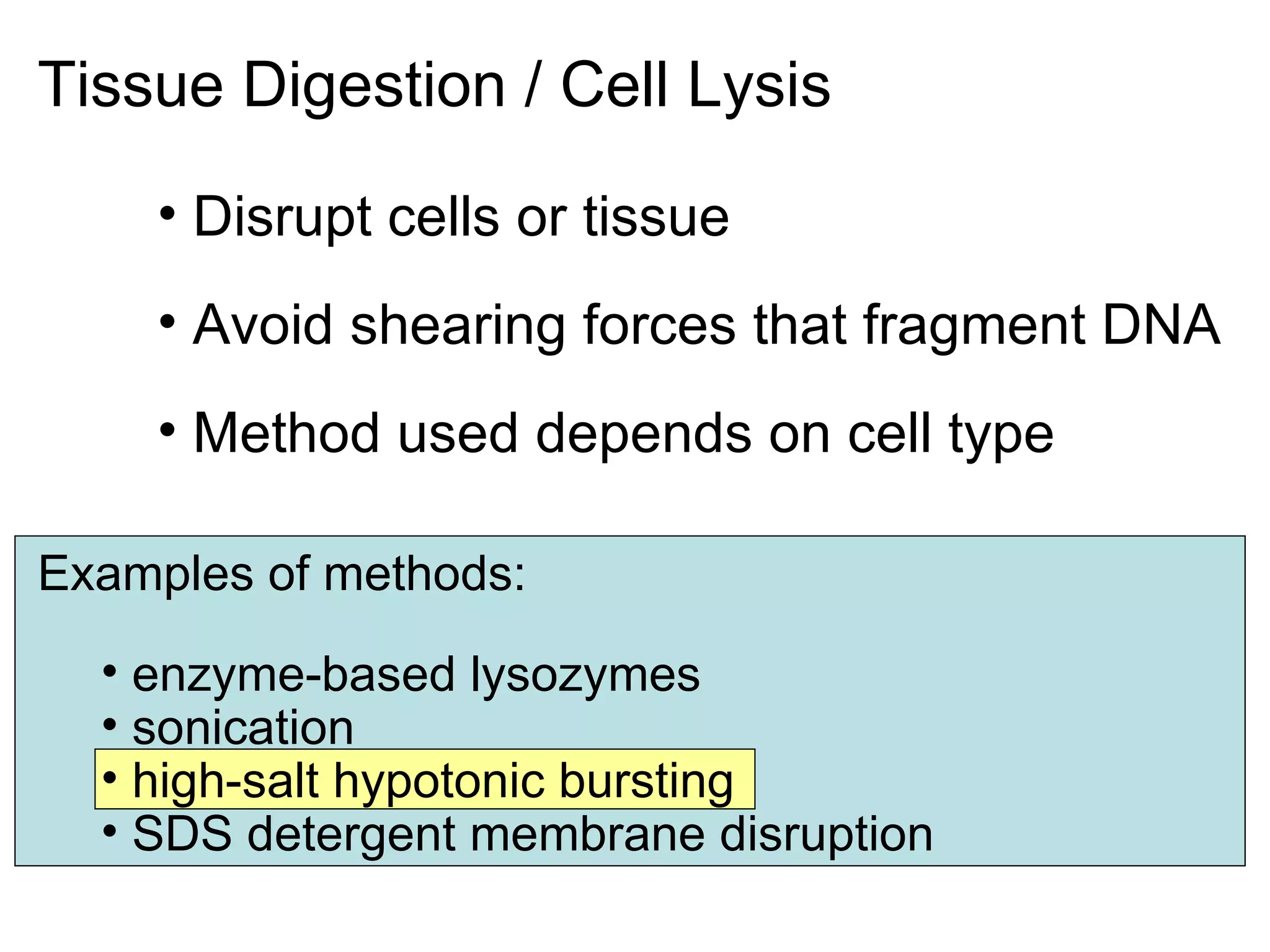
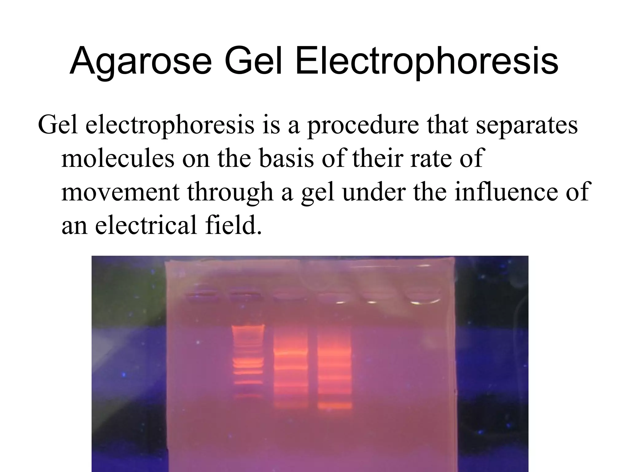
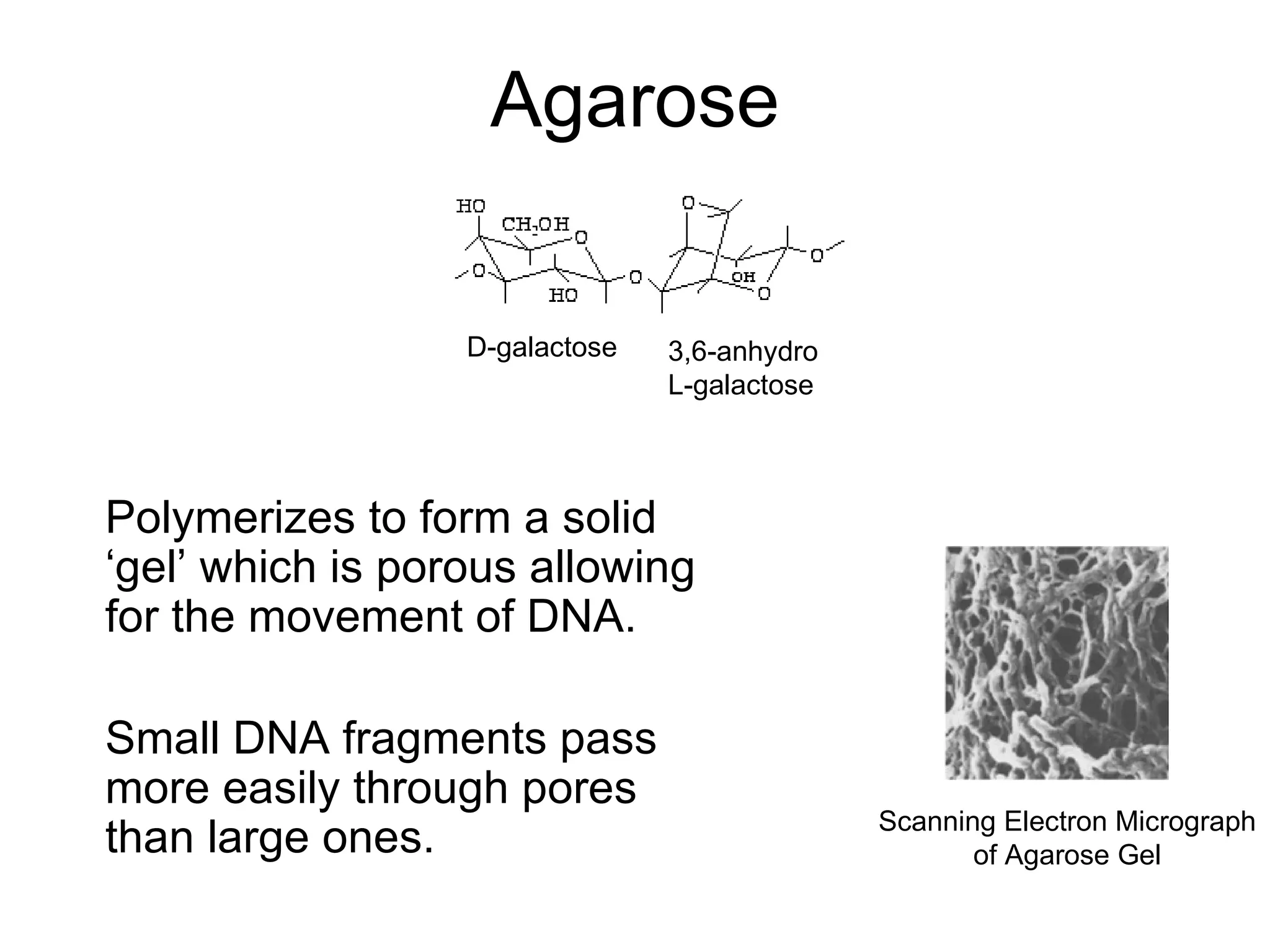
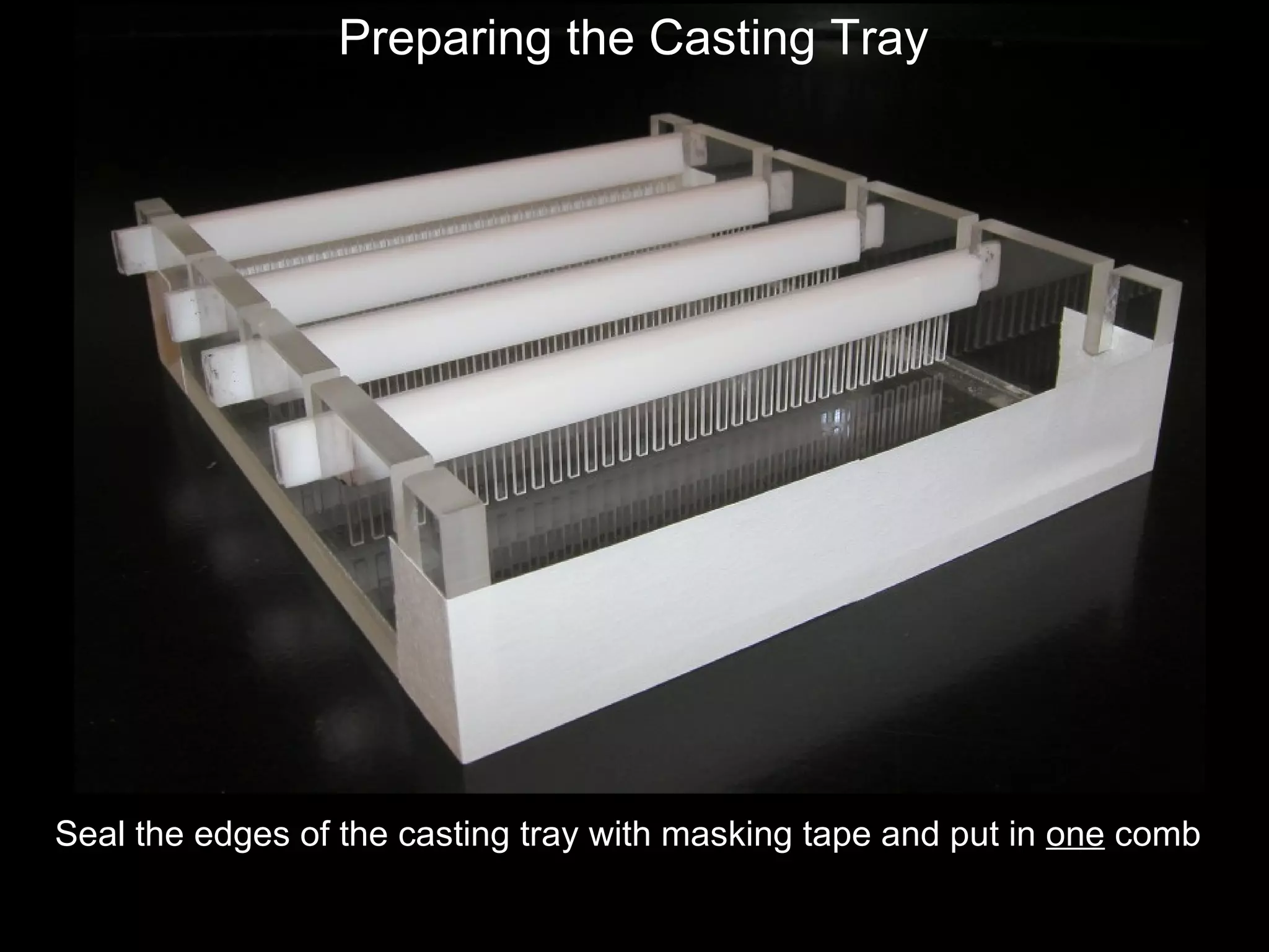
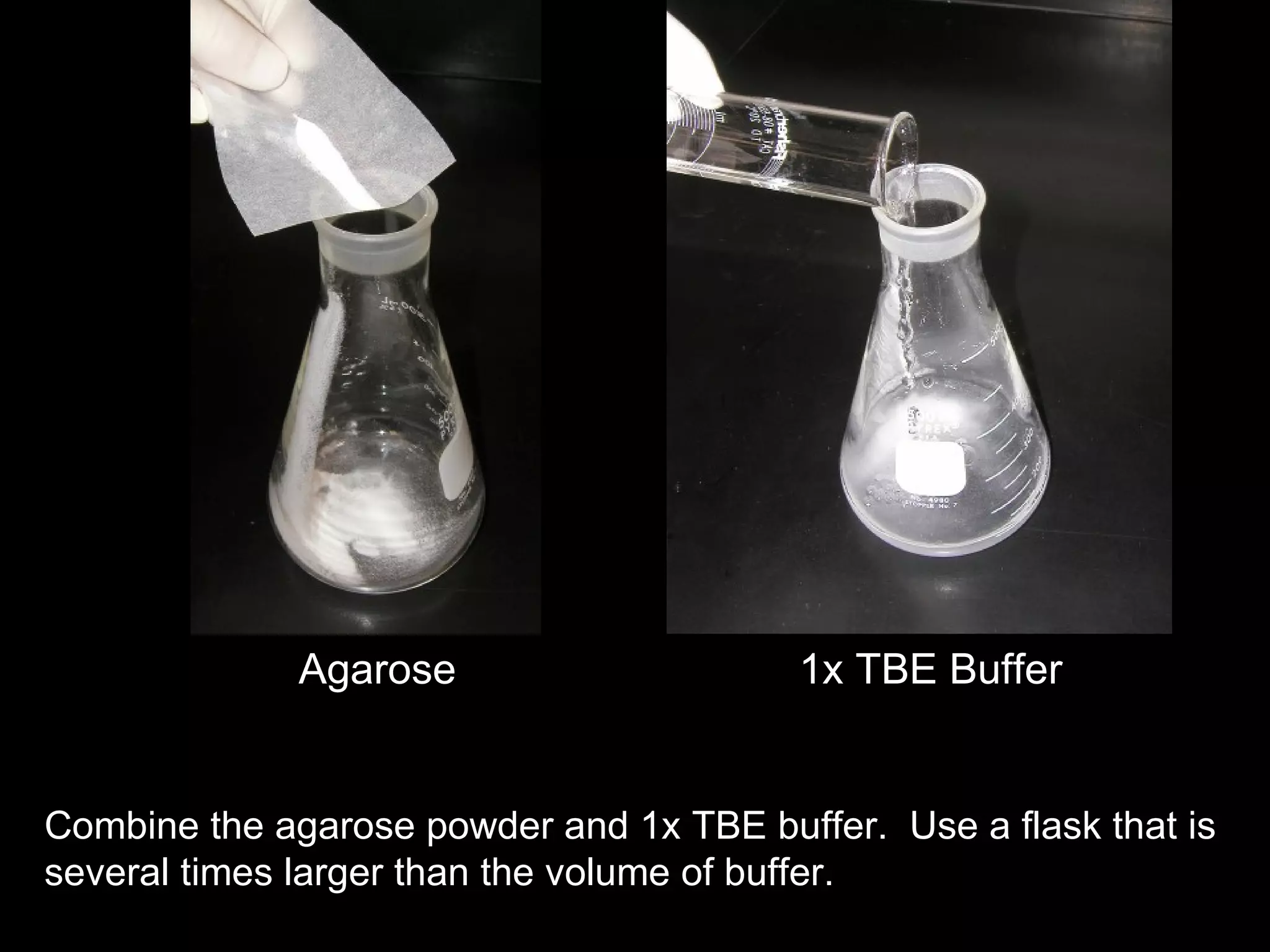
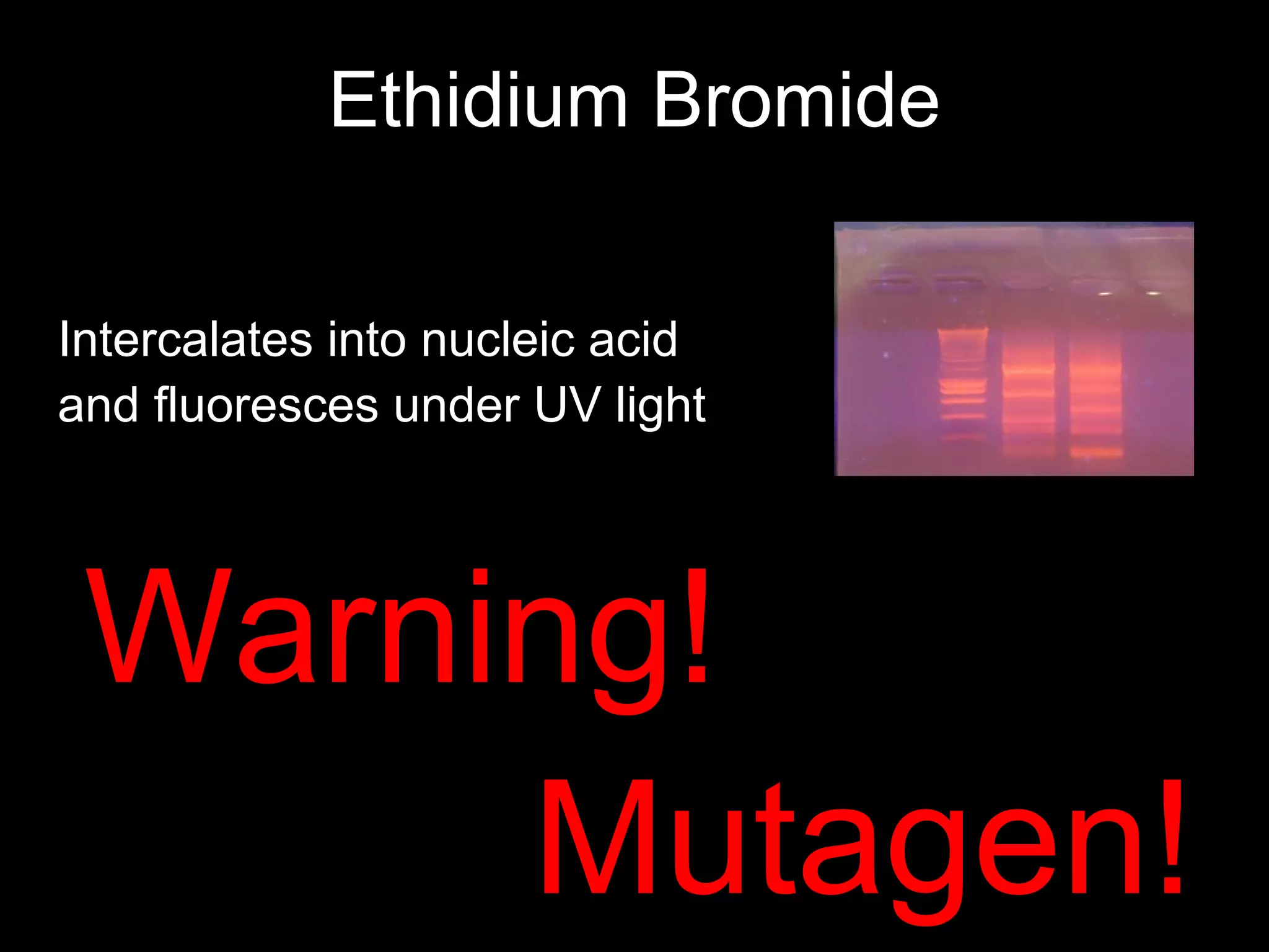
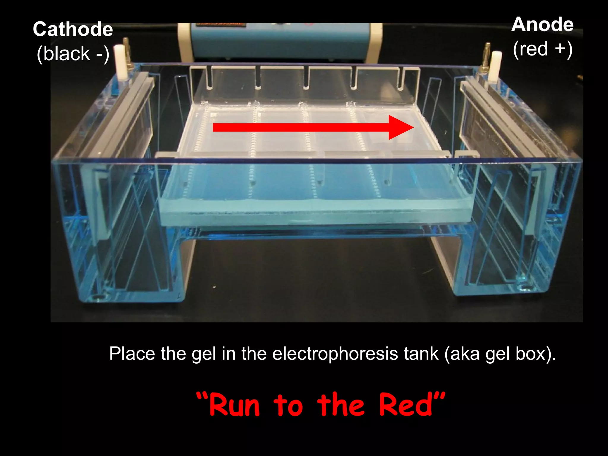
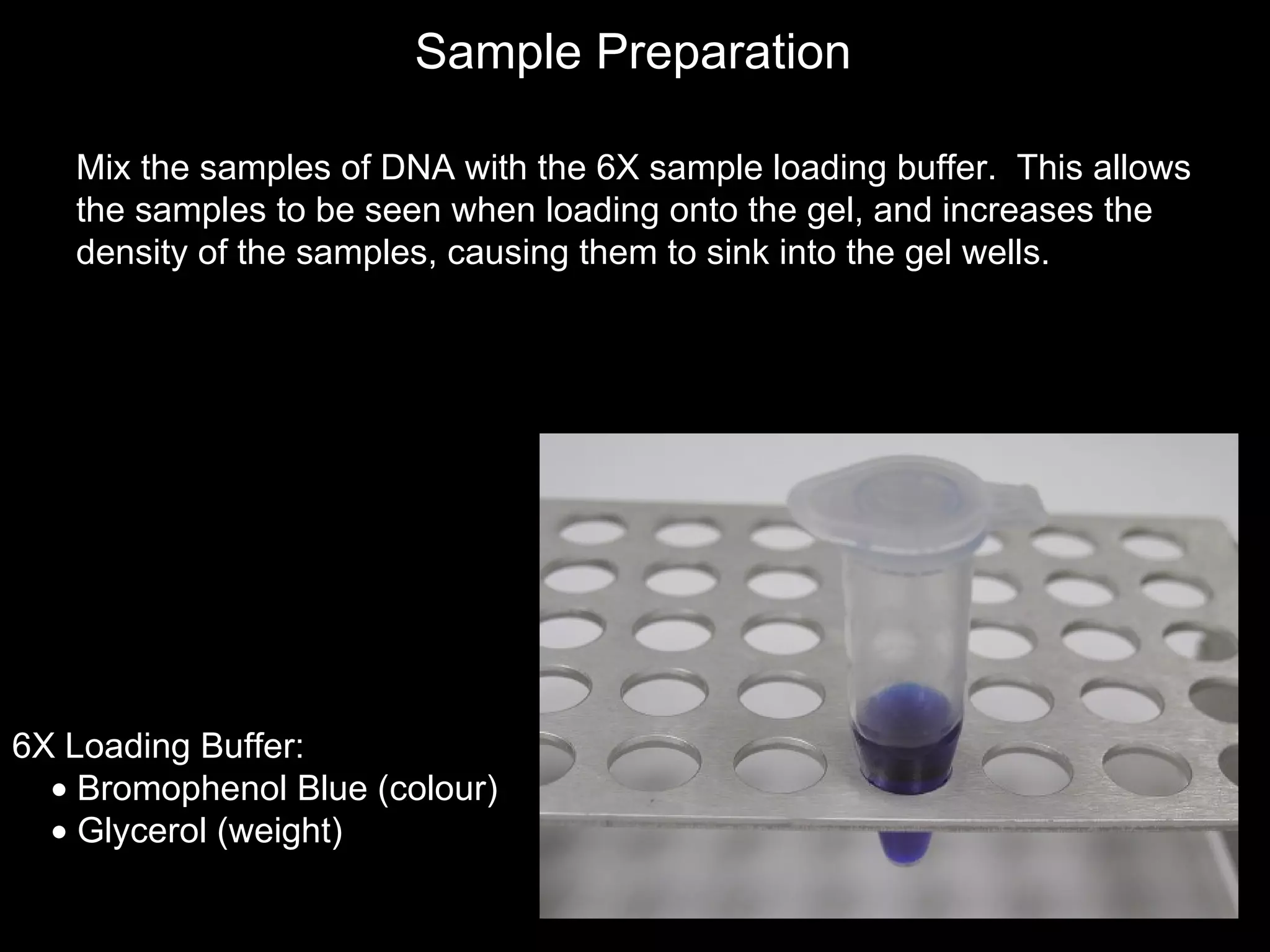
DNA Isolation & Agarose Electrophoresis Lecture 2 Source: www.flickr.com/photos/62558149@N00/171512923
Nucleic Acid purification can be performed starting from various biological materials:   Blood Tissues Bacteria Fungi Animal cells Plant cells Dung/Stool Agarose gels
Type of starting material Expected yield Sample age (I.e. quality) Time required for procedure Cost Quality control issues Technique used depends on …
General Extraction Protocols Include: Tissue Digestion / Cell Lysis Chelating and Proteinase Agents Nucleic Acid and Protein Separation Clean-up
Tissue Digestion / Cell Lysis Disrupt cells or tissue Avoid shearing forces that fragment DNA Method used depends on cell type Examples of methods: enzyme-based lysozymes sonication high-salt hypotonic bursting SDS detergent membrane disruption
QIAgen DNeasy DNA Kit Treat with Proteinase K - digest proteins and inhibit nucleases Lysis: QIAgen Buffer AL (guanidium-HCl) - hypertonic high salt solution Precipitation of DNA with EtOH Spin column - DNA binds to silica  membrane Wash Elution 1 and 2, with warm Buffer AE  (Tris-EDTA)
Agarose Gel Electrophoresis Gel electrophoresis is a procedure that separates molecules on the basis of their rate of movement through a gel under the influence of an electrical field.
DNA will be attracted to the positively charged pole ( RED ) and repelled from negatively charged pole (BLACK) “ Run to the Red” DNA is negatively charged because of the sugar-phosphate backbone.
Agarose Polymerizes to form a solid ‘gel’ which is porous allowing for the movement of DNA.  Small DNA fragments pass more easily through pores than large ones. D-galactose 3,6-anhydro L-galactose Scanning Electron Micrograph of Agarose Gel
An agarose gel is prepared by combining agarose powder and a buffer solution. Agarose   TBE Buffer  Flask for boiling  Special thanks to Michael Clark  (University of Rochester) for these images.
Casting tray  Gel combs  Power supply  Gel tank   Cover Electrical leads  Electrophoresis Equipment
Seal the edges of the casting tray with masking tape and put in  one  comb Preparing the Casting Tray
Agarose 1x TBE Buffer Combine the agarose powder and 1x TBE buffer.  Use a flask that is several times larger than the volume of buffer.
Melting the Agarose Insoluble at room temp Soluble once boiled  Once the gel has reaches about 55  o C,  Add 4-5  µ l of Ethidium Bromide dye to pre-stain.
Ethidium Bromide Warning! Mutagen! Intercalates into nucleic acid and fluoresces under UV light
Carefully pour the melted agarose solution into the casting tray.  Avoid air bubbles. Pouring the gel
The gel comb should be submerged in the melted agarose solution.
When cooled (> 20 mins), the agarose polymerizes, forming a flexible gel. Carefully remove the combs and tape.
Place the gel in the electrophoresis tank (aka gel box). “ Run to the Red” Cathode (black -) Anode (red +)
TBE buffer    Add enough 1x TBE buffer to cover the gel to a depth of at least 1 mm.  Make sure each well is filled with buffer.
6X Loading Buffer:    Bromophenol Blue (colour)    Glycerol (weight) Sample Preparation Mix the samples of DNA with the 6X sample loading buffer.  This allows the samples to be seen when loading onto the gel, and increases the density of the samples, causing them to sink into the gel wells.
Loading the Gel Carefully place the pipette tip inside a well and  gently/slowly  expel the sample.  The sample should sink into the well.
Place the cover on the electrophoresis chamber, connecting the electrical leads.  Connect the electrical leads to the power supply. When the power is turned on, bubbles should form on the electrodes wires in the electrophoresis chamber. Running the Gel
   wells    Bromophenol Blue Cathode (-) Anode (+) After the current is applied, make sure the Gel is running in the correct direction.  Bromophenol blue will run in the same direction as the DNA. DNA (-) 
Visualizing the DNA under UV light 1  2  3  4 2.0 0.6
Eric’s Quote of the Week ‘ Never throw away your samples until the end of the experiment.’  Dumpster diving SUCKS! Source: www.flickr.com/photos/50262392@N00/45684291

Biol2 Lecture 2 Dna Isolation And Agarose Gel

  • 1.
    DNA Isolation &Agarose Electrophoresis Lecture 2 Source: www.flickr.com/photos/62558149@N00/171512923
  • 2.
    Nucleic Acid purificationcan be performed starting from various biological materials: Blood Tissues Bacteria Fungi Animal cells Plant cells Dung/Stool Agarose gels
  • 3.
    Type of startingmaterial Expected yield Sample age (I.e. quality) Time required for procedure Cost Quality control issues Technique used depends on …
  • 4.
    General Extraction ProtocolsInclude: Tissue Digestion / Cell Lysis Chelating and Proteinase Agents Nucleic Acid and Protein Separation Clean-up
  • 5.
    Tissue Digestion /Cell Lysis Disrupt cells or tissue Avoid shearing forces that fragment DNA Method used depends on cell type Examples of methods: enzyme-based lysozymes sonication high-salt hypotonic bursting SDS detergent membrane disruption
  • 6.
    QIAgen DNeasy DNAKit Treat with Proteinase K - digest proteins and inhibit nucleases Lysis: QIAgen Buffer AL (guanidium-HCl) - hypertonic high salt solution Precipitation of DNA with EtOH Spin column - DNA binds to silica membrane Wash Elution 1 and 2, with warm Buffer AE (Tris-EDTA)
  • 7.
    Agarose Gel ElectrophoresisGel electrophoresis is a procedure that separates molecules on the basis of their rate of movement through a gel under the influence of an electrical field.
  • 8.
    DNA will beattracted to the positively charged pole ( RED ) and repelled from negatively charged pole (BLACK) “ Run to the Red” DNA is negatively charged because of the sugar-phosphate backbone.
  • 9.
    Agarose Polymerizes toform a solid ‘gel’ which is porous allowing for the movement of DNA. Small DNA fragments pass more easily through pores than large ones. D-galactose 3,6-anhydro L-galactose Scanning Electron Micrograph of Agarose Gel
  • 10.
    An agarose gelis prepared by combining agarose powder and a buffer solution. Agarose  TBE Buffer  Flask for boiling  Special thanks to Michael Clark (University of Rochester) for these images.
  • 11.
    Casting tray Gel combs  Power supply  Gel tank   Cover Electrical leads  Electrophoresis Equipment
  • 12.
    Seal the edgesof the casting tray with masking tape and put in one comb Preparing the Casting Tray
  • 13.
    Agarose 1x TBEBuffer Combine the agarose powder and 1x TBE buffer. Use a flask that is several times larger than the volume of buffer.
  • 14.
    Melting the AgaroseInsoluble at room temp Soluble once boiled Once the gel has reaches about 55 o C, Add 4-5 µ l of Ethidium Bromide dye to pre-stain.
  • 15.
    Ethidium Bromide Warning!Mutagen! Intercalates into nucleic acid and fluoresces under UV light
  • 16.
    Carefully pour themelted agarose solution into the casting tray. Avoid air bubbles. Pouring the gel
  • 17.
    The gel combshould be submerged in the melted agarose solution.
  • 18.
    When cooled (>20 mins), the agarose polymerizes, forming a flexible gel. Carefully remove the combs and tape.
  • 19.
    Place the gelin the electrophoresis tank (aka gel box). “ Run to the Red” Cathode (black -) Anode (red +)
  • 20.
    TBE buffer  Add enough 1x TBE buffer to cover the gel to a depth of at least 1 mm. Make sure each well is filled with buffer.
  • 21.
    6X Loading Buffer: Bromophenol Blue (colour)  Glycerol (weight) Sample Preparation Mix the samples of DNA with the 6X sample loading buffer. This allows the samples to be seen when loading onto the gel, and increases the density of the samples, causing them to sink into the gel wells.
  • 22.
    Loading the GelCarefully place the pipette tip inside a well and gently/slowly expel the sample. The sample should sink into the well.
  • 23.
    Place the coveron the electrophoresis chamber, connecting the electrical leads. Connect the electrical leads to the power supply. When the power is turned on, bubbles should form on the electrodes wires in the electrophoresis chamber. Running the Gel
  • 24.
    wells  Bromophenol Blue Cathode (-) Anode (+) After the current is applied, make sure the Gel is running in the correct direction. Bromophenol blue will run in the same direction as the DNA. DNA (-) 
  • 25.
    Visualizing the DNAunder UV light 1 2 3 4 2.0 0.6
  • 26.
    Eric’s Quote ofthe Week ‘ Never throw away your samples until the end of the experiment.’ Dumpster diving SUCKS! Source: www.flickr.com/photos/50262392@N00/45684291