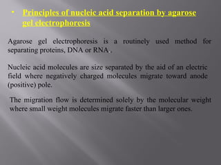
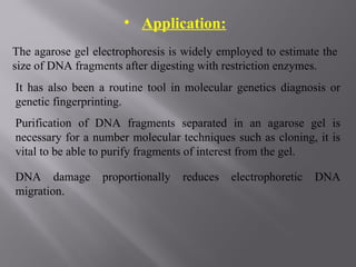
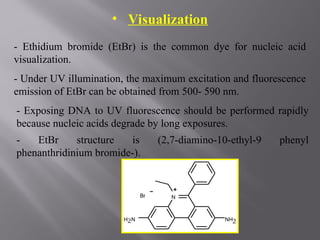
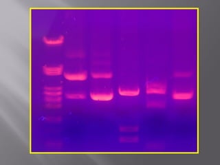
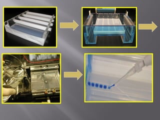
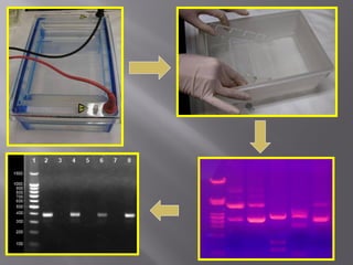
AGAROSE GEL
ELECTROPHORESIS
OUTLINE
• Principle.
• Applications.
•Visualization.
•Preparation of gel.
•Factors affecting DNA migration.
•Materials.
•Methods.
•Advantages.
• Principles of nucleic acid separation by agarose
gel electrophoresis
Agarose gel electrophoresis is a routinely used method for
separating proteins, DNA or RNA..
Nucleic acid molecules are size separated by the aid of an electric
field where negatively charged molecules migrate toward anode
(positive) pole.
The migration flow is determined solely by the molecular weight
where small weight molecules migrate faster than larger ones.
• Application:
The agarose gel electrophoresis is widely employed to estimate the
size of DNA fragments after digesting with restriction enzymes.
It has also been a routine tool in molecular genetics diagnosis or
genetic fingerprinting.
Purification of DNA fragments separated in an agarose gel is
necessary for a number molecular techniques such as cloning, it is
vital to be able to purify fragments of interest from the gel.
DNA damage proportionally reduces electrophoretic DNA
migration.
• Visualization
- Ethidium bromide (EtBr) is the common dye for nucleic acid
visualization.
- Under UV illumination, the maximum excitation and fluorescence
emission of EtBr can be obtained from 500- 590 nm.
- Exposing DNA to UV fluorescence should be performed rapidly
because nucleic acids degrade by long exposures.
- EtBr structure is (2,7-diamino-10-ethyl-9 phenyl
phenanthridinium bromide-).
- An alternative DNA stain is SYBR Green I, is more expensive,
and 25 times more sensitive than Ethidium bromide.
- It has been shown to have low levels of mutagenicity and toxicity
compared with Ethidium bromide .
- EtBr is an irritant to the skin, eyes, mouth, and upper respiratory
tract.
• Preparing and running standard agarose DNA gels
- Several electrophoresis buffers can be used for fractionating nucleic
acid such as, Tris acetate- EDTA (TAE) or Tris-borate-EDTA (TBE).
- For gel preparation, agarose powder electrophoresis grade is mixed
with electrophoresis buffer to the desired concentrations then heated
in a microwave oven until completely dissolved.
- The mixture is cooled and poured into the casting tray for
solidification, after the gel solidification the comb is removed.
- PCR product mixed with loading dye is placed in the wells, wells
should be placed towards the negative electrode.
- Ethidium bromide migrates in the reverse direction, meets and
couples with DNA fragments.
• Factors affecting DNA Migration in agarose
gels:
1. Agarose concentration.
2. Voltage.
3. Electrophoresis buffer.
4. Effect of Ethidium bromide.
1. Agarose concentration:
- Agarose gel electrophoresis can be used for the separation of
DNA fragments ranging from 50 base pair to several mega
bases (millions of bases) using specialized apparatus.
- Increasing the agarose concentration of a gel reduces the
migration speed and enables separation of smaller DNA
molecules.
- For large DNA fragments use low agarose conc. About 0.7 % and for
small DNA fragments use high agarose conc. (2%).
- Higher concentrations have the disadvantage of long run times.
2. Voltage.
- Migration of fragments in an agarose gel depends on the difference
in electric current.
- Different optimal voltages are required for different fragment sizes.
3. Electrophoresis buffer.
Various buffers are used for agarose electrophoresis.
The two most common buffers for nucleic acids are TAE and TBE.
Buffers not only establish an ideal pH, but provide ions to support
conductivity.
In general, the ideal buffer should produce less heat, have a long life
and a good conductivity.
TAE is a good choice for large DNA fragments, has low ionic
strength but TBE gives better resolution for separation of small (<1
kb) fragments of DNA. It has a high ionic strength and buffering
capacity.
4. Effect of Ethidium bromide
Ethidium bromide is a fluorescent dye and it intercalates between
nucleic acids bases and provides opportunity to easily detect nucleic
acid fragments in gels.
The samples are mixed well with 6x loading dye before being loaded.
It contains two different dyes (bromo phenol blue and xylene cyanol
FF) for visual tracking of DNA migration during electrophoresis.
The presence of glycerol ensures that the DNA in the ladder and
sample forms a layer at the bottom of the well.
• Materials
1- DNA samples.
2- Electrophoresis buffer.
3- Agarose.
4- 6x loading buffer( dye).
5- Ethidium bromide.
6- UV illumination system.
• Method
1- Making an agarose gel:
- Put agarose in electrophoresis buffer at the concentration you need.
- Heat the mixture in microwave at high temperature for few minutes.
- Let the mix to be cooled until reach 60°c .
- Prepare your gel mold and fix the comb you need.
- Pour the melted gel , be sure that the gel not too hot or too cold.
- Allow the gel to solidify well, then remove the comb.
2- After gel solidification , put it in the electrophoresis device.
3- Cover the gel with electrophoresis buffer.
4- Mix the sample with loading dye then load it in arrange.
5- Close the lid of the gel tank and attach the electrical leads so that
the DNA will migrate toward the positive anode.
6- Adjust the voltage and time, watch DNA migration by watching
bromo phenol blue and xylene cyanol FF migration to the end of the
gel.
7- When the DNA samples have migrated for a sufficient distance
through the gel, turn off the electric current.
8- Immerse the gel in Ethidium bromide box for 20-45 minutes at
room temperature then examine it under UV light .
• Advantages
Agarose gel electrophoresis provides multiple advantages
that make it widely popular: .
1- Nucleic acids are not chemically altered during the size
separation process.
2- Agarose gels can easily be viewed and handled.
3- samples can be recovered and extracted from the gels
easily for further studies.
Lab5_Gel electrophoresispppppp000000.ppt

Lab5_Gel electrophoresispppppp000000.ppt

  • 1.
  • 2.
    OUTLINE • Principle. • Applications. •Visualization. •Preparationof gel. •Factors affecting DNA migration. •Materials. •Methods. •Advantages.
  • 3.
    • Principles ofnucleic acid separation by agarose gel electrophoresis Agarose gel electrophoresis is a routinely used method for separating proteins, DNA or RNA.. Nucleic acid molecules are size separated by the aid of an electric field where negatively charged molecules migrate toward anode (positive) pole. The migration flow is determined solely by the molecular weight where small weight molecules migrate faster than larger ones.
  • 4.
    • Application: The agarosegel electrophoresis is widely employed to estimate the size of DNA fragments after digesting with restriction enzymes. It has also been a routine tool in molecular genetics diagnosis or genetic fingerprinting. Purification of DNA fragments separated in an agarose gel is necessary for a number molecular techniques such as cloning, it is vital to be able to purify fragments of interest from the gel. DNA damage proportionally reduces electrophoretic DNA migration.
  • 5.
    • Visualization - Ethidiumbromide (EtBr) is the common dye for nucleic acid visualization. - Under UV illumination, the maximum excitation and fluorescence emission of EtBr can be obtained from 500- 590 nm. - Exposing DNA to UV fluorescence should be performed rapidly because nucleic acids degrade by long exposures. - EtBr structure is (2,7-diamino-10-ethyl-9 phenyl phenanthridinium bromide-).
  • 6.
    - An alternativeDNA stain is SYBR Green I, is more expensive, and 25 times more sensitive than Ethidium bromide. - It has been shown to have low levels of mutagenicity and toxicity compared with Ethidium bromide . - EtBr is an irritant to the skin, eyes, mouth, and upper respiratory tract.
  • 7.
    • Preparing andrunning standard agarose DNA gels - Several electrophoresis buffers can be used for fractionating nucleic acid such as, Tris acetate- EDTA (TAE) or Tris-borate-EDTA (TBE). - For gel preparation, agarose powder electrophoresis grade is mixed with electrophoresis buffer to the desired concentrations then heated in a microwave oven until completely dissolved. - The mixture is cooled and poured into the casting tray for solidification, after the gel solidification the comb is removed. - PCR product mixed with loading dye is placed in the wells, wells should be placed towards the negative electrode. - Ethidium bromide migrates in the reverse direction, meets and couples with DNA fragments.
  • 8.
    • Factors affectingDNA Migration in agarose gels: 1. Agarose concentration. 2. Voltage. 3. Electrophoresis buffer. 4. Effect of Ethidium bromide.
  • 9.
    1. Agarose concentration: -Agarose gel electrophoresis can be used for the separation of DNA fragments ranging from 50 base pair to several mega bases (millions of bases) using specialized apparatus. - Increasing the agarose concentration of a gel reduces the migration speed and enables separation of smaller DNA molecules. - For large DNA fragments use low agarose conc. About 0.7 % and for small DNA fragments use high agarose conc. (2%). - Higher concentrations have the disadvantage of long run times.
  • 10.
    2. Voltage. - Migrationof fragments in an agarose gel depends on the difference in electric current. - Different optimal voltages are required for different fragment sizes. 3. Electrophoresis buffer. Various buffers are used for agarose electrophoresis. The two most common buffers for nucleic acids are TAE and TBE. Buffers not only establish an ideal pH, but provide ions to support conductivity. In general, the ideal buffer should produce less heat, have a long life and a good conductivity. TAE is a good choice for large DNA fragments, has low ionic strength but TBE gives better resolution for separation of small (<1 kb) fragments of DNA. It has a high ionic strength and buffering capacity.
  • 11.
    4. Effect ofEthidium bromide Ethidium bromide is a fluorescent dye and it intercalates between nucleic acids bases and provides opportunity to easily detect nucleic acid fragments in gels. The samples are mixed well with 6x loading dye before being loaded. It contains two different dyes (bromo phenol blue and xylene cyanol FF) for visual tracking of DNA migration during electrophoresis. The presence of glycerol ensures that the DNA in the ladder and sample forms a layer at the bottom of the well.
  • 12.
    • Materials 1- DNAsamples. 2- Electrophoresis buffer. 3- Agarose. 4- 6x loading buffer( dye). 5- Ethidium bromide. 6- UV illumination system.
  • 13.
    • Method 1- Makingan agarose gel: - Put agarose in electrophoresis buffer at the concentration you need. - Heat the mixture in microwave at high temperature for few minutes. - Let the mix to be cooled until reach 60°c . - Prepare your gel mold and fix the comb you need. - Pour the melted gel , be sure that the gel not too hot or too cold. - Allow the gel to solidify well, then remove the comb.
  • 16.
    2- After gelsolidification , put it in the electrophoresis device. 3- Cover the gel with electrophoresis buffer. 4- Mix the sample with loading dye then load it in arrange. 5- Close the lid of the gel tank and attach the electrical leads so that the DNA will migrate toward the positive anode. 6- Adjust the voltage and time, watch DNA migration by watching bromo phenol blue and xylene cyanol FF migration to the end of the gel. 7- When the DNA samples have migrated for a sufficient distance through the gel, turn off the electric current. 8- Immerse the gel in Ethidium bromide box for 20-45 minutes at room temperature then examine it under UV light .
  • 19.
    • Advantages Agarose gelelectrophoresis provides multiple advantages that make it widely popular: . 1- Nucleic acids are not chemically altered during the size separation process. 2- Agarose gels can easily be viewed and handled. 3- samples can be recovered and extracted from the gels easily for further studies.