SEVERESEVERE COMBINE
SEVERE COMBINED
IMMUNODEFICIENCY
SYNDROME
INTRODUCTION
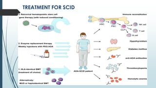
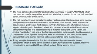
 Severe combined immunodeficiency syndrome (SCID), also known as
alymphocytosis, Glanzmann-Riniker syndrome, severe mixed
immunodeficiency syndrome and thymic alymphoplasia, is a primary
immune deficiency. The defining characteristics is usually a severe defect in
both T and B lymphocytes system . This normally results in onset of one or
more infections within first few month of life. The infections are usually life
threatening and may include pneumonia, meningitis or bloodstream
infections.
 It is generally heritable.
 There are 13 different genetic defects that can cause SCID.
 It is often called as “bubble boy disease” because it victims are extremely
vulnerable to infectious disease and some of them ,such as David Vitter, a
boy with x-linked SCID, lived for 12 years in a plastic, germ-free bubble.
 SCID is the result of an immune system so highly compromised that it’s
consider almost absent.
TYPES
1. X-LINKED SEVERE COMBINED IMMUNODEFICIENCY
2. ADENOSINE DEAMINASE DEFICIENCY
3. PURINE NUCLEOSIDE PHOSPHORYLASE DEFICIENCY
4. RETICULAR DYSGENESIS
5. OMENN SYNDROME
6. BARE LYMPHOCYTE SYNDROME
7. JANUS KINASE-3 (JAK3)
8. ARTEMIS/DCLRE1C
X-LINKED SEVERE COMBINED IMMUONDEFICIENCY
It is the most common form of scid ,affecting nearly 45% of all cases. It arises due
to a mutation in a gene on the X-chromosome that encodes for a component called
common gamma chain, this component is required for development of T-cell
growth factor receptor and other growth factor receptors (interleukins), lack of this
component causes very low T-lymphocyte and NK lymphocyte count B-
lymphocytes are non-functional but have higher count . This deficiency is inherited
as x-linked recessive trait.
ADENOSINE DEAMINASE DEFICIENCY(ADA)
ADA deficiency is the second most common form of scid accounting for 15% of all
cases . ADA is essential for metabolic functions of a variety of body cells especially
T-cells. Lack of this enzyme leads to an accumulations of toxic metabolic by-
products (dATP) within lymphocytes that causes the cells to die. Babies with this
type of scid have the lowest total lymphocyte count including low T,B,NK-
lymphocytes count.
This deficiency is inherited as autosomal recessive disorder.
PURINE NUCLEOSIDE PHOSPHORYLASE
DEFICIENCY
An autosomal recessive disorder involving mutations of
the purine nucleoside phosphorylase (PNP) gene. PNP is
a key enzyme in the purine salvage pathway.
Impairment of this enzyme causes elevateddGTP levels
resulting in T-cell toxicity and deficiency.
RETICULAR DYSGENESIS
Inability of granulocyte precursors to form granules .
SYMPTOMS AND DETECTION
 SCID patients are usually affected by severe bacterial, viral and fungal
infections in early life and often present with interstitial lung disease,
chronic diarrhea, and failure to thrive. Ear infections, recurrent
pneumocystis jirovecii (previously carinii) pneumonia, and profuse oral
candidis commonly occur. These babies if untreated usually die within 1
year due to severe recurrent infections unless they have undergone
successful hematopoietic stem cell transplantation.
 Several US states are performing pilot studies to diagnose scid in new
born through T-cell recombinant excision circle i.e. “newborn screening”.
 In a positive family history detection by sequencing fetal DNA is also
helpful. Despite this efforts a standard testing for scid is not currently
available due to the diversity of the genetic defects.
 Otherwise scid is not diagnose until six month, usually indicated by
recurrent infections. The delay in detection is because newborn carry
their mother’s antibodies for the first few weeks of their life and scid
babies look normal.
TREATMENT FOR SCID
TREATMENT FOR SCID
 The most common treatment for scid is BONE MARROW TRANSPLANTATION, which
has been successful using either a matched related or unrelated donor, or a half matched
donor, who would be either parent.
 The half matched type of transplant is called haploidentical. Haploidentical bone marrow
transplant requires the donor marrow to be depleted of all mature T-cells to avoid the
occurrence of graft-versus-host-disease (GVHD). Consequently, a functional immune
system takes longer to develop a patient who receives a haploidentical bone marrow
transplant compared to a patient receiving a matched transplant. David Vetter, the
original “bubble boy” had one of the first transplantation but eventually died because of a
unscreened virus, Epstein- Barr (tests were not available at that time), in his newly
transplanted bone marrow from his sister, an unmatched bone marrow donor.
 Today transplants done in the first 3 months have a high success rate. Inutero
transplants, which is done before the child is born, have also some success. However
complications such as GVHD are difficult to treat if they were to occur.
TREATMENT FOR SCID
 GENE THERAPY has been attempted as an alternative to bone marrow transplant.
Transduction of the missing gene to hematopoietic stem cells using viral vectors is being
tested in ADAscid an X-linked scid. In 1990 a 4- year old Ashante-DeSilva became the first
patient to undergo successful gene therapy. Researches collected samples of Ashanti's
blood, isolated some of her WBCs, and used a retrovirus to inject a healthy adenosine
deaminase gene into them. These cells were injected back into her body, and began to
express a normal enzyme. This, augmented by weekly injection of ADA ,corrected her
deficiency.
 However these trials with gene therapy were stopped, when it was discovered that some of
the patients have developed leukemia resulting from insertion of the gene carrying retrovirus
near the oncogene. No leukemia cases has yet been seen in trials of ADA-SCID, which
does not involve the gamma c gene that may be oncogenic when expressed by a retrovirus.
 A non- curative treatment for patient with ADA-SCID is enzyme replacement therapy in
which the patient is injected with polyethyleneglycol-coupled adenosine deaminase (PEG-
ADA) which metabolizes the toxic substrate (dATP) of the ADA enzyme, and prevents their
accumulations, thus restoring the T-cell functions.
SCID IN ANIMALS
(Non-human SCID)
 SCID mice were and still used in disease, vaccine,
and transplant research, especially as animal
models for testing the safety of new vaccines or
therapeutic agents in people with weakened immune system.
 An animal variation of the disease, an autosomal recessive gene
with clinical signs similar, to human condition, also affects the
Arabian horses.
 Another animal with well characterized SCID pathology is the dog.
There are 2 forms, an X-linked SCID that has similar ontology to X-
linked SCID in human, and an autosomal recessive form similar to
SCID in Arabian horses and mice.
Thank you

severe combined immunodeficiency syndrome

  • 1.
  • 2.
    INTRODUCTION  Severe combinedimmunodeficiency syndrome (SCID), also known as alymphocytosis, Glanzmann-Riniker syndrome, severe mixed immunodeficiency syndrome and thymic alymphoplasia, is a primary immune deficiency. The defining characteristics is usually a severe defect in both T and B lymphocytes system . This normally results in onset of one or more infections within first few month of life. The infections are usually life threatening and may include pneumonia, meningitis or bloodstream infections.  It is generally heritable.  There are 13 different genetic defects that can cause SCID.  It is often called as “bubble boy disease” because it victims are extremely vulnerable to infectious disease and some of them ,such as David Vitter, a boy with x-linked SCID, lived for 12 years in a plastic, germ-free bubble.  SCID is the result of an immune system so highly compromised that it’s consider almost absent.
  • 3.
    TYPES 1. X-LINKED SEVERECOMBINED IMMUNODEFICIENCY 2. ADENOSINE DEAMINASE DEFICIENCY 3. PURINE NUCLEOSIDE PHOSPHORYLASE DEFICIENCY 4. RETICULAR DYSGENESIS 5. OMENN SYNDROME 6. BARE LYMPHOCYTE SYNDROME 7. JANUS KINASE-3 (JAK3) 8. ARTEMIS/DCLRE1C
  • 4.
    X-LINKED SEVERE COMBINEDIMMUONDEFICIENCY It is the most common form of scid ,affecting nearly 45% of all cases. It arises due to a mutation in a gene on the X-chromosome that encodes for a component called common gamma chain, this component is required for development of T-cell growth factor receptor and other growth factor receptors (interleukins), lack of this component causes very low T-lymphocyte and NK lymphocyte count B- lymphocytes are non-functional but have higher count . This deficiency is inherited as x-linked recessive trait. ADENOSINE DEAMINASE DEFICIENCY(ADA) ADA deficiency is the second most common form of scid accounting for 15% of all cases . ADA is essential for metabolic functions of a variety of body cells especially T-cells. Lack of this enzyme leads to an accumulations of toxic metabolic by- products (dATP) within lymphocytes that causes the cells to die. Babies with this type of scid have the lowest total lymphocyte count including low T,B,NK- lymphocytes count. This deficiency is inherited as autosomal recessive disorder.
  • 5.
    PURINE NUCLEOSIDE PHOSPHORYLASE DEFICIENCY Anautosomal recessive disorder involving mutations of the purine nucleoside phosphorylase (PNP) gene. PNP is a key enzyme in the purine salvage pathway. Impairment of this enzyme causes elevateddGTP levels resulting in T-cell toxicity and deficiency. RETICULAR DYSGENESIS Inability of granulocyte precursors to form granules .
  • 6.
    SYMPTOMS AND DETECTION SCID patients are usually affected by severe bacterial, viral and fungal infections in early life and often present with interstitial lung disease, chronic diarrhea, and failure to thrive. Ear infections, recurrent pneumocystis jirovecii (previously carinii) pneumonia, and profuse oral candidis commonly occur. These babies if untreated usually die within 1 year due to severe recurrent infections unless they have undergone successful hematopoietic stem cell transplantation.  Several US states are performing pilot studies to diagnose scid in new born through T-cell recombinant excision circle i.e. “newborn screening”.  In a positive family history detection by sequencing fetal DNA is also helpful. Despite this efforts a standard testing for scid is not currently available due to the diversity of the genetic defects.  Otherwise scid is not diagnose until six month, usually indicated by recurrent infections. The delay in detection is because newborn carry their mother’s antibodies for the first few weeks of their life and scid babies look normal.
  • 7.
  • 8.
    TREATMENT FOR SCID The most common treatment for scid is BONE MARROW TRANSPLANTATION, which has been successful using either a matched related or unrelated donor, or a half matched donor, who would be either parent.  The half matched type of transplant is called haploidentical. Haploidentical bone marrow transplant requires the donor marrow to be depleted of all mature T-cells to avoid the occurrence of graft-versus-host-disease (GVHD). Consequently, a functional immune system takes longer to develop a patient who receives a haploidentical bone marrow transplant compared to a patient receiving a matched transplant. David Vetter, the original “bubble boy” had one of the first transplantation but eventually died because of a unscreened virus, Epstein- Barr (tests were not available at that time), in his newly transplanted bone marrow from his sister, an unmatched bone marrow donor.  Today transplants done in the first 3 months have a high success rate. Inutero transplants, which is done before the child is born, have also some success. However complications such as GVHD are difficult to treat if they were to occur.
  • 9.
    TREATMENT FOR SCID GENE THERAPY has been attempted as an alternative to bone marrow transplant. Transduction of the missing gene to hematopoietic stem cells using viral vectors is being tested in ADAscid an X-linked scid. In 1990 a 4- year old Ashante-DeSilva became the first patient to undergo successful gene therapy. Researches collected samples of Ashanti's blood, isolated some of her WBCs, and used a retrovirus to inject a healthy adenosine deaminase gene into them. These cells were injected back into her body, and began to express a normal enzyme. This, augmented by weekly injection of ADA ,corrected her deficiency.  However these trials with gene therapy were stopped, when it was discovered that some of the patients have developed leukemia resulting from insertion of the gene carrying retrovirus near the oncogene. No leukemia cases has yet been seen in trials of ADA-SCID, which does not involve the gamma c gene that may be oncogenic when expressed by a retrovirus.  A non- curative treatment for patient with ADA-SCID is enzyme replacement therapy in which the patient is injected with polyethyleneglycol-coupled adenosine deaminase (PEG- ADA) which metabolizes the toxic substrate (dATP) of the ADA enzyme, and prevents their accumulations, thus restoring the T-cell functions.
  • 10.
    SCID IN ANIMALS (Non-humanSCID)  SCID mice were and still used in disease, vaccine, and transplant research, especially as animal models for testing the safety of new vaccines or therapeutic agents in people with weakened immune system.  An animal variation of the disease, an autosomal recessive gene with clinical signs similar, to human condition, also affects the Arabian horses.  Another animal with well characterized SCID pathology is the dog. There are 2 forms, an X-linked SCID that has similar ontology to X- linked SCID in human, and an autosomal recessive form similar to SCID in Arabian horses and mice.
  • 11.