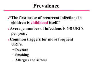
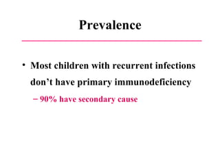
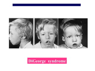
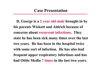
Primary Immunodeficiency  Diseases ( PID )   MBBS.weebly.com
Objectives What will I learn? Characteristics of immune  development in children classification and  clinical manifestation   Diagnosis Treatment
D.Deorge Wiskott Aldrich
Case Presentation D. George is a  2 year old male  brought in by his parents Wiskott and Aldrich because of concerns about  recurrent infections .  They state he has been sick many times over the last two years.  He has been in the hospital twice with some sort of infection.  He has also had  frequent  upper respiratory infections and has had Otitis Media  7  times in the last two years.
The parents of D. George are very concerned.  They wonder is there something wrong with him.  ●  Is it normal to have so many infections?   ●  Could there be something wrong with his   immune system?   ●  How are you going to figure this out?   ●   Does he need testing?
Questions ● What other information should we try to get from D. George and the family? ●   Are there clues we could be missing in the history? ●   Are there clues in the physical?
Immunology  review (development and features of IM)
Organs of the Immune System
Immune system Specific  Nospecific ILs IFNγ TNFα IgM IgG1 ~ 4 IgA1 、 2 IgD 、 IgE phagocytic cells   Macrophages (MC/MΦ) Neutrophils T  cells B   cells ( organs,  cells and molecules   ) complement system
Immunology Review ●   Have Lymphoid Progenitor for Lymphocytes Becomes T cell in Thymus Important in Cellular Immunity Develop into CD4, CD8 or other cells Secretes cytokines, interleukins, etc. Assists B cells in making immunoglobulins Becomes B cell in Bone Marrow Begins with IgM Matures to form other immunoglobulins  IgA, IgE, or IgG (with subclasses IgG1-4) Mature cell is Plasma cell Immunoglobulins used to surround antigens for phagocytosis Responsible for Specific immunity (and memory)
SL SC ProB CFU MØ PMN Plet RBC PreB PT T THYRUM Epi. BM CD3 + IFN- γ 、 IL-2 IL-4  、 5   、 8 、 9 、 10 、 13 Development of Immune cell B Plasma IgM B Plasma IgA IgA IgM B Plasma IgG IgG B Plasma IgE IgE CD19 + CD20   + CD8 + CD4 + TH1 TH2 CTL
More Immunology Review? ● Neutrophils and Macrophages Surround and gobble up organisms, often those surrounded by immunoglobulins (Phagocytosis and Opsonization). Part of natural or innate or nonspecific immunity. ●   Complement system Cascade of plasma proteins which aid in chemotaxis and opsonization.
 
Characteristic of immune development in children T cell and cytokine CTL ↓ —— susceptibility to infections T H 2 ↑ ——  allergic diseases   B cell and antibody antibody production↓ , all kinds of Ig↓ MC/MΦ function insufficient PMN function insufficient Complement system Other immune molecules Mannose-binding lectin
Development of Immunoglobulin  6M birth 100% IgG level of Infant IgG from mother IgG of infant
Age dependent changes of serum Igs levels(g/L ) Ages IgG IgA IgM Neonate 6.46-17.74 0.004-0.017 0.05-0.27 1m- 2.75-7.50 0.05-0.60 0.10-0.70 4m- 3.70-8.30 0.14-0.50 0.33-1.25 7m- 3.50-8.90 0.06-0.54 0.36-1.20 1y- 5.52-11.46 0.06-0.74 0.60-2.12 3y- 4.95-12.74 0.33-0.89 0.65-2.01 7y- 6.09-12.85 0.52-2.16 0.67-2.46 12y- 6.98-14.26 0.92-2.50 0.56-2.18 15-18y 7.54-16.02 0.89-3.24 0.72-2.28
Schematic diagram of the exposure of microorganism during early life fetus Full tern 6M Day care pathogens probiotics
Period of susceptible children premature Full term 6M Day care
Prevalence ● “ The first cause of recurrent infections in children is  childhood  itself.” ●   Average number of infections is 6-8 URI’s per year. ●   Common triggers for more frequent URI’s. Daycare Smoking Allergies and asthma
Prevalence Most children with recurrent infections don’t have primary immunodeficiency 90% have secondary cause
Secondary Causes of Recurrent Infections HIV, HIV, and HIV Anatomic Foreign Body Central line Eustacian Tube Obstruction Sinus tract/fistula Sacral Dimple Cribiform Plate Disruption  Lung sequestration Hypotonia causing aspiration Vesicoureteral Reflux Medications Allergy or Asthma   Cystic Fibrosis GERD Malnutrition Sickle Cell Asplenia Diabetes Cancer Colonization with resistant organism (i.e. MRSA)
Primary Immunodeficiency Disease A group of disorders characterized by an  impaired ability to produce normal immune response.  Most of these disorders are caused  by mutations in genes  involved in the development and function of immune organs, cells, and molecules. Clinical features : Recurrent infection ,  high risk of autoimmune diseases, allergy and malignancy
50% 20% 10% 18% 2% Antibody Complement Phagocyte Cell medi ated Combined Distribution of PID  Up to 2007 more then 200 kinds of PID reported
 
Classification(new) Combined Immunodeficiency  (B and T cells) Predominantly antibody deficiency  (B cells  and Ab) Predominantly T-cell deficiency  (T cells) Immunodeficiency syndromes Phagocyte deficiency  ( PMN’s) Complement deficiency Others Note :--  There is significant overlap among syndromes. --Great variability in expression of disorders for all categories  from mild to severe/fatal.
Combined immunodeficiencies ( 1 ) 1.  Severe  combined immunodeficiency ( SCID ) X-linked (γc deficiency) Autosomal recessive (Jak3 deficiency) RAG1/RAG2 deficiency  Adenosine deaminase (ADA) deficiency Reticular dysgenesis   T  –  B + T  -  B  -
Severe Combined Immunodeficiency , SCID (Bubble boy)
Combined immunodeficiencies   ( 2 ) 2.  Hyper-IgM syndrome 3. Purine nucleoside phosphorylase (PNP) deficiency 4. MHC class Ⅱ deficiency
Clinical features of combined immunodeficiency  Onset age at early infants(4 - 5 months)  Recurrent infection with fungi, virus, bacteria, mycobacterium, protozoa  Opportunistic infections  Poor prognosis, early infant deaths  Severe infection after live virus vaccine and BCG  GVHD after blood transfusion  High risk of malignancy
Humoral / B-cell Defects
Predominantly antibody defects ●   Panhypogammaglobulinemia X-linked agammaglobulinaemia ( Bruton  disease ) Common variable immunodeficiency ( CVID ) Transient hypogammaglobulinaemia of infancy  (ITHG) ●   Selective Ig deficiency Ig heavy chain deficiency IgA deficiency Selective IgG subclass deficiency
— Bruton disease —  mutations in btk — maturation disorder of  pre-B cell
CVID—variable ( lack of signals from T cells ) ITHG—delayed maturation of T H  function
Predominantly antibody defects Common clinical manifestations: ●   Recurrent bacterial infections (sepsis and meningitis) ●   Viral ,fungal or protozoan infections  rare ●   Lymphatic system hypoplasia-  tonsils, lymph node  ( except CVID ) ●   Autoimmune disease
Predominantly antibody defects Laboratory test   ● Serum  Ig ↓ (< 3 ~ 4g/L ) ●   Natural antibody ↓  ( hemagglutinin titers < 1∶4 ) ●   Common antibody ↓ ,> 2 A , ASO < 1 ∶10 ●   Antibody responses to vaccine antigens ↓ ●   Circulating  B cell  ( CD 19 + 、 CD 20 +) ↓,  bearing Ig cell ↓
                                                                                               .   Cell-Mediated/T cell Immunity
Predominantly  T-cell defects 1.  CD 4 +   deficiency 2. CD 7 +  deficiency   3. IL-2  deficiency 4. multiple cytokines  deficiency   ( IL-2 、 -4 、 -5 ) Not completely understood ??
Destination  serum Ig  B-cells  T-cells  genetic defect  clinical findings ●   Wiskott-  IgM↓  Normal  Progressive ↓  XL  Thrombocytopenia Aldrich Syn  Mutation in WAS   eczema  lymphoma ●   Ataxia-  IgA, E, G↓ Normal  ↓  ATM  Ataxia,  Telangiectasia  IgM ↑  telangiectasia  ●   DiGeorge  Normal or ↓   Normal  ↓or normal  Deletion of  Hypoparathyroidism Syn  chromosome  conotruncal defect 22q11.2-pter  abnormal facies Immunodeficiency syndrome deficiencies
Wiskott-Aldrich Syndrome X-linked Recessive Gene defect of WAS protein  B and T cell dysfunction Triad of  Thrombocytopenia Eczema Recurrent pyogenic infections Treatment – Stem cell or Bone Marrow transplant Prognosis - Average life expectancy 11 years
( eczema )
Ataxia-Telangiectasia Autosomal Recessive Have both B and T cell dysfunction  more characteristics of B cell dysfunction Associated Symptoms Ataxia from early age – progressive Telangiectasia develop after 2 yrs High risk for various malignancies Endocrine abnormalities – many with Diabetes Liver Dysfunction Treatment – supportive Prognosis – death often in early childhood
Ataxia
telangiectasi a
DiGeorge  Syndrome
DiGeorge Syndrome Deletion of chromosome 22q11.2 Defective development of 3 rd  and 4 th  pharyngeal pouches Absence of Thymus Therefore low or absent T cells No B cell abnormalities except in more severe forms. Associated Anomalies Conotruncal Cardiac Defects  VSD Tetralogy of Fallot Interrupted Aortic Arch Parathyroid Hypoplasia  Low Calcium Tetany
DiGeorge Syndrome Other Anomalies Cleft Palate Velocardiofacial Syndrome Esophageal abnormalities Ocular anomalies Renal anomalies Increased incidence of Autoimmune disease Diagnosis – FISH Will often have decreased CD3 T cells Treatment IVIG and antibiotic prophylaxis Should be on TMP/SFA for PCP prophylaxis Thymic transplant or Bone marrow transplant
DiGeorge anormaly
Hypertelorism hooded eyelids   short philtrum with  fish-mouth appearance   ,  micrognathia   Low set ears telecanthus with short  palpebral fissures   Facial features of children with DiGeorge syndrome
DiGeorge  syndrome
Phagocytic Disorders                                                                
Congenital defects of phagocytic number  and/or function ●   Sever congenital neutropenia  ( SCN , Kostmann syndrome ) ● Chronic granulomatous disease ●   Chediak-Hiashi syndrome
Bacteria phagosome Bacteria Phagosome Neutrophil NADPH  H + NADPH  H + e -   +O 2 O 2 - H + H 2 O 2 Normsal phagocyte Dysfunction of phagocyte Chronic granulomatous disease
Chronic Granulomatous Disease Rare – 20 cases/year in the US Genetics 70 % X-linked recessive Defect in NADPH oxidase Can’t form reactive oxygen species to destroy micro-organisms Symptoms Pneumonia, Abscesses, Adenitis, Osteomyelitis Uniquely susceptible to Aspergillosis
Chronic Granulomatous Disease Associated Symptoms Severe Acne Excessive Granulomata – often in GI tract  Lupus Chorioretinitis Diagnosis – Nitroblue Tetrazolium Test (NBT) Treatment Antibacterial and antifungal prophylaxis Interferon Gamma Stem cell or Bone Marrow Transplant
Complement Disorders
Complement deficiency Defects  Inheritance  Clinical findings ●   Classical pathway  Infections , (C1q 、 r 、 s 、 C 2 、 C 4 )  AR  Autoimmune   disease C 1  inhibitor  AD  Hereditary   angioedema ●   Alternaive  pathway  Recurrent  pyogenic  infection  (C 3 、 FactorⅠ 、 FactorH)  AR ●   Others  Neisseria infection (C 5  ~ 8 、 properdin 、 factor D)   AR  Lupus-link syndrome C 9   AR  Asymptomatic
Common clinical manifestations PID ●  Infection  recurrent ▼ Age  >50 %  younger than 3 yrs ▼ Location  respiratory tract  , GI tract… ▼ Pathogen  ▼ Course ●  Malignancy and autoimmune disease ●  Tendency of inheritance  <15yrs  80 % male ●  Others
Table 1. Characteristic infections of the primary immunodeficiencies component primary pathogen primary site clinical example T-cells intracellular, bacteria viruses, protozoa, fungi, non-specific SCID, DiGeorge B-cells pneumococcus, streptococcus,  haemophilus lung, skin, CNS IgG, IgM deficiency IgG, IgM deficiency enteric bacteria and viruses GI, nasal, eye IgA deficiency phagocytes Staphylococcal, Klebsiella Pseudomonas, lung, skin, regional lymph node Chronic granulomatous disease (CGD) complement neisseria, Haemophilus, pneumococcus, streptococcus CNS lung skin C3, Factors I and H,  late C omponents
Approach to the patients with suspected immunodeficiency ●   The medical history in immunodeficiency ●  Physical examination ●  Laboratory investigation
Key History ●  Get history of infections Location Organism Frequency Response to therapy Seriousness (i.e. hospitalization) ●   Family History – Including Consanguinity   ●   Growth Pattern
From INFO4PI.ORG
Physical finding ● Failure to thrive ●   Dysmorphism(abnormal facial features) ●   Skin  and mucosa Eczema,  petechiae Abscesses,  pyoderma Telangiectasia Delayed umbilical separation ●   Respiratory tract ………
Diagnostic Work Up ●   Antibody Defects Quantitative - Immunoglobulin levels Functional - Antibody Titers to immunizations ●   T cell Quantitative – CBC, Abs lymphocyte count Functional – Skin tests for antigens (Mumps, candida, etc.) Chest  x-ray ●   Phagocyte  Quantitative – CBC, Abs neutrophil count Functional – NBT test ●   Complement Quantitative – C3, C4 Functional – CH50
Initial and advanced laboratory tests for immunodeficiency
From INFO4PI.ORG
Management of PID ●   General treatment ●   Replacement therapy ●   Immune reconstruction ●   Gene therapy
General management of PID ●   Diet ●   Avoidance of pathogens (“germ-free” care) ●   Antibiotics Use in acute illness Prophylactic  ●   Avoid whole blood transfusion in combined immunodeficiency disorder(GVHR) ●   Avoid live virus vaccines and BCG
Bubble Boy
Immunoglobulin replacement Treatment of severe antibody disorders ● IVIG  400 ~ 600mg/kg/m  iv  drip ●   Frozen plasma  10ml/kg/month ◎  Caution with  administration of blood production if selective  IgA deficiency
How to get out of the  bubble ?
Specific treatment for cellular deficiency  ●   Bone marrow transplantation ●   Replacement therapy Enzyme replacement Gene therapy Thymic hormones Cytokines ●   Fetal thymus transplantation
A new  hope for gene therapy of  immunodeficency how to get out of the bubble?
Specific treatment of phagocytic disorders ●   Interferon gamma for CGD ●   Granulocyte transfusion
Case Presentation D. George is a  2 year old male  brought in by his parents Wiskott and Aldrich because of concerns about  recurrent infections .  They state he has been sick many times over the last two years.  He has been in the hospital twice with some sort of infection.  He has also had frequent upper respiratory infections and has had Otitis Media  7 times  in the last two years.
Questions ● What other history should we get? ●   Does the child need work up for immunodeficiency? Depends on history What immunodeficiency should we worry about? What work up should be done?
 
 
Related website http://www.info4pi.org/aboutPIin/ http://elearning.sysu.edu.cn/webapps/login/
Thanks you for  your attention
Mini case discussion
Case presentation (1) A  3-month -old  boy  born of non-consanguineous marriage  presented with  recurrent diarrhea  and  failure to thrive  requiring 3 hospitalizations in past.  born at full term with a birth weight of 3.25 kg and was on exclusive breast feeds.  received BCG and 1st dose of OPV and DPT.  had two elder sisters. The eldest of them was small for gestational age, had chronic diarrhea, failure to thrive and  died  at 2 months of age. The other sister  died  at 9 months of age due to pneumonia.
Case presentation (2) His weight was 2.7 kg and length was 49 cm.  He had triangular facies,  tonsils were absent  and  no BCG scar  was seen.  There was  no  rash,  lymphadenopathy  or organomegaly.
Case presentation (3) His hemogram showed total leucocyte count of 6,300 cells/cumm with absolute lymphocyte count of  1575 cells/cumm .  X-ray chest showed  no evidence of thymus .  HIV ELISA was negative.
characteristics 3 month old boy recurrent infections with failure to thrive  absent tonsils borderline lymphocyte counts  X-ray chest showed no evidence of thymus
Questions What kinds of diseases did the baby suffer? What kinds of Lab tests would you order? How can you  save his life? How about his prognosis?
Diagnosis T-B+ SCID

14 Primary Immunodeficiency Diseases

  • 1.
    Primary Immunodeficiency Diseases ( PID ) MBBS.weebly.com
  • 2.
    Objectives What willI learn? Characteristics of immune development in children classification and clinical manifestation Diagnosis Treatment
  • 3.
  • 4.
    Case Presentation D.George is a 2 year old male brought in by his parents Wiskott and Aldrich because of concerns about recurrent infections . They state he has been sick many times over the last two years. He has been in the hospital twice with some sort of infection. He has also had frequent upper respiratory infections and has had Otitis Media 7 times in the last two years.
  • 5.
    The parents ofD. George are very concerned. They wonder is there something wrong with him. ● Is it normal to have so many infections? ● Could there be something wrong with his immune system? ● How are you going to figure this out? ● Does he need testing?
  • 6.
    Questions ● Whatother information should we try to get from D. George and the family? ● Are there clues we could be missing in the history? ● Are there clues in the physical?
  • 7.
    Immunology review(development and features of IM)
  • 8.
    Organs of theImmune System
  • 9.
    Immune system Specific Nospecific ILs IFNγ TNFα IgM IgG1 ~ 4 IgA1 、 2 IgD 、 IgE phagocytic cells Macrophages (MC/MΦ) Neutrophils T cells B cells ( organs, cells and molecules ) complement system
  • 10.
    Immunology Review ● Have Lymphoid Progenitor for Lymphocytes Becomes T cell in Thymus Important in Cellular Immunity Develop into CD4, CD8 or other cells Secretes cytokines, interleukins, etc. Assists B cells in making immunoglobulins Becomes B cell in Bone Marrow Begins with IgM Matures to form other immunoglobulins IgA, IgE, or IgG (with subclasses IgG1-4) Mature cell is Plasma cell Immunoglobulins used to surround antigens for phagocytosis Responsible for Specific immunity (and memory)
  • 11.
    SL SC ProBCFU MØ PMN Plet RBC PreB PT T THYRUM Epi. BM CD3 + IFN- γ 、 IL-2 IL-4 、 5 、 8 、 9 、 10 、 13 Development of Immune cell B Plasma IgM B Plasma IgA IgA IgM B Plasma IgG IgG B Plasma IgE IgE CD19 + CD20 + CD8 + CD4 + TH1 TH2 CTL
  • 12.
    More Immunology Review?● Neutrophils and Macrophages Surround and gobble up organisms, often those surrounded by immunoglobulins (Phagocytosis and Opsonization). Part of natural or innate or nonspecific immunity. ● Complement system Cascade of plasma proteins which aid in chemotaxis and opsonization.
  • 13.
  • 14.
    Characteristic of immunedevelopment in children T cell and cytokine CTL ↓ —— susceptibility to infections T H 2 ↑ —— allergic diseases B cell and antibody antibody production↓ , all kinds of Ig↓ MC/MΦ function insufficient PMN function insufficient Complement system Other immune molecules Mannose-binding lectin
  • 15.
    Development of Immunoglobulin 6M birth 100% IgG level of Infant IgG from mother IgG of infant
  • 16.
    Age dependent changesof serum Igs levels(g/L ) Ages IgG IgA IgM Neonate 6.46-17.74 0.004-0.017 0.05-0.27 1m- 2.75-7.50 0.05-0.60 0.10-0.70 4m- 3.70-8.30 0.14-0.50 0.33-1.25 7m- 3.50-8.90 0.06-0.54 0.36-1.20 1y- 5.52-11.46 0.06-0.74 0.60-2.12 3y- 4.95-12.74 0.33-0.89 0.65-2.01 7y- 6.09-12.85 0.52-2.16 0.67-2.46 12y- 6.98-14.26 0.92-2.50 0.56-2.18 15-18y 7.54-16.02 0.89-3.24 0.72-2.28
  • 17.
    Schematic diagram ofthe exposure of microorganism during early life fetus Full tern 6M Day care pathogens probiotics
  • 18.
    Period of susceptiblechildren premature Full term 6M Day care
  • 19.
    Prevalence ● “The first cause of recurrent infections in children is childhood itself.” ● Average number of infections is 6-8 URI’s per year. ● Common triggers for more frequent URI’s. Daycare Smoking Allergies and asthma
  • 20.
    Prevalence Most childrenwith recurrent infections don’t have primary immunodeficiency 90% have secondary cause
  • 21.
    Secondary Causes ofRecurrent Infections HIV, HIV, and HIV Anatomic Foreign Body Central line Eustacian Tube Obstruction Sinus tract/fistula Sacral Dimple Cribiform Plate Disruption Lung sequestration Hypotonia causing aspiration Vesicoureteral Reflux Medications Allergy or Asthma Cystic Fibrosis GERD Malnutrition Sickle Cell Asplenia Diabetes Cancer Colonization with resistant organism (i.e. MRSA)
  • 22.
    Primary Immunodeficiency DiseaseA group of disorders characterized by an impaired ability to produce normal immune response. Most of these disorders are caused by mutations in genes involved in the development and function of immune organs, cells, and molecules. Clinical features : Recurrent infection , high risk of autoimmune diseases, allergy and malignancy
  • 23.
    50% 20% 10%18% 2% Antibody Complement Phagocyte Cell medi ated Combined Distribution of PID Up to 2007 more then 200 kinds of PID reported
  • 24.
  • 25.
    Classification(new) Combined Immunodeficiency (B and T cells) Predominantly antibody deficiency (B cells and Ab) Predominantly T-cell deficiency (T cells) Immunodeficiency syndromes Phagocyte deficiency ( PMN’s) Complement deficiency Others Note :-- There is significant overlap among syndromes. --Great variability in expression of disorders for all categories from mild to severe/fatal.
  • 26.
    Combined immunodeficiencies (1 ) 1. Severe combined immunodeficiency ( SCID ) X-linked (γc deficiency) Autosomal recessive (Jak3 deficiency) RAG1/RAG2 deficiency Adenosine deaminase (ADA) deficiency Reticular dysgenesis T – B + T - B -
  • 27.
    Severe Combined Immunodeficiency, SCID (Bubble boy)
  • 28.
    Combined immunodeficiencies ( 2 ) 2. Hyper-IgM syndrome 3. Purine nucleoside phosphorylase (PNP) deficiency 4. MHC class Ⅱ deficiency
  • 29.
    Clinical features ofcombined immunodeficiency Onset age at early infants(4 - 5 months) Recurrent infection with fungi, virus, bacteria, mycobacterium, protozoa Opportunistic infections Poor prognosis, early infant deaths Severe infection after live virus vaccine and BCG GVHD after blood transfusion High risk of malignancy
  • 30.
  • 31.
    Predominantly antibody defects● Panhypogammaglobulinemia X-linked agammaglobulinaemia ( Bruton disease ) Common variable immunodeficiency ( CVID ) Transient hypogammaglobulinaemia of infancy (ITHG) ● Selective Ig deficiency Ig heavy chain deficiency IgA deficiency Selective IgG subclass deficiency
  • 32.
    — Bruton disease— mutations in btk — maturation disorder of pre-B cell
  • 33.
    CVID—variable ( lackof signals from T cells ) ITHG—delayed maturation of T H function
  • 34.
    Predominantly antibody defectsCommon clinical manifestations: ● Recurrent bacterial infections (sepsis and meningitis) ● Viral ,fungal or protozoan infections rare ● Lymphatic system hypoplasia- tonsils, lymph node ( except CVID ) ● Autoimmune disease
  • 35.
    Predominantly antibody defectsLaboratory test ● Serum Ig ↓ (< 3 ~ 4g/L ) ● Natural antibody ↓ ( hemagglutinin titers < 1∶4 ) ● Common antibody ↓ ,> 2 A , ASO < 1 ∶10 ● Antibody responses to vaccine antigens ↓ ● Circulating B cell ( CD 19 + 、 CD 20 +) ↓, bearing Ig cell ↓
  • 36.
  • 37.
    Predominantly T-celldefects 1. CD 4 + deficiency 2. CD 7 + deficiency 3. IL-2 deficiency 4. multiple cytokines deficiency ( IL-2 、 -4 、 -5 ) Not completely understood ??
  • 38.
    Destination serumIg B-cells T-cells genetic defect clinical findings ● Wiskott- IgM↓ Normal Progressive ↓ XL Thrombocytopenia Aldrich Syn Mutation in WAS eczema lymphoma ● Ataxia- IgA, E, G↓ Normal ↓ ATM Ataxia, Telangiectasia IgM ↑ telangiectasia ● DiGeorge Normal or ↓ Normal ↓or normal Deletion of Hypoparathyroidism Syn chromosome conotruncal defect 22q11.2-pter abnormal facies Immunodeficiency syndrome deficiencies
  • 39.
    Wiskott-Aldrich Syndrome X-linkedRecessive Gene defect of WAS protein B and T cell dysfunction Triad of Thrombocytopenia Eczema Recurrent pyogenic infections Treatment – Stem cell or Bone Marrow transplant Prognosis - Average life expectancy 11 years
  • 40.
  • 41.
    Ataxia-Telangiectasia Autosomal RecessiveHave both B and T cell dysfunction more characteristics of B cell dysfunction Associated Symptoms Ataxia from early age – progressive Telangiectasia develop after 2 yrs High risk for various malignancies Endocrine abnormalities – many with Diabetes Liver Dysfunction Treatment – supportive Prognosis – death often in early childhood
  • 42.
  • 43.
  • 44.
  • 45.
    DiGeorge Syndrome Deletionof chromosome 22q11.2 Defective development of 3 rd and 4 th pharyngeal pouches Absence of Thymus Therefore low or absent T cells No B cell abnormalities except in more severe forms. Associated Anomalies Conotruncal Cardiac Defects VSD Tetralogy of Fallot Interrupted Aortic Arch Parathyroid Hypoplasia Low Calcium Tetany
  • 46.
    DiGeorge Syndrome OtherAnomalies Cleft Palate Velocardiofacial Syndrome Esophageal abnormalities Ocular anomalies Renal anomalies Increased incidence of Autoimmune disease Diagnosis – FISH Will often have decreased CD3 T cells Treatment IVIG and antibiotic prophylaxis Should be on TMP/SFA for PCP prophylaxis Thymic transplant or Bone marrow transplant
  • 47.
  • 48.
    Hypertelorism hooded eyelids short philtrum with fish-mouth appearance , micrognathia Low set ears telecanthus with short palpebral fissures Facial features of children with DiGeorge syndrome
  • 49.
  • 50.
    Phagocytic Disorders                                                               
  • 51.
    Congenital defects ofphagocytic number and/or function ● Sever congenital neutropenia ( SCN , Kostmann syndrome ) ● Chronic granulomatous disease ● Chediak-Hiashi syndrome
  • 52.
    Bacteria phagosome BacteriaPhagosome Neutrophil NADPH H + NADPH H + e - +O 2 O 2 - H + H 2 O 2 Normsal phagocyte Dysfunction of phagocyte Chronic granulomatous disease
  • 53.
    Chronic Granulomatous DiseaseRare – 20 cases/year in the US Genetics 70 % X-linked recessive Defect in NADPH oxidase Can’t form reactive oxygen species to destroy micro-organisms Symptoms Pneumonia, Abscesses, Adenitis, Osteomyelitis Uniquely susceptible to Aspergillosis
  • 54.
    Chronic Granulomatous DiseaseAssociated Symptoms Severe Acne Excessive Granulomata – often in GI tract Lupus Chorioretinitis Diagnosis – Nitroblue Tetrazolium Test (NBT) Treatment Antibacterial and antifungal prophylaxis Interferon Gamma Stem cell or Bone Marrow Transplant
  • 55.
  • 56.
    Complement deficiency Defects Inheritance Clinical findings ● Classical pathway Infections , (C1q 、 r 、 s 、 C 2 、 C 4 ) AR Autoimmune disease C 1 inhibitor AD Hereditary angioedema ● Alternaive pathway Recurrent pyogenic infection (C 3 、 FactorⅠ 、 FactorH) AR ● Others Neisseria infection (C 5 ~ 8 、 properdin 、 factor D) AR Lupus-link syndrome C 9 AR Asymptomatic
  • 57.
    Common clinical manifestationsPID ● Infection recurrent ▼ Age >50 % younger than 3 yrs ▼ Location respiratory tract , GI tract… ▼ Pathogen ▼ Course ● Malignancy and autoimmune disease ● Tendency of inheritance <15yrs 80 % male ● Others
  • 58.
    Table 1. Characteristicinfections of the primary immunodeficiencies component primary pathogen primary site clinical example T-cells intracellular, bacteria viruses, protozoa, fungi, non-specific SCID, DiGeorge B-cells pneumococcus, streptococcus, haemophilus lung, skin, CNS IgG, IgM deficiency IgG, IgM deficiency enteric bacteria and viruses GI, nasal, eye IgA deficiency phagocytes Staphylococcal, Klebsiella Pseudomonas, lung, skin, regional lymph node Chronic granulomatous disease (CGD) complement neisseria, Haemophilus, pneumococcus, streptococcus CNS lung skin C3, Factors I and H, late C omponents
  • 59.
    Approach to thepatients with suspected immunodeficiency ● The medical history in immunodeficiency ● Physical examination ● Laboratory investigation
  • 60.
    Key History ● Get history of infections Location Organism Frequency Response to therapy Seriousness (i.e. hospitalization) ● Family History – Including Consanguinity ● Growth Pattern
  • 61.
  • 62.
    Physical finding ●Failure to thrive ● Dysmorphism(abnormal facial features) ● Skin and mucosa Eczema, petechiae Abscesses, pyoderma Telangiectasia Delayed umbilical separation ● Respiratory tract ………
  • 63.
    Diagnostic Work Up● Antibody Defects Quantitative - Immunoglobulin levels Functional - Antibody Titers to immunizations ● T cell Quantitative – CBC, Abs lymphocyte count Functional – Skin tests for antigens (Mumps, candida, etc.) Chest x-ray ● Phagocyte Quantitative – CBC, Abs neutrophil count Functional – NBT test ● Complement Quantitative – C3, C4 Functional – CH50
  • 64.
    Initial and advancedlaboratory tests for immunodeficiency
  • 65.
  • 66.
    Management of PID● General treatment ● Replacement therapy ● Immune reconstruction ● Gene therapy
  • 67.
    General management ofPID ● Diet ● Avoidance of pathogens (“germ-free” care) ● Antibiotics Use in acute illness Prophylactic ● Avoid whole blood transfusion in combined immunodeficiency disorder(GVHR) ● Avoid live virus vaccines and BCG
  • 68.
  • 69.
    Immunoglobulin replacement Treatmentof severe antibody disorders ● IVIG 400 ~ 600mg/kg/m iv drip ● Frozen plasma 10ml/kg/month ◎ Caution with administration of blood production if selective IgA deficiency
  • 70.
    How to getout of the bubble ?
  • 71.
    Specific treatment forcellular deficiency ● Bone marrow transplantation ● Replacement therapy Enzyme replacement Gene therapy Thymic hormones Cytokines ● Fetal thymus transplantation
  • 72.
    A new hope for gene therapy of immunodeficency how to get out of the bubble?
  • 73.
    Specific treatment ofphagocytic disorders ● Interferon gamma for CGD ● Granulocyte transfusion
  • 74.
    Case Presentation D.George is a 2 year old male brought in by his parents Wiskott and Aldrich because of concerns about recurrent infections . They state he has been sick many times over the last two years. He has been in the hospital twice with some sort of infection. He has also had frequent upper respiratory infections and has had Otitis Media 7 times in the last two years.
  • 75.
    Questions ● Whatother history should we get? ● Does the child need work up for immunodeficiency? Depends on history What immunodeficiency should we worry about? What work up should be done?
  • 76.
  • 77.
  • 78.
    Related website http://www.info4pi.org/aboutPIin/http://elearning.sysu.edu.cn/webapps/login/
  • 79.
    Thanks you for your attention
  • 80.
  • 81.
    Case presentation (1)A 3-month -old boy born of non-consanguineous marriage presented with recurrent diarrhea and failure to thrive requiring 3 hospitalizations in past. born at full term with a birth weight of 3.25 kg and was on exclusive breast feeds. received BCG and 1st dose of OPV and DPT. had two elder sisters. The eldest of them was small for gestational age, had chronic diarrhea, failure to thrive and died at 2 months of age. The other sister died at 9 months of age due to pneumonia.
  • 82.
    Case presentation (2)His weight was 2.7 kg and length was 49 cm. He had triangular facies, tonsils were absent and no BCG scar was seen. There was no rash, lymphadenopathy or organomegaly.
  • 83.
    Case presentation (3)His hemogram showed total leucocyte count of 6,300 cells/cumm with absolute lymphocyte count of 1575 cells/cumm . X-ray chest showed no evidence of thymus . HIV ELISA was negative.
  • 84.
    characteristics 3 monthold boy recurrent infections with failure to thrive absent tonsils borderline lymphocyte counts X-ray chest showed no evidence of thymus
  • 85.
    Questions What kindsof diseases did the baby suffer? What kinds of Lab tests would you order? How can you save his life? How about his prognosis?
  • 86.