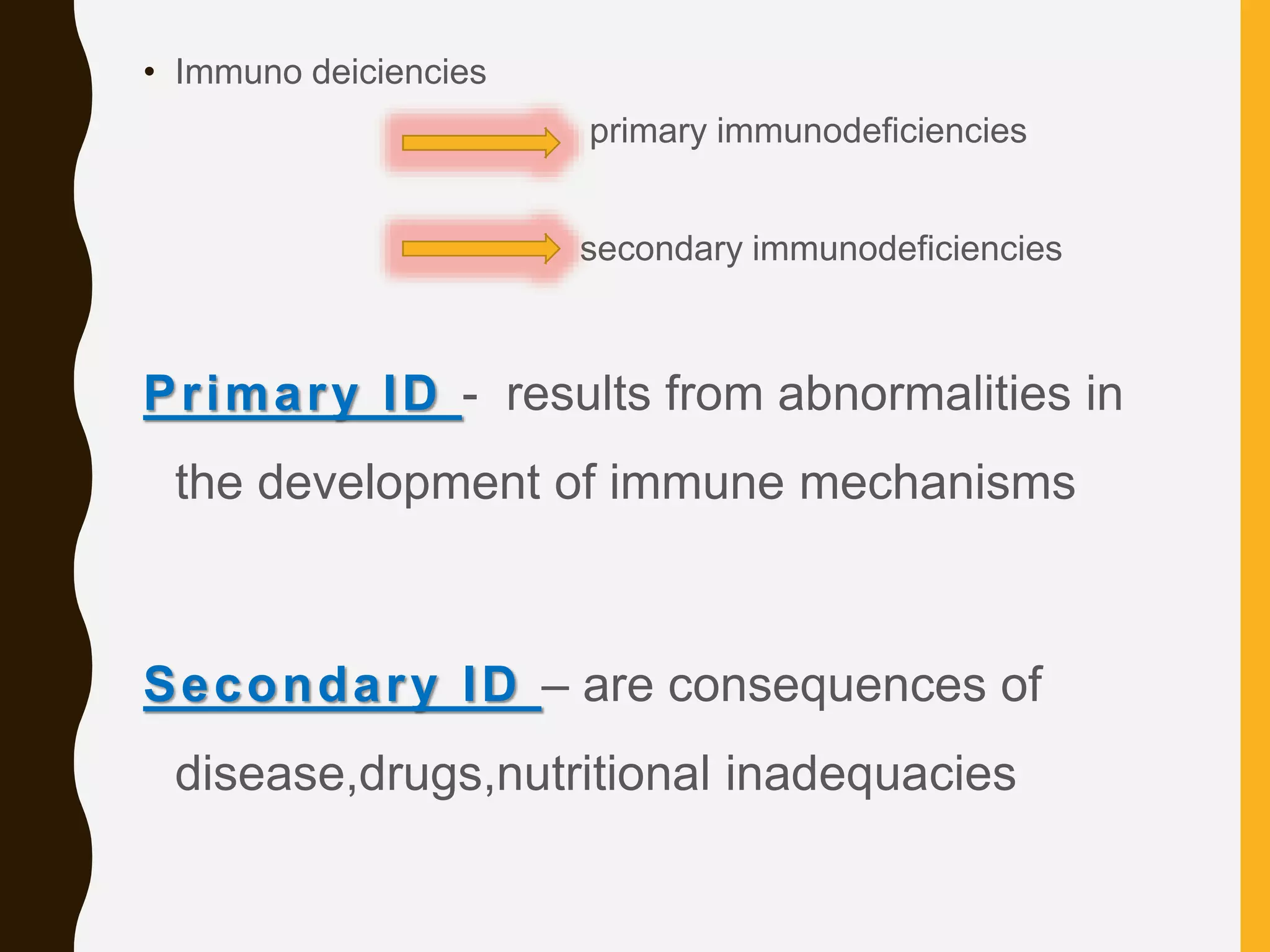
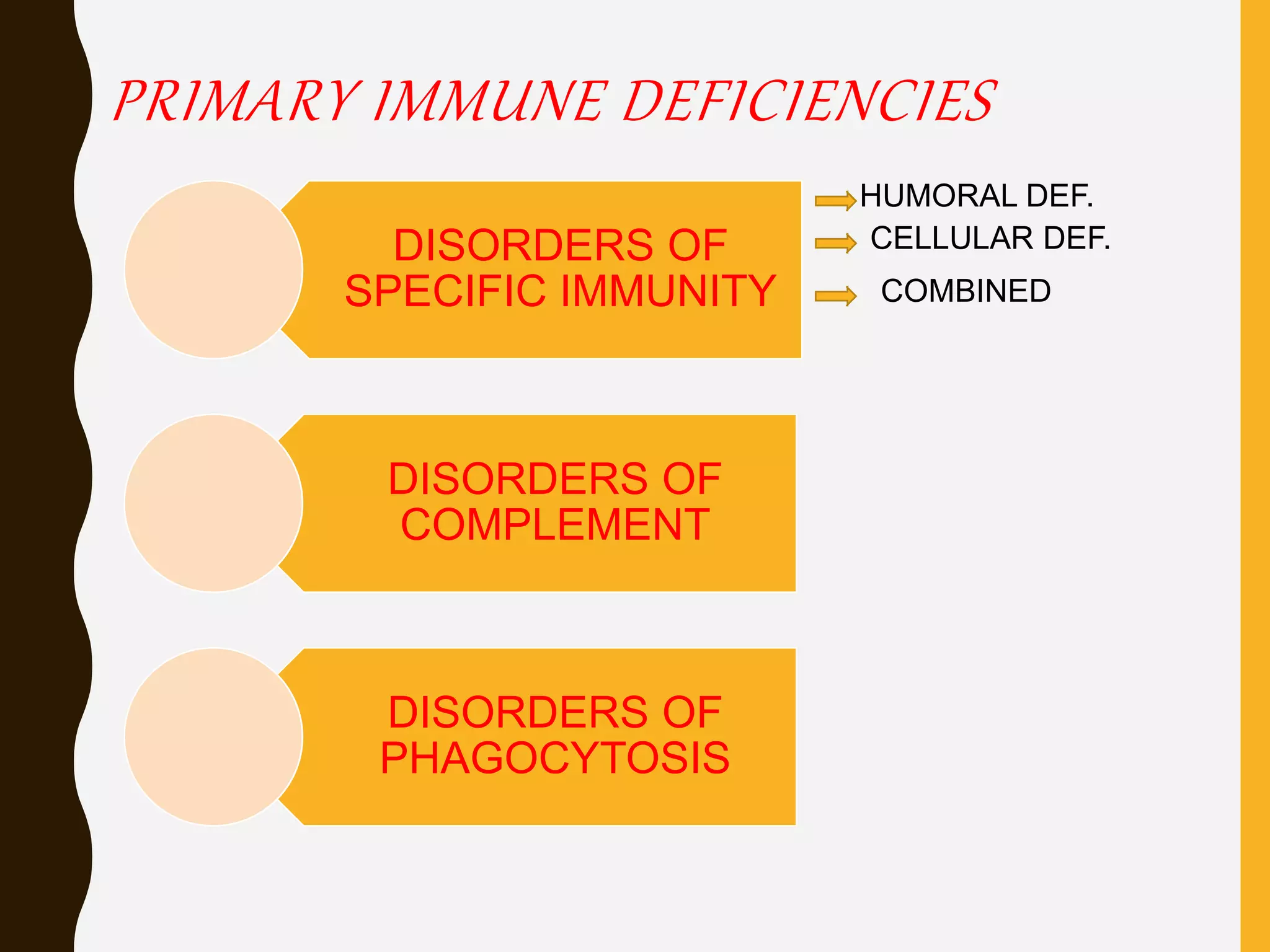
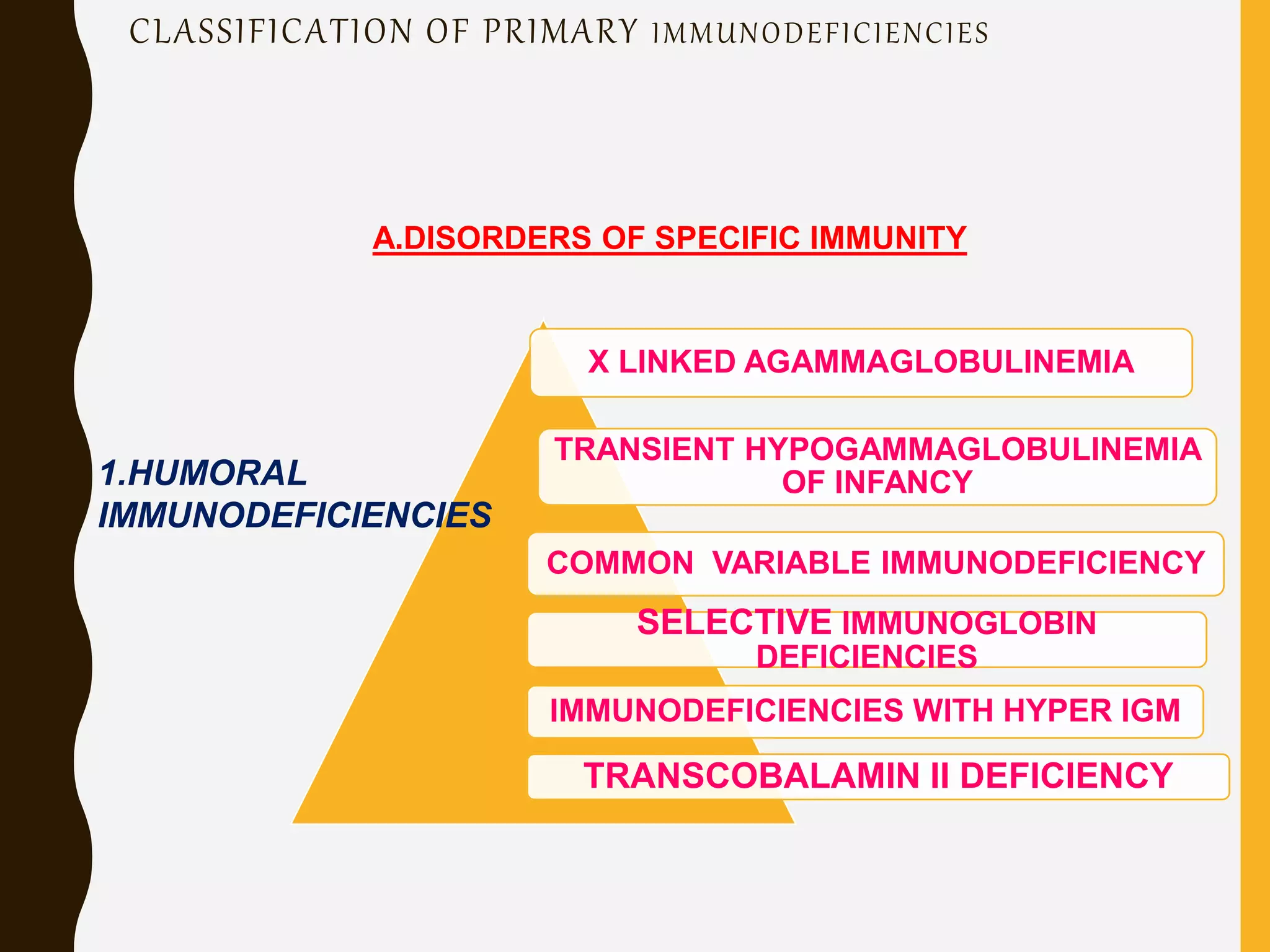
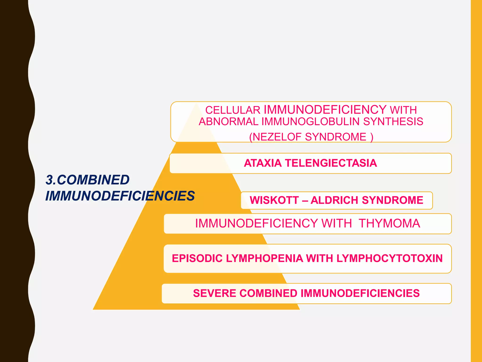
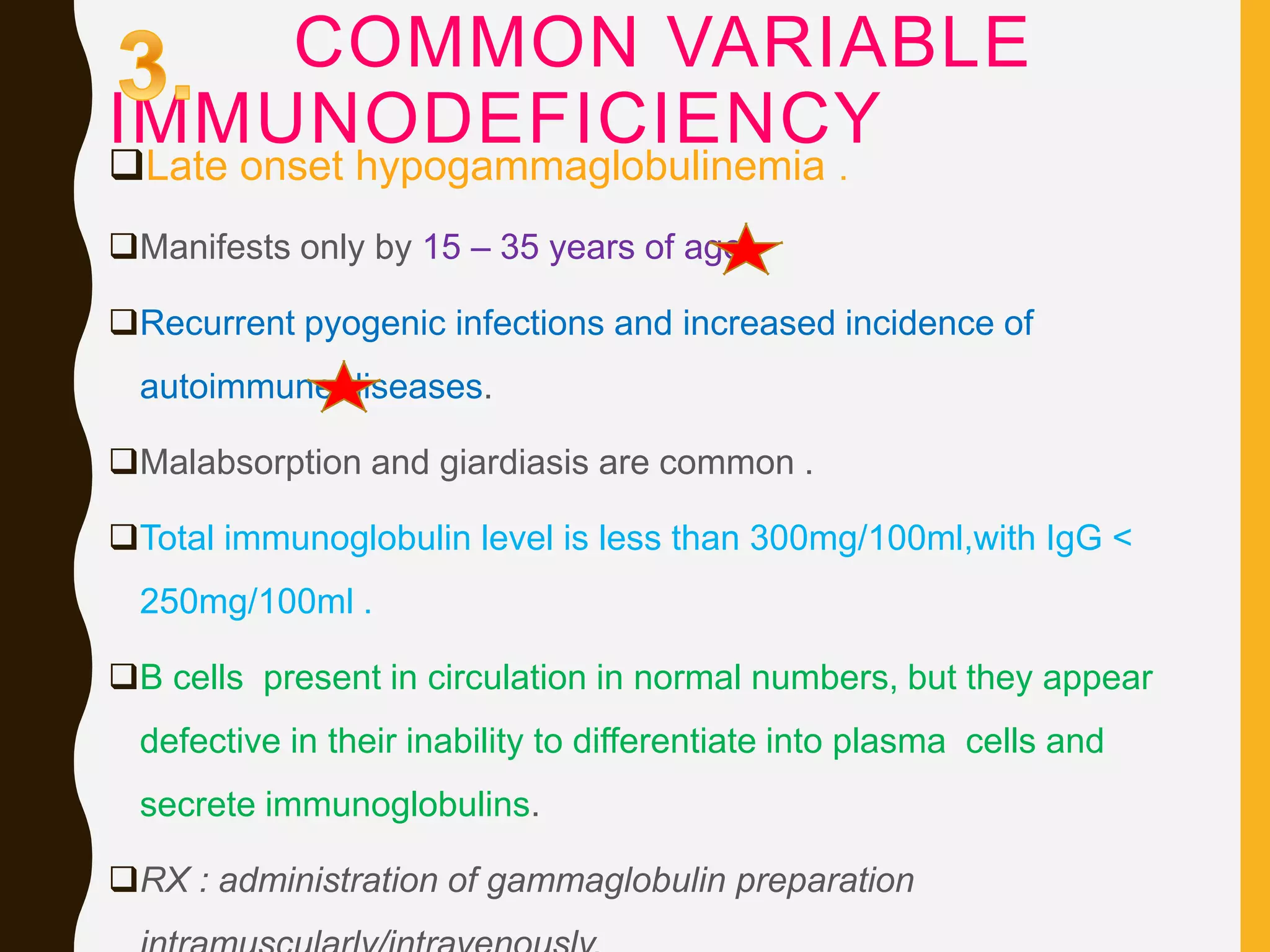
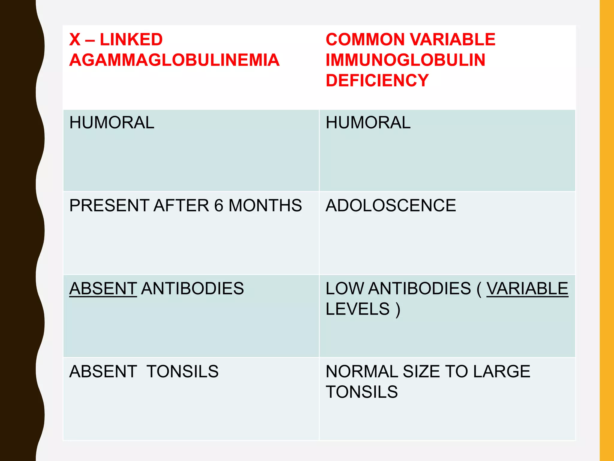
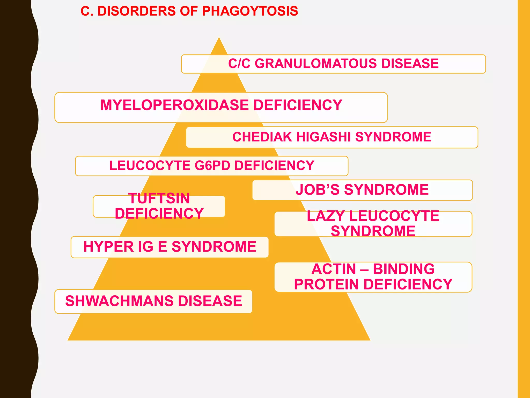
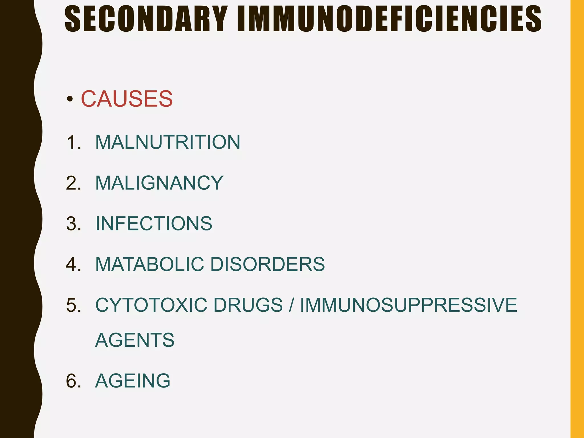
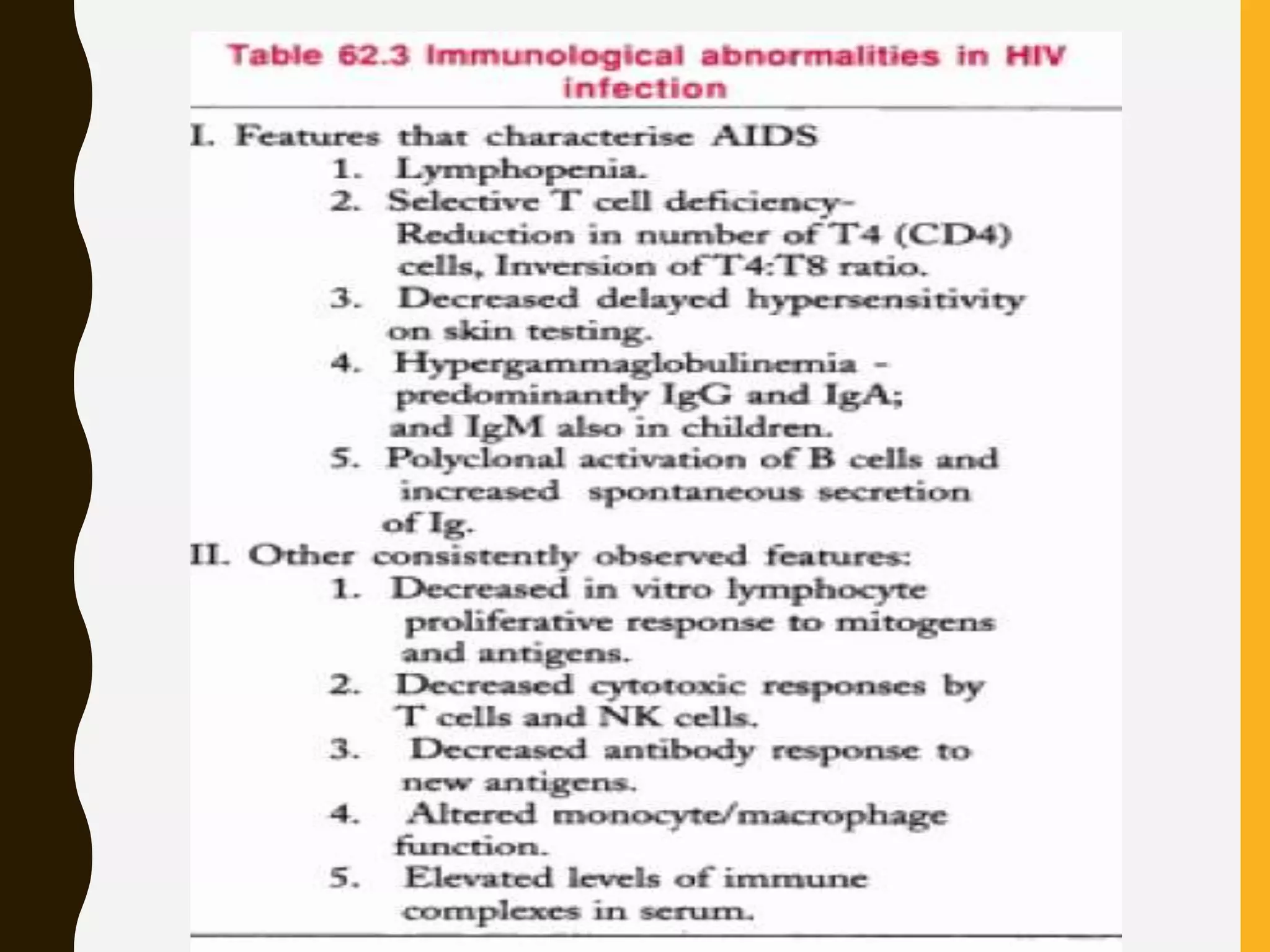
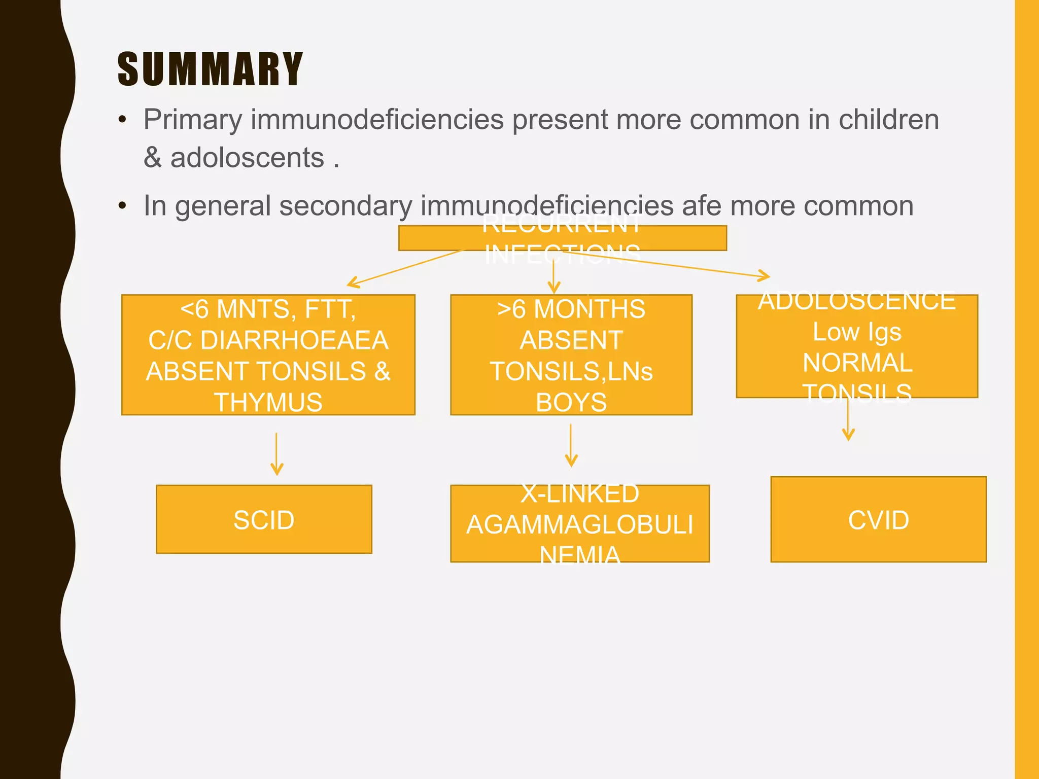
IMMUNODEFICIENCY
DISEASES
LEARNING OBJECTIVES
• Types of immunodeficiencies
Primary
secondary
• Classification of primary immunedeficiencies
• Secondary immune deficiencies
• AIDS
• Immunodeficiency deficiency diseases are
conditions where the defense mechanisms of the
body are impaired .
• Deficiencies of defense mechanisms may involve
SPECIFIC IMMUNE FNS – humoral immunity,cell
mediated immunity or both – or NON SPECIFIC
MECHANISMS such as phagocytosis &
complement, which augment and act in
conjunction with specific immune processes .
• Immuno deiciencies
primary immunodeficiencies
secondary immunodeficiencies
Primary ID - results from abnormalities in
the development of immune mechanisms
Secondary ID – are consequences of
disease,drugs,nutritional inadequacies
PRIMARY IMMUNE DEFICIENCIES
DISORDERS OF
SPECIFIC IMMUNITY
DISORDERS OF
COMPLEMENT
DISORDERS OF
PHAGOCYTOSIS
HUMORAL DEF.
CELLULAR DEF.
COMBINED
CLASSIFICATION OF PRIMARY IMMUNODEFICIENCIES
X LINKED AGAMMAGLOBULINEMIA
TRANSIENT HYPOGAMMAGLOBULINEMIA
OF INFANCY
COMMON VARIABLE IMMUNODEFICIENCY
SELECTIVE IMMUNOGLOBIN
DEFICIENCIES
IMMUNODEFICIENCIES WITH HYPER IGM
TRANSCOBALAMIN II DEFICIENCY
1.HUMORAL
IMMUNODEFICIENCIES
A.DISORDERS OF SPECIFIC IMMUNITY
THYMIC HYPOPLASIA
CHRONIC
MUCOCUTANEOUS
CANDIDIASIS
PURINE NUCLEOSIDE
PHOSPHORYLASE
DEFICIENCY
2.CELLULAR
IMMUNODEFICIENCIES
CELLULAR IMMUNODEFICIENCY WITH
ABNORMAL IMMUNOGLOBULIN SYNTHESIS
(NEZELOF SYNDROME )
ATAXIA TELENGIECTASIA
WISKOTT – ALDRICH SYNDROME
IMMUNODEFICIENCY WITH THYMOMA
EPISODIC LYMPHOPENIA WITH LYMPHOCYTOTOXIN
SEVERE COMBINED IMMUNODEFICIENCIES
3.COMBINED
IMMUNODEFICIENCIES
COMPLEMENT
COMPONENT
DEFICIENCIES
COMPLEMENT
INHIBITOR
DEFICIENCIES
B. DISORDERS OF COMPLEMENT
C/C GRANULOMATOUS DISEASE
MYELOPEROXIDASE DEFICIENCY
CHEDIAK HIGASHI SYNDROME
LEUCOCYTE G6PD DEFICIENCY
JOB’S SYNDROME
TUFTSIN
DEFICIENCY LAZY LEUCOCYTE
SYNDROME
HYPER IG E SYNDROME
ACTIN – BINDING
PROTEIN DEFICIENCY
SHWACHMANS DISEASE
C. DISORDERS OF PHAGOYTOSIS
X LINKED AGAMMAGLOBULINEMIA
TRANSIENT HYPOGAMMAGLOBULINEMIA
OF INFANCY
COMMON VARIABLE IMMUNODEFICIENCY
SELECTIVE IMMUNOGLOBIN
DEFICIENCIES
IMMUNODEFICIENCIES WITH HYPER IGM
TRANSCOBALAMIN II DEFICIENCY
1.HUMORAL
IMMUNODEFICIENCIES
A.DISORDERS OF SPECIFIC IMMUNITY
X LINKED
AGAMMAGLOBULINEMIA• First immunodeficiency disease to have
been recognised .
• Described by BRUTON ( 1952 ) .
• Due to Bruton Tyrosine Kinase
mutation
• Seen only in MALE infants .
• Manifestations not apparent till about 6
months of age .
NAÏVE B CELLS
PLASMA CELLS
IgG,IgM,IgA,IgE
BTK
• Clinical pesentation :
 Recurrent serious infns
(pneumococci,streptococci,meningococci,ps
eudomonas & h.influenzae)
 No tonsils or palpable lymph node
• Avoid Live microbial vaccines
• All classes of immunoglobulins are grossly
depleted in serum .
• IgG level being less than a tenth,IgA and
IgM less than a hundredth of the normal
level .
• Marked decrease in proportion of B cells in
circulation.
• CMI not affected .
• MX : 300mg/kg of gammaglobulin per kg of
bodyweight in 3 doses foll. by monthly injns
of 100mg/kg .
TRANSIENT
HYPOGAMMAGLOBULINEMIA OF
INFANCYDue to abnormal delay in the initiation of IgG
synthesis in some infants .
Presents b/w 6 -12 months of age .
When there is a delay, immunodeficiency occurs .
Recurrent otitis media & respiratory infectios are
common .
Spontaneous recovery occurs b/w 18 & 30 months
of age .
Found in infants of both sexes .
Rx : may be req in some cases, but
prophylactical administration not
recommended .
M = F
COMMON VARIABLE
IMMUNODEFICIENCY
Late onset hypogammaglobulinemia .
Manifests only by 15 – 35 years of age .
Recurrent pyogenic infections and increased incidence of
autoimmune diseases.
Malabsorption and giardiasis are common .
Total immunoglobulin level is less than 300mg/100ml,with IgG <
250mg/100ml .
B cells present in circulation in normal numbers, but they appear
defective in their inability to differentiate into plasma cells and
secrete immunoglobulins.
RX : administration of gammaglobulin preparation
X – LINKED
AGAMMAGLOBULINEMIA
COMMON VARIABLE
IMMUNOGLOBULIN
DEFICIENCY
HUMORAL HUMORAL
PRESENT AFTER 6 MONTHS ADOLOSCENCE
ABSENT ANTIBODIES LOW ANTIBODIES ( VARIABLE
LEVELS )
ABSENT TONSILS NORMAL SIZE TO LARGE
TONSILS
SELECTIVE IMMUNOGLOBULIN
DEFICIENCIES• Selective deficiency of one or more immunoglobulin
classes.
• These dysgammaglobulinemias are common.
• Isolated IgA deficiency is the most common .
• Often accompanied by atopic disorders .
• Increased susceptibility to respiratory infection &
steatorrhea.
• Selective IgM deficiency has been ass with septicemia .
• Selective IgG deficiency has been ass with c/c
progressive bronchiectasis .
IMMUNODEFICIENCIES
WITH HYPER IgM• XL , AR .
• Low IgA & IgG, IgE levels are seen with elevated IgM .
• Mutations in the genes that code for CD40 LIGAND
• IgM molecules appear to have normal structure and possess
antibody activity .
• Enhanced susceptibility to infns & autoimmune processes such
as thrombocytopenia,neutropenia,hemolytic anemia & renal
lesions.
• Some develop malignant infiltration with IgM producing cells.
• DD : Congenital Rubella
• RX: IgG REPLACEMENT THERAPY
TRANSCOBALAMIN II
DEFICIENCY
• AR
• Pts show metabolic effects of vit B12 def.
including megaloblastic anemia and intestinal
villous atrophy .
• Immunological defects : depleted plasma
cells,diminished immunoglobulin levels,
impaired phagocytosis .
• Rx : vit B12 .
GENERAL PRESENTATION
T – CELL
DEFECTS
B – CELL
DEFECTS
PRESENT
AFTER
BIRTH
PRESENT
AT 6
MONTHS
OR
OLDER
COMBINED
DEFECTS
ARE MORE
SEVERE
 VIRAL INFNS
 FUNGAL INFNS
 MYCOBACTERI
A
 PCP
BACTERIAL INFNS
PROTOZOAL
INFNS
ENTERO VIRUS
THYMIC HYPOPLASIA
CHRONIC
MUCOCUTANEOUS
CANDIDIASIS
PURINE NUCLEOSIDE
PHOSPHORYLASE
DEFICIENCY
2.CELLULAR
IMMUNODEFICIENCIES
THYMIC HYPOPLASIA/DIGEORGE
SYNDROME
 Failure of the third and fourth pharyngeal pouch .
 Due to 22 q 11 mutation
 Lack of thymus  T cell deficiency
 Lack of parathyroid  hypocalcemia
 Abnormalities of heart,great vessels
 Abnormal facial features
CHRONIC MUCOCUTANEOUS CANDIDIASIS
 Abnormal immunological response to candida
albicans.
 Severe chronic candidiasis of mucosa,skin &
nails.
 No increased susceptibility to other infns.
 CMI to candida is deficient.
RX
Transfer factor
therapy along with
Amphotericin B
PURINE NUCLEOSIDE
PHOSPHORYLASE DEFICIENCY
 AR
 Decreased CMI
 Recurrent or chronic infns
 Low serum uric acid points to the diagnosis
Hypoplstic anemia
Recurrent pneumonia
Diarrhoea
Candidiasis
CELLULAR IMMUNODEFICIENCY WITH ABNORMAL
IMMUNOGLOBULIN SYNTHESIS
(NEZELOF SYNDROME )
ATAXIA TELENGIECTASIA
WISKOTT – ALDRICH SYNDROME
IMMUNODEFICIENCY WITH THYMOMA
EPISODIC LYMPHOPENIA WITH LYMPHOCYTOTOXIN
SEVERE COMBINED IMMUNODEFICIENCIES
3.COMBINED
IMMUNODEFICIENCIES
CELLULAR IMMUNODEFICIENCY WITH
ABNORMAL IMMUNOGLOBULIN
SYNTHESIS
(NEZELOF SYNDROME)
• Group of disorders
• Depressed CMI with selectively elevated,decreased or normal
levels of immunoglobulin
• Marked T cell def with varyind B cell def .
• In spite of normal level of Igs,antigenic stimuli do not induce ab
fmn.
Recurrent
fungal,bacterial &
viral infns
Rx
BMT
Transfer factor
Thymus transplantn
Antimicrobials
ATAXIA TELENGIECTASIA
• AR
• Ataxia telengiectasia mutated (ATM ) gene at 11q22 -
23
• 23 ( AUTOSOMAL RECESSIVE ) .
• Affects T and B cell .
• Thymic hypoplasia
CLINICAL PRESENTATION
 Ataxic gait in second yr of life .
 Inability to ambulate independently @
10 yrs of age .
 Telengectasia ( dilated blood vessels ) .
 Ovarian dysgenesis
 Risk of malignancy
Diagnosis
ELEVATED AFP
DECR. IgA AND IgE
MRI: CEREBRAL ATROPHY &
VENTRICULAR ENLARGEMENT
Rx
IVIG
ANTIBIOTICS
WISKOTT ALDRICH SYNDROME
• XLR
• ONLY BOYS
• Mutn IN WASp
COMMON PRESENTATION
• Eczema
• Bloody diarrhea
• Atopic dermatitis
• Infn with encapsulated bacteria
Eczema
Thrombocytopenia
Small platelets
Recurrent infns – T
cells affected
Diagnosis
Decr. T cell number &
function
Low IgM
Prenatal- CVS
- amniocentesis
Rx
Skin care
IVIG and/or corticosteroids
Platelet transfusion
splenectomy
AATAXIA TELENGECTASIA FRIEDREICH’S ATAXIA
AR AR
Chr 11 CHR 9
Ataxia Ataxia
Age 1 yr Age 8 -15 yrs
require wheel chair by 10/11 yrs Loss of ambulation 15 yrs after the
onset
intact sensation & negative
rhombergs sign
Spinal or sensory ataxia & positiv
rhombergs sign
telengectasia No
T& B cells affected No
COMPLEMENT
COMPONENT
DEFICIENCIES
COMPLEMENT
INHIBITOR
DEFICIENCIES
B. DISORDERS OF COMPLEMENT
COMPLEMENT COMPONENT
DEFICIENCIES
• AR
• Freq ass with SLE
• Recurrent pyogenic infns with C3 deficiency
• Neisserial infns with C6,C7 & C8
COMPLEMENT INHIBITOR
DEFICIENCIES
• AD
• Hereditary angioneurotic edema – C1 inhibitor def
• Chronic recurrent pyogenic infections – C3b inactivator def
C/C GRANULOMATOUS DISEASE
MYELOPEROXIDASE DEFICIENCY
CHEDIAK HIGASHI SYNDROME
LEUCOCYTE G6PD DEFICIENCY
JOB’S SYNDROME
TUFTSIN
DEFICIENCY LAZY LEUCOCYTE
SYNDROME
HYPER IG E SYNDROME
ACTIN – BINDING
PROTEIN DEFICIENCY
SHWACHMANS DISEASE
C. DISORDERS OF PHAGOYTOSIS
C/C GRANULOMATOUS DISEASE
&MYELOPEROXIDASE DEFICIENCY
AR
XLR
NBT
CHEDIAK HIGASHI SYNDROME
Clinical features
AR
Albinism,photophobia,nystagmus
Frequent and severe pyogenic infns
Mild bleeding diathesis
Recurrent infections
DIAGNOSIS
LARGE
CYTOPLASMIC
GRANULES( IB ) IN
ALL NUCLEATED
CELLS
GRANULES ARE
PEROXIDASE
POSITIVE
RX
BMT
LEUCOCYTE G6PD DEFICIENCY
• Leucocytes deficient in gluc 6 phosphate dehydrogenase
• Diminished bactericidal activity after phagocytosis
• NBT test normal
JOB’S SYNDROME
• Primary defect in phagocytic function
• Serum immunoglobulins normal except for IgE
TUFTSIN DEFICIENCY
Prone to local & systemic bacterial infns
LAZY LEUCOCYTE SYNDROME
Basic defect in chemotaxis and neutrophil mobility.
HYPER IgE SYNDROME
Cellular and humoral immune mechanisms are normal
But IgE levels aremore
SECONDARY IMMUNODEFICIENCIES
• CAUSES
1. MALNUTRITION
2. MALIGNANCY
3. INFECTIONS
4. MATABOLIC DISORDERS
5. CYTOTOXIC DRUGS / IMMUNOSUPPRESSIVE
AGENTS
6. AGEING
• DEFICIENCIES in humoral and cellular immune responses occur
secondarily during course of many disease processes .
• Secondary immune deficiency is more common than the primary
immune deficiency
• AIDS, Aquired immune deficiency syndrome is
the most common .
• Humoral deficiency  lymphoid malignancy ( CLL )
 nephrotic syndrome
 exfoliative skin disease
 protein losing enteropathioes
 multiple myeloma
• Cellular deficiency  lymphoreticular malignancies
 lymphatic obstruction or lymphorrheas
 lepromatous leprosy
 measles
AIDS
• HIV 1
• Infects helper T cells (TH ) cells
• Normal count – 1200/mm³
• Aids - < 200/mm³ of blood
• Monocytes also get infected .
RX
HIV
ANTIVIRAL
S
SUMMARY
• Primary immunodeficiencies present more common in children
& adoloscents .
• In general secondary immunodeficiencies afe more common
<6 MNTS, FTT,
C/C DIARRHOEAEA
ABSENT TONSILS &
THYMUS
>6 MONTHS
ABSENT
TONSILS,LNs
BOYS
ADOLOSCENCE
Low Igs
NORMAL
TONSILS
RECURRENT
INFECTIONS
SCID
X-LINKED
AGAMMAGLOBULI
NEMIA
CVID
RECURRENT
INFNS
Staph aureus
Pneumatocele
Coarse facial features
Eczema
PCP infns
Mouth ulcers
Neutropenia
Recurrent gi infns
Recurrent sinusitis
Low IgA
Hyper IgE syndrome Hyper IgM
syndrome
IgA deficiency
Immuno deficiency diseases- primary and secondary

Immuno deficiency diseases- primary and secondary

  • 2.
  • 3.
    LEARNING OBJECTIVES • Typesof immunodeficiencies Primary secondary • Classification of primary immunedeficiencies • Secondary immune deficiencies • AIDS
  • 4.
    • Immunodeficiency deficiencydiseases are conditions where the defense mechanisms of the body are impaired . • Deficiencies of defense mechanisms may involve SPECIFIC IMMUNE FNS – humoral immunity,cell mediated immunity or both – or NON SPECIFIC MECHANISMS such as phagocytosis & complement, which augment and act in conjunction with specific immune processes .
  • 5.
    • Immuno deiciencies primaryimmunodeficiencies secondary immunodeficiencies Primary ID - results from abnormalities in the development of immune mechanisms Secondary ID – are consequences of disease,drugs,nutritional inadequacies
  • 6.
    PRIMARY IMMUNE DEFICIENCIES DISORDERSOF SPECIFIC IMMUNITY DISORDERS OF COMPLEMENT DISORDERS OF PHAGOCYTOSIS HUMORAL DEF. CELLULAR DEF. COMBINED
  • 7.
    CLASSIFICATION OF PRIMARYIMMUNODEFICIENCIES X LINKED AGAMMAGLOBULINEMIA TRANSIENT HYPOGAMMAGLOBULINEMIA OF INFANCY COMMON VARIABLE IMMUNODEFICIENCY SELECTIVE IMMUNOGLOBIN DEFICIENCIES IMMUNODEFICIENCIES WITH HYPER IGM TRANSCOBALAMIN II DEFICIENCY 1.HUMORAL IMMUNODEFICIENCIES A.DISORDERS OF SPECIFIC IMMUNITY
  • 8.
  • 9.
    CELLULAR IMMUNODEFICIENCY WITH ABNORMALIMMUNOGLOBULIN SYNTHESIS (NEZELOF SYNDROME ) ATAXIA TELENGIECTASIA WISKOTT – ALDRICH SYNDROME IMMUNODEFICIENCY WITH THYMOMA EPISODIC LYMPHOPENIA WITH LYMPHOCYTOTOXIN SEVERE COMBINED IMMUNODEFICIENCIES 3.COMBINED IMMUNODEFICIENCIES
  • 10.
  • 11.
    C/C GRANULOMATOUS DISEASE MYELOPEROXIDASEDEFICIENCY CHEDIAK HIGASHI SYNDROME LEUCOCYTE G6PD DEFICIENCY JOB’S SYNDROME TUFTSIN DEFICIENCY LAZY LEUCOCYTE SYNDROME HYPER IG E SYNDROME ACTIN – BINDING PROTEIN DEFICIENCY SHWACHMANS DISEASE C. DISORDERS OF PHAGOYTOSIS
  • 12.
    X LINKED AGAMMAGLOBULINEMIA TRANSIENTHYPOGAMMAGLOBULINEMIA OF INFANCY COMMON VARIABLE IMMUNODEFICIENCY SELECTIVE IMMUNOGLOBIN DEFICIENCIES IMMUNODEFICIENCIES WITH HYPER IGM TRANSCOBALAMIN II DEFICIENCY 1.HUMORAL IMMUNODEFICIENCIES A.DISORDERS OF SPECIFIC IMMUNITY
  • 13.
    X LINKED AGAMMAGLOBULINEMIA• Firstimmunodeficiency disease to have been recognised . • Described by BRUTON ( 1952 ) . • Due to Bruton Tyrosine Kinase mutation • Seen only in MALE infants . • Manifestations not apparent till about 6 months of age . NAÏVE B CELLS PLASMA CELLS IgG,IgM,IgA,IgE BTK
  • 14.
    • Clinical pesentation:  Recurrent serious infns (pneumococci,streptococci,meningococci,ps eudomonas & h.influenzae)  No tonsils or palpable lymph node
  • 15.
    • Avoid Livemicrobial vaccines • All classes of immunoglobulins are grossly depleted in serum . • IgG level being less than a tenth,IgA and IgM less than a hundredth of the normal level . • Marked decrease in proportion of B cells in circulation. • CMI not affected . • MX : 300mg/kg of gammaglobulin per kg of bodyweight in 3 doses foll. by monthly injns of 100mg/kg .
  • 16.
    TRANSIENT HYPOGAMMAGLOBULINEMIA OF INFANCYDue toabnormal delay in the initiation of IgG synthesis in some infants . Presents b/w 6 -12 months of age . When there is a delay, immunodeficiency occurs . Recurrent otitis media & respiratory infectios are common . Spontaneous recovery occurs b/w 18 & 30 months of age . Found in infants of both sexes . Rx : may be req in some cases, but prophylactical administration not recommended . M = F
  • 17.
    COMMON VARIABLE IMMUNODEFICIENCY Late onsethypogammaglobulinemia . Manifests only by 15 – 35 years of age . Recurrent pyogenic infections and increased incidence of autoimmune diseases. Malabsorption and giardiasis are common . Total immunoglobulin level is less than 300mg/100ml,with IgG < 250mg/100ml . B cells present in circulation in normal numbers, but they appear defective in their inability to differentiate into plasma cells and secrete immunoglobulins. RX : administration of gammaglobulin preparation
  • 18.
    X – LINKED AGAMMAGLOBULINEMIA COMMONVARIABLE IMMUNOGLOBULIN DEFICIENCY HUMORAL HUMORAL PRESENT AFTER 6 MONTHS ADOLOSCENCE ABSENT ANTIBODIES LOW ANTIBODIES ( VARIABLE LEVELS ) ABSENT TONSILS NORMAL SIZE TO LARGE TONSILS
  • 19.
    SELECTIVE IMMUNOGLOBULIN DEFICIENCIES• Selectivedeficiency of one or more immunoglobulin classes. • These dysgammaglobulinemias are common. • Isolated IgA deficiency is the most common . • Often accompanied by atopic disorders . • Increased susceptibility to respiratory infection & steatorrhea. • Selective IgM deficiency has been ass with septicemia . • Selective IgG deficiency has been ass with c/c progressive bronchiectasis .
  • 20.
    IMMUNODEFICIENCIES WITH HYPER IgM•XL , AR . • Low IgA & IgG, IgE levels are seen with elevated IgM . • Mutations in the genes that code for CD40 LIGAND • IgM molecules appear to have normal structure and possess antibody activity . • Enhanced susceptibility to infns & autoimmune processes such as thrombocytopenia,neutropenia,hemolytic anemia & renal lesions. • Some develop malignant infiltration with IgM producing cells. • DD : Congenital Rubella • RX: IgG REPLACEMENT THERAPY
  • 22.
    TRANSCOBALAMIN II DEFICIENCY • AR •Pts show metabolic effects of vit B12 def. including megaloblastic anemia and intestinal villous atrophy . • Immunological defects : depleted plasma cells,diminished immunoglobulin levels, impaired phagocytosis . • Rx : vit B12 .
  • 23.
    GENERAL PRESENTATION T –CELL DEFECTS B – CELL DEFECTS PRESENT AFTER BIRTH PRESENT AT 6 MONTHS OR OLDER COMBINED DEFECTS ARE MORE SEVERE  VIRAL INFNS  FUNGAL INFNS  MYCOBACTERI A  PCP BACTERIAL INFNS PROTOZOAL INFNS ENTERO VIRUS
  • 24.
  • 25.
    THYMIC HYPOPLASIA/DIGEORGE SYNDROME  Failureof the third and fourth pharyngeal pouch .  Due to 22 q 11 mutation  Lack of thymus  T cell deficiency  Lack of parathyroid  hypocalcemia  Abnormalities of heart,great vessels  Abnormal facial features
  • 26.
    CHRONIC MUCOCUTANEOUS CANDIDIASIS Abnormal immunological response to candida albicans.  Severe chronic candidiasis of mucosa,skin & nails.  No increased susceptibility to other infns.  CMI to candida is deficient. RX Transfer factor therapy along with Amphotericin B
  • 27.
    PURINE NUCLEOSIDE PHOSPHORYLASE DEFICIENCY AR  Decreased CMI  Recurrent or chronic infns  Low serum uric acid points to the diagnosis Hypoplstic anemia Recurrent pneumonia Diarrhoea Candidiasis
  • 28.
    CELLULAR IMMUNODEFICIENCY WITHABNORMAL IMMUNOGLOBULIN SYNTHESIS (NEZELOF SYNDROME ) ATAXIA TELENGIECTASIA WISKOTT – ALDRICH SYNDROME IMMUNODEFICIENCY WITH THYMOMA EPISODIC LYMPHOPENIA WITH LYMPHOCYTOTOXIN SEVERE COMBINED IMMUNODEFICIENCIES 3.COMBINED IMMUNODEFICIENCIES
  • 29.
    CELLULAR IMMUNODEFICIENCY WITH ABNORMALIMMUNOGLOBULIN SYNTHESIS (NEZELOF SYNDROME) • Group of disorders • Depressed CMI with selectively elevated,decreased or normal levels of immunoglobulin • Marked T cell def with varyind B cell def . • In spite of normal level of Igs,antigenic stimuli do not induce ab fmn. Recurrent fungal,bacterial & viral infns Rx BMT Transfer factor Thymus transplantn Antimicrobials
  • 30.
    ATAXIA TELENGIECTASIA • AR •Ataxia telengiectasia mutated (ATM ) gene at 11q22 - 23 • 23 ( AUTOSOMAL RECESSIVE ) . • Affects T and B cell . • Thymic hypoplasia CLINICAL PRESENTATION  Ataxic gait in second yr of life .  Inability to ambulate independently @ 10 yrs of age .  Telengectasia ( dilated blood vessels ) .  Ovarian dysgenesis  Risk of malignancy Diagnosis ELEVATED AFP DECR. IgA AND IgE MRI: CEREBRAL ATROPHY & VENTRICULAR ENLARGEMENT Rx IVIG ANTIBIOTICS
  • 31.
    WISKOTT ALDRICH SYNDROME •XLR • ONLY BOYS • Mutn IN WASp COMMON PRESENTATION • Eczema • Bloody diarrhea • Atopic dermatitis • Infn with encapsulated bacteria Eczema Thrombocytopenia Small platelets Recurrent infns – T cells affected Diagnosis Decr. T cell number & function Low IgM Prenatal- CVS - amniocentesis Rx Skin care IVIG and/or corticosteroids Platelet transfusion splenectomy
  • 32.
    AATAXIA TELENGECTASIA FRIEDREICH’SATAXIA AR AR Chr 11 CHR 9 Ataxia Ataxia Age 1 yr Age 8 -15 yrs require wheel chair by 10/11 yrs Loss of ambulation 15 yrs after the onset intact sensation & negative rhombergs sign Spinal or sensory ataxia & positiv rhombergs sign telengectasia No T& B cells affected No
  • 34.
  • 36.
    COMPLEMENT COMPONENT DEFICIENCIES • AR •Freq ass with SLE • Recurrent pyogenic infns with C3 deficiency • Neisserial infns with C6,C7 & C8
  • 37.
    COMPLEMENT INHIBITOR DEFICIENCIES • AD •Hereditary angioneurotic edema – C1 inhibitor def • Chronic recurrent pyogenic infections – C3b inactivator def
  • 38.
    C/C GRANULOMATOUS DISEASE MYELOPEROXIDASEDEFICIENCY CHEDIAK HIGASHI SYNDROME LEUCOCYTE G6PD DEFICIENCY JOB’S SYNDROME TUFTSIN DEFICIENCY LAZY LEUCOCYTE SYNDROME HYPER IG E SYNDROME ACTIN – BINDING PROTEIN DEFICIENCY SHWACHMANS DISEASE C. DISORDERS OF PHAGOYTOSIS
  • 39.
  • 40.
  • 41.
    Clinical features AR Albinism,photophobia,nystagmus Frequent andsevere pyogenic infns Mild bleeding diathesis Recurrent infections DIAGNOSIS LARGE CYTOPLASMIC GRANULES( IB ) IN ALL NUCLEATED CELLS GRANULES ARE PEROXIDASE POSITIVE RX BMT
  • 42.
    LEUCOCYTE G6PD DEFICIENCY •Leucocytes deficient in gluc 6 phosphate dehydrogenase • Diminished bactericidal activity after phagocytosis • NBT test normal JOB’S SYNDROME • Primary defect in phagocytic function • Serum immunoglobulins normal except for IgE TUFTSIN DEFICIENCY Prone to local & systemic bacterial infns LAZY LEUCOCYTE SYNDROME Basic defect in chemotaxis and neutrophil mobility. HYPER IgE SYNDROME Cellular and humoral immune mechanisms are normal But IgE levels aremore
  • 43.
    SECONDARY IMMUNODEFICIENCIES • CAUSES 1.MALNUTRITION 2. MALIGNANCY 3. INFECTIONS 4. MATABOLIC DISORDERS 5. CYTOTOXIC DRUGS / IMMUNOSUPPRESSIVE AGENTS 6. AGEING
  • 44.
    • DEFICIENCIES inhumoral and cellular immune responses occur secondarily during course of many disease processes . • Secondary immune deficiency is more common than the primary immune deficiency • AIDS, Aquired immune deficiency syndrome is the most common . • Humoral deficiency  lymphoid malignancy ( CLL )  nephrotic syndrome  exfoliative skin disease  protein losing enteropathioes  multiple myeloma • Cellular deficiency  lymphoreticular malignancies  lymphatic obstruction or lymphorrheas  lepromatous leprosy  measles
  • 45.
    AIDS • HIV 1 •Infects helper T cells (TH ) cells • Normal count – 1200/mm³ • Aids - < 200/mm³ of blood • Monocytes also get infected .
  • 47.
  • 48.
    SUMMARY • Primary immunodeficienciespresent more common in children & adoloscents . • In general secondary immunodeficiencies afe more common <6 MNTS, FTT, C/C DIARRHOEAEA ABSENT TONSILS & THYMUS >6 MONTHS ABSENT TONSILS,LNs BOYS ADOLOSCENCE Low Igs NORMAL TONSILS RECURRENT INFECTIONS SCID X-LINKED AGAMMAGLOBULI NEMIA CVID
  • 49.
    RECURRENT INFNS Staph aureus Pneumatocele Coarse facialfeatures Eczema PCP infns Mouth ulcers Neutropenia Recurrent gi infns Recurrent sinusitis Low IgA Hyper IgE syndrome Hyper IgM syndrome IgA deficiency