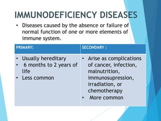
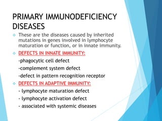
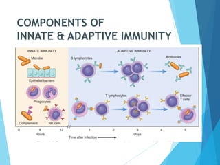
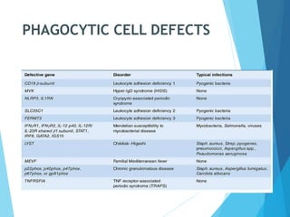
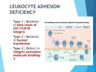
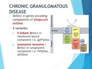
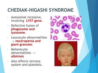
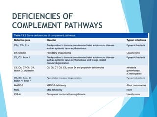
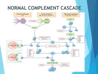
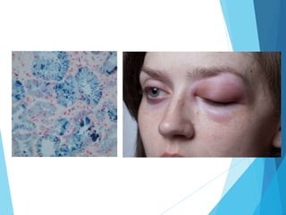
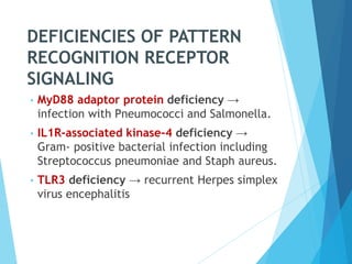
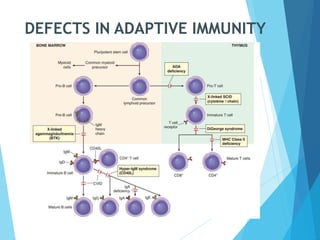
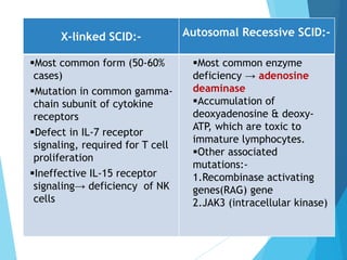
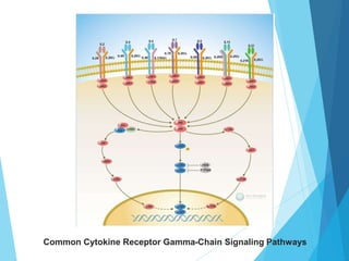
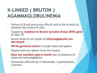
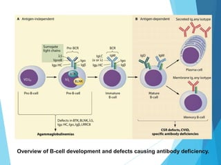
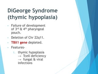
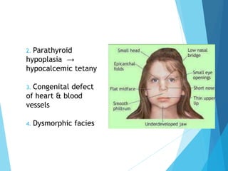
Speaker - Dr Rajesh kumar
IMMUNODEFICIENCY DISEASES
PRIMARY: SECONDARY :
• Usually hereditary
• 6 months to 2 years of
life
• Less common
• Arise as complications
of cancer, infection,
malnutrition,
immunosupression,
irradiation, or
chemotherapy
• More common
• Diseases caused by the absence or failure of
normal function of one or more elements of
immune system.
PRIMARY IMMUNODEFICIENCY
DISEASES
 These are the diseases caused by inherited
mutations in genes involved in lymphocyte
maturation or function, or in innate immunity.
 DEFECTS IN INNATE IMMUNITY:
-phagocytic cell defect
-complement system defect
-defect in pattern recognition receptor
 DEFECTS IN ADAPTIVE IMMUNITY:
- lymphocyte maturation defect
- lymphocyte activation defect
- associated with systemic diseases
COMPONENTS OF
INNATE & ADAPTIVE IMMUNITY
PHAGOCYTIC CELL DEFECTS
LEUKOCYTE ADHESION
DEFICIENCY
• Type 1 : Mutation
in beta chain of
CD11/CD18
integrin
• Type 2 : Mutation
in fucosyl
transferase
• Type 3 : Defect in
integrin activation
molecule kindling-
3
CHRONIC GRANULOMATOUS
DISEASE
• Defect in genes encoding
components of phagocyte
oxidase
• 2 variants:
1) X linked: Defect in
membrane bound
component i.e. gp91phox
2) Autosomal recessive :
Defect in cytoplasmic
component i.e. P47phox,
p67phox
CHEDIAK-HIGASHI SYNDROME
• Autosomal recessive,
involving LYST gene.
• Defective fusion of
phagosome and
lysosome.
• Leucocyte abnormalities
→ neutropenia and
giant granules
• Melanocyte
abnormalities →
albinism
• Also affects nervous
system and platelets.
DEFICIENCIES OF
COMPLEMENT PATHWAYS
NORMAL COMPLEMENT CASCADE
DEFICIENCIES OF PATTERN
RECOGNITION RECEPTOR
SIGNALING
• MyD88 adaptor protein deficiency →
infection with Pneumococci and Salmonella.
• IL1R-associated kinase-4 deficiency →
Gram- positive bacterial infection including
Streptococcus pneumoniae and Staph aureus.
• TLR3 deficiency → recurrent Herpes simplex
virus encephalitis
DEFECTS IN ADAPTIVE IMMUNITY
SEVERE COMBINED
IMMUNODEFICIENCY
X-linked SCID:- Autosomal Recessive SCID:-
Most common form (50-60%
cases)
Mutation in common gamma-
chain subunit of cytokine
receptors
Defect in IL-7 receptor
signaling, required for T cell
proliferation
Ineffective IL-15 receptor
signaling→ deficiency of NK
cells
Most common enzyme
deficiency → adenosine
deaminase
Accumulation of
deoxyadenosine & deoxy-
ATP, which are toxic to
immature lymphocytes.
Other associated
mutations:-
1.Recombinase activating
genes(RAG) gene
2.JAK3 (intracellular kinase)
Common Cytokine Receptor Gamma-Chain Signaling Pathways
X-LINKED ( BRUTON )
AGAMMAGLOBULINEMIA
• Failure of B-cell precursors (Pro-B cells & Pre-B cells) to
develop into mature B cells.
• Caused by mutation in Bruton tyrosine kinase (BTK) gene
at Xq21.22
• Serum levels of all classes of immunoglobulins are
decreased.
• HP-No germinal centre in lymph node and spleen.
• Plasma cells are absent from the tissues.
• Does not manifest upto 6 month due to presence of
maternal immunoglobulin.
• Commonly affected by H.influenzae, S.pneumoniae,
S.aureus.
Overview of B-cell development and defects causing antibody deficiency.
DiGeorge Syndrome
(thymic hypoplasia)
• Failure of development
of 3rd & 4th pharyngeal
pouch.
• Deletion of Chr 22q11.
• TBX1 gene depleted.
• Features-
1. thymic hypoplasia
→ Tcell deficiency
→ fungal & viral
infections
2. Parathyroid
hypoplasia →
hypocalcemic tetany
3. Congenital defect
of heart & blood
vessels
4. Dysmorphic facies
Bare lymphocyte syndrome
• Mutation in transcription factors required
for class II MHC expression.
• No development of CD4+ T cell → defect
in cellular immunity.
• Humoral immunity also affected.
HYPER-IgM SYNDROME
• Production of IgM without IgA, IgG, or IgE
antibodies.
• 2 patterns:-
a) X-linked recessive (70% cases) :
mutation in CD40L gene
(located on Xq26)
b) Autosomal recessive :
mutation in CD40 or activation
induced deaminase (AID)
Clinical features:-
• Recurrent bacterial infections due to lack of
opsonizing IgG antibodies.
• Increased susceptibility to Pneumocystis
jiroveci.
• IgM antibodies act against blood cells→
autoimmune hemolytic anaemia, neutropenia
or thrombocytopenia.
COMMON VARIABLE
IMMUNODEFICIENCY
• Hypogammaglobulinemia, generally affecting
all the antibody classes, but sometimes only
IgG.
• Relatives have high incidence of selective IgA
deficiency.
• Normal or near- normal B cells, but unable to
differentiate into plasma cells.
• Mutations:
 Receptor for a cytokine BAFF
 ICOS ( inducible costimulator)
Features:-
• Typically present with recurrent sino-
pulmonary pyogenic infections.
• Recurrent Herpes virus infections (20% cases)
• Persistent diarrhoea due to Giardia lamblia.
• Affects both sexes equally and onset is late
• Hyperplastic B cell zones
• Increase incidence of autoimmune disease
and lymphoid malignancy
ISOLATED IgA DEFICIENCY
• Extremely low level of both serum and
secretory IgA.
• Impaired differentiation of naive B
lymphocyte to IgA producing plasma cell.
• Molecular basis not known. Probably due to
defect in receptor for BAFF.
• Mucosal immunity mostly affected.
• Recurrent sino-pulmonary infections &
diarrhoea.
• Increase incidence of allergy & autoimmune
disorders.
X LINKED
LYMPHOPROLIFERATIVE
SYNDROME
• Mutation in gene encoding SLAM
associated protein.
• Diminished NK & T cell activation.
• Inability to eliminate EB virus leading to
fulminant Infectious mononucleosis.
WISKOTT ALDRICH SYNDROME
• X linked disease.
• Triad of thrombocytopenia, eczema and recurrent
infections.
• Mutation in gene for WASP located at Xp11.23
• WASP protein involved in cytoskeleton dependent
responses including cell migration & signal
transduction.
 Features :-
• Normal thymus but loss of T lymphocyte in
peripheral blood and lymph node.
• No antibody production to polysaccharides and
poor response to protein antigens.
• IgM ↓↓, IgG – Normal,
IgA & IgE -↑↑
• Increased incidence of B cell lymphoma.
ATAXIA TELANGIECTASIA
• Autosomal recessive
• Mutation of gene encoding ATM on Chr. 11
• ATM protein is a sensor of DNA damage →
activates P53 to activate cell cycle checkpoints
→ apoptosis in cells with damaged DNA.
• ATM contributes to the stability of DNA double-
strand break complexes during V(D)J
recombination.
• Characterized by
1) ataxia
2) vascular
malformation
3) neurological deficit
4) increase incidence
of tumor and
immunodeficiency
• Both T & B cell affected.
• Humoral immunity
predominantly affected.
• Defective production of
IgA & IgG2.
APPROACH TO PATIENTS WITH
SUSPECTED IMMUNODEFICIENCY
• Detailed history
• Physical examination
• Investigations
KEY HISTORY
• History of infections: location,
organism, frequency, response to
therapy, hospitalization
• Family history – consanguinity
• History of allergy
• Developmental delays
10 warning signs of
Immunodeficiency
1
Eight or more new ear
infections within one year
Recurrent deep skin
or organ abscess
6
2
Two or more serious sinus
infections within one year
Persistent thrush in mouth or
elsewhere on skin after age
one.
7
3
Two or more months on
antibiotics with little effect
Need for I/V antibiotics to
clear infections
8
4
Two or more pneumonias
within one year
Two or more deep seated
infections
9
5
Failure of an infant to gain
weight or grow naturally
Family history of primary
immunodeficiency
10
PHYSICAL FINDINGS
• Failure to thrive
• Abnormal facial features
• Skin and mucosa: eczema, petechiae, abscess,
pyoderma, telangiectasia, delayed umbilical
cord separation
• Respiratory tract: bronchial breath sound, fine
crepitations
INVESTIGATIONS
 CBC:-Absolute lymphocyte count
-Absolute neutrophil count
-Platelet count
 Peripheral smear
 Delayed-type hypersensitivity skin
tests
 Chest x-ray
 serum immunoglobulins
 lymphocyte subset analysis by
flowcytometry
 antibody response to
-known exposure
-known immunizations
 enzyme assays: ADA,NADPH OXIDASE
 chemotaxis assays
 nitroblue tetrazolium slide test
 dihydro rhodamine flowcytometry
 Complement level:C3,C5,CH50
 biopsies: skin,lymphnode,thymus
 cytogenetic analysis: FISH for 22q11
mutation analysis: CD40L,JAK3,ZAP70
Nitroblue Tetrazolium Slide Test
 Neutrophils make
reactive oxygen species
which reduces colorless
NBT dye into a blue
colored formazan salt
 In CGD these reactive
oxygen species are not
formed, so dye is not
reduced to blue color
TREATMENT
• SUPPORTIVE THERAPY- antibiotics
• REPLACEMENT THERAPY- IV
immunoglobulins, enzymes
• DEFINITIVE THERAPY- fetal thymic grafts,
bone marrow transplantation
• GENE THERAPY
immuno.pptx

immuno.pptx

  • 1.
    Speaker - DrRajesh kumar
  • 2.
    IMMUNODEFICIENCY DISEASES PRIMARY: SECONDARY: • Usually hereditary • 6 months to 2 years of life • Less common • Arise as complications of cancer, infection, malnutrition, immunosupression, irradiation, or chemotherapy • More common • Diseases caused by the absence or failure of normal function of one or more elements of immune system.
  • 3.
    PRIMARY IMMUNODEFICIENCY DISEASES  Theseare the diseases caused by inherited mutations in genes involved in lymphocyte maturation or function, or in innate immunity.  DEFECTS IN INNATE IMMUNITY: -phagocytic cell defect -complement system defect -defect in pattern recognition receptor  DEFECTS IN ADAPTIVE IMMUNITY: - lymphocyte maturation defect - lymphocyte activation defect - associated with systemic diseases
  • 4.
    COMPONENTS OF INNATE &ADAPTIVE IMMUNITY
  • 5.
  • 6.
    LEUKOCYTE ADHESION DEFICIENCY • Type1 : Mutation in beta chain of CD11/CD18 integrin • Type 2 : Mutation in fucosyl transferase • Type 3 : Defect in integrin activation molecule kindling- 3
  • 7.
    CHRONIC GRANULOMATOUS DISEASE • Defectin genes encoding components of phagocyte oxidase • 2 variants: 1) X linked: Defect in membrane bound component i.e. gp91phox 2) Autosomal recessive : Defect in cytoplasmic component i.e. P47phox, p67phox
  • 9.
    CHEDIAK-HIGASHI SYNDROME • Autosomalrecessive, involving LYST gene. • Defective fusion of phagosome and lysosome. • Leucocyte abnormalities → neutropenia and giant granules • Melanocyte abnormalities → albinism • Also affects nervous system and platelets.
  • 10.
  • 11.
  • 13.
    DEFICIENCIES OF PATTERN RECOGNITIONRECEPTOR SIGNALING • MyD88 adaptor protein deficiency → infection with Pneumococci and Salmonella. • IL1R-associated kinase-4 deficiency → Gram- positive bacterial infection including Streptococcus pneumoniae and Staph aureus. • TLR3 deficiency → recurrent Herpes simplex virus encephalitis
  • 14.
  • 15.
  • 16.
    X-linked SCID:- AutosomalRecessive SCID:- Most common form (50-60% cases) Mutation in common gamma- chain subunit of cytokine receptors Defect in IL-7 receptor signaling, required for T cell proliferation Ineffective IL-15 receptor signaling→ deficiency of NK cells Most common enzyme deficiency → adenosine deaminase Accumulation of deoxyadenosine & deoxy- ATP, which are toxic to immature lymphocytes. Other associated mutations:- 1.Recombinase activating genes(RAG) gene 2.JAK3 (intracellular kinase)
  • 17.
    Common Cytokine ReceptorGamma-Chain Signaling Pathways
  • 18.
    X-LINKED ( BRUTON) AGAMMAGLOBULINEMIA • Failure of B-cell precursors (Pro-B cells & Pre-B cells) to develop into mature B cells. • Caused by mutation in Bruton tyrosine kinase (BTK) gene at Xq21.22 • Serum levels of all classes of immunoglobulins are decreased. • HP-No germinal centre in lymph node and spleen. • Plasma cells are absent from the tissues. • Does not manifest upto 6 month due to presence of maternal immunoglobulin. • Commonly affected by H.influenzae, S.pneumoniae, S.aureus.
  • 19.
    Overview of B-celldevelopment and defects causing antibody deficiency.
  • 20.
    DiGeorge Syndrome (thymic hypoplasia) •Failure of development of 3rd & 4th pharyngeal pouch. • Deletion of Chr 22q11. • TBX1 gene depleted. • Features- 1. thymic hypoplasia → Tcell deficiency → fungal & viral infections
  • 21.
    2. Parathyroid hypoplasia → hypocalcemictetany 3. Congenital defect of heart & blood vessels 4. Dysmorphic facies
  • 22.
    Bare lymphocyte syndrome •Mutation in transcription factors required for class II MHC expression. • No development of CD4+ T cell → defect in cellular immunity. • Humoral immunity also affected.
  • 23.
    HYPER-IgM SYNDROME • Productionof IgM without IgA, IgG, or IgE antibodies. • 2 patterns:- a) X-linked recessive (70% cases) : mutation in CD40L gene (located on Xq26) b) Autosomal recessive : mutation in CD40 or activation induced deaminase (AID)
  • 25.
    Clinical features:- • Recurrentbacterial infections due to lack of opsonizing IgG antibodies. • Increased susceptibility to Pneumocystis jiroveci. • IgM antibodies act against blood cells→ autoimmune hemolytic anaemia, neutropenia or thrombocytopenia.
  • 26.
    COMMON VARIABLE IMMUNODEFICIENCY • Hypogammaglobulinemia,generally affecting all the antibody classes, but sometimes only IgG. • Relatives have high incidence of selective IgA deficiency. • Normal or near- normal B cells, but unable to differentiate into plasma cells. • Mutations:  Receptor for a cytokine BAFF  ICOS ( inducible costimulator)
  • 27.
    Features:- • Typically presentwith recurrent sino- pulmonary pyogenic infections. • Recurrent Herpes virus infections (20% cases) • Persistent diarrhoea due to Giardia lamblia. • Affects both sexes equally and onset is late • Hyperplastic B cell zones • Increase incidence of autoimmune disease and lymphoid malignancy
  • 28.
    ISOLATED IgA DEFICIENCY •Extremely low level of both serum and secretory IgA. • Impaired differentiation of naive B lymphocyte to IgA producing plasma cell. • Molecular basis not known. Probably due to defect in receptor for BAFF. • Mucosal immunity mostly affected. • Recurrent sino-pulmonary infections & diarrhoea. • Increase incidence of allergy & autoimmune disorders.
  • 29.
    X LINKED LYMPHOPROLIFERATIVE SYNDROME • Mutationin gene encoding SLAM associated protein. • Diminished NK & T cell activation. • Inability to eliminate EB virus leading to fulminant Infectious mononucleosis.
  • 30.
    WISKOTT ALDRICH SYNDROME •X linked disease. • Triad of thrombocytopenia, eczema and recurrent infections.
  • 31.
    • Mutation ingene for WASP located at Xp11.23 • WASP protein involved in cytoskeleton dependent responses including cell migration & signal transduction.  Features :- • Normal thymus but loss of T lymphocyte in peripheral blood and lymph node. • No antibody production to polysaccharides and poor response to protein antigens. • IgM ↓↓, IgG – Normal, IgA & IgE -↑↑ • Increased incidence of B cell lymphoma.
  • 32.
    ATAXIA TELANGIECTASIA • Autosomalrecessive • Mutation of gene encoding ATM on Chr. 11 • ATM protein is a sensor of DNA damage → activates P53 to activate cell cycle checkpoints → apoptosis in cells with damaged DNA. • ATM contributes to the stability of DNA double- strand break complexes during V(D)J recombination.
  • 33.
    • Characterized by 1)ataxia 2) vascular malformation 3) neurological deficit 4) increase incidence of tumor and immunodeficiency • Both T & B cell affected. • Humoral immunity predominantly affected. • Defective production of IgA & IgG2.
  • 34.
    APPROACH TO PATIENTSWITH SUSPECTED IMMUNODEFICIENCY • Detailed history • Physical examination • Investigations
  • 35.
    KEY HISTORY • Historyof infections: location, organism, frequency, response to therapy, hospitalization • Family history – consanguinity • History of allergy • Developmental delays
  • 36.
    10 warning signsof Immunodeficiency 1 Eight or more new ear infections within one year Recurrent deep skin or organ abscess 6 2 Two or more serious sinus infections within one year Persistent thrush in mouth or elsewhere on skin after age one. 7 3 Two or more months on antibiotics with little effect Need for I/V antibiotics to clear infections 8 4 Two or more pneumonias within one year Two or more deep seated infections 9 5 Failure of an infant to gain weight or grow naturally Family history of primary immunodeficiency 10
  • 37.
    PHYSICAL FINDINGS • Failureto thrive • Abnormal facial features • Skin and mucosa: eczema, petechiae, abscess, pyoderma, telangiectasia, delayed umbilical cord separation • Respiratory tract: bronchial breath sound, fine crepitations
  • 38.
    INVESTIGATIONS  CBC:-Absolute lymphocytecount -Absolute neutrophil count -Platelet count  Peripheral smear  Delayed-type hypersensitivity skin tests  Chest x-ray
  • 39.
     serum immunoglobulins lymphocyte subset analysis by flowcytometry  antibody response to -known exposure -known immunizations
  • 40.
     enzyme assays:ADA,NADPH OXIDASE  chemotaxis assays  nitroblue tetrazolium slide test  dihydro rhodamine flowcytometry  Complement level:C3,C5,CH50  biopsies: skin,lymphnode,thymus  cytogenetic analysis: FISH for 22q11 mutation analysis: CD40L,JAK3,ZAP70
  • 41.
    Nitroblue Tetrazolium SlideTest  Neutrophils make reactive oxygen species which reduces colorless NBT dye into a blue colored formazan salt  In CGD these reactive oxygen species are not formed, so dye is not reduced to blue color
  • 42.
    TREATMENT • SUPPORTIVE THERAPY-antibiotics • REPLACEMENT THERAPY- IV immunoglobulins, enzymes • DEFINITIVE THERAPY- fetal thymic grafts, bone marrow transplantation • GENE THERAPY