
Recommended
More Related Content
What's hot
What's hot (20)
Similar to Digestive system
Similar to Digestive system (20)
Recently uploaded
Recently uploaded (20)
Digestive system
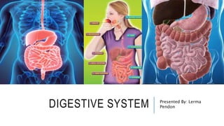
- 1. DIGESTIVE SYSTEM Presented By: Lerma Pendon
- 2. WHAT IS DIGESTIVE SYSTEM? • A group of organs working together to convert food into energy • Breakdown of food into small molecules • Absorbs nutrients and water to feed the entire body
- 3. TWO MAJOR PARTS: 1. Alimentary Canal/Gastrointestinal Tract: Food passes through a long tube inside the body known as the alimentary canal or the gastrointestinal tract (GI tract). It has two openings: the mouth and the anus. The alimentary canal is made up of the mouth, pharynx, esophagus, stomach, small intestines, large intestines, rectum and anus.
- 5. TWO MAJOR PARTS: 2. Accessory Organs (the Glands) • Not part of the path of food, but play a critical role. • Include: Liver Gall bladder Pancreas
- 6. ALIMENTARY CANAL / GASTROINTESTINAL TRACK
- 7. MOUTH • Teeth mechanically break down food into small pieces. • Tongue mixes food with saliva (contains amylase, which helps break down starch). • Responsible for mastication. Mastication is the process of chewing. Also known as mechanical digestion. • Bolus is the product mechanical digestion.
- 8. PHARYNX • Also called the throat. • The portion of the digestive tract that receives the food from your mouth. • Its muscular walls function in the process of swallowing, and it serves as a pathway for the movement of food from the mouth to the esophagus. • Epiglottis is a flap-like structure at the back of the throat that closes windpipe over the trachea preventing food from entering it. It is located in the pharynx. • It has 3 divisions.
- 9. • Nasopharynx – located superior and posterior to the soft palate. Contains the pharyngeal tonsils and tubal tonsils. • Oropharynx – Contains palatine and lingual tonsils. The tonsils remove pathogens that enter the pharynx. • Laryngopharynx – inferior to the epiglottis and posterior to the larynx. This division opens into the esophagus and larynx.
- 10. ESOPHAGUS • A muscular tube connecting the pharynx to the stomach. Approximately 10 inches long. • It moves the food from the throat to the stomach using muscle movement called peristalsis. • Peristalsis is a wavelike contraction from the esophagus to large intestine. • At the inferior end of the esophagus is muscular ring called cardiac sphincter or cardioesophageal sphincter. • The function of this sphincter is to close the end of the esophagus and prevent the backflow of food from the stomach to esophagus.
- 11. STOMACH • A muscular sac that is located on the left side of the abdominal cavity, just inferior to the diaphragm. • This major organ acts as a storage tank for food so that the body has time to digest large meals properly. • Mixes food with digestive juices that contain enzymes such as lipase to break down Proteins and Lipids, the process is called chemical digestion. • The product of chemical digestion is called Chyme. It is a the pulpy acidic fluid which passes from the stomach to the small intestine, consisting of gastric juices and partly digested food. • The pH of gastric acid is 1.5 to 3.5 in the human stomach lumen. • The stomach also contains hydrochloric acid (HCl) that kills bacteria.
- 12. SMALL INTESTINE • A long, thin tube about 1 inch in diameter and about 10 feet long. • The entire small intestine is coiled like a hose and the inside surface is full of many ridges and folds. These folds are used to maximize the digestion of food and absorption of nutrients. • By the time food leaves the small intestine, around 90% of all nutrients have been extracted from the food that entered it. Absorbs: 80% water Vitamins Minerals Carbohydrates Proteins Lipids • Nutrients from the food pass into the bloodstream through the small
- 13. THREE (3) PARTS OF THE SMALL INTESTINE 1. Duodenum - the first part of the small intestine. After foods mix with stomach acid, they move into the duodenum, where they mix with bile from the gallbladder and digestive juices from the pancreas. 2. Jejunum - the middle segment of the small intestine. Most of the nutrients present in food are absorbed by the jejunum before being passed on to the ileum for further absorption. 3. Ileum - The function of the ileum is mainly to absorb vitamin B12 and bile salts and whatever products of digestion were not absorbed by the jejunum
- 15. LARGE INTESTINE • A long, thick tube about 2.5 inches in diameter and about 5 feet long. It is located just inferior to the stomach and wraps around the superior and lateral border of the small intestine. • The large intestine absorbs water and contains many symbiotic bacteria that aid in the breaking down of wastes to extract some small amounts of nutrients. • It is where the feces are formed. •It is also called “Colon"
- 16. THREE (3) PARTS OF THE LARGE INTESTINE 1. Ascending Colon – the beginning part of the colon which carries feces from the cecum to the transverse colon. 2. Transverse Colon – the longest region of the colon where much of the absorption and feces formation takes place. 3. Descending Colon – the walls of the descending colon absorb water as well as remaining nutrients and vitamins from the feces and depositing these substances into our bloodstream before they are eliminated from the body.
- 17. RECTUM • The final straight portion of the large intestine, approximately 6 inches long. • The rectum is a continuation of the large intestine and connects to the anus. • For temporary storage of feces. • To expel solid and gaseous waste from the gastrointestinal tract.
- 18. ANUS • The anus is the last part of the digestive tract. It is a 2-inch long canal consisting of the pelvic floor muscles and the two anal sphincters (internal and external). • The lining of the upper anus is specialized to detect rectal contents. It lets you know whether the contents are liquid, gas, or solid. • The internal sphincter is always tight, except when stool enters the rectum. It keeps us continent when we are asleep or otherwise unaware of the presence of stool. • When we get an urge to go to the bathroom, we rely on our external sphincter to hold the stool until reaching a toilet, where it then relaxes to release the contents.
- 20. SPHINCTERS • A ring-like muscles that contract or close a bodily opening or passage in the alimentary canal to prevent backflow. • 3 Sphincters in the Alimentary Canal: 1. Cardioesophageal Sphincter – prevents the backflow of food from the stomach to espophagus. 2. Pyloric Sphincter – prevents the backflow of food from the small intestine to stomach. 3. Anal Sphincter – prevents the release of feces/wastes.
- 22. LIVER • The liver weighs about 3 pounds and is the second largest organ in the body. • The liver has many different functions in the body, but the main function of the liver in digestion is the production of bile and its secretion into the small intestine. • Bile is a yellowish substance for fat digestion. • Liver filters out toxins and waste including drugs and alcohol and poisons.
- 23. GALL BLADDER • A small, pear-shaped organ located just posterior to the liver. • The gallbladder is used to store and recycle excess bile from the small intestine so that it can be reused for the digestion of subsequent meals. • Stores bile from the liver, releases it into the small intestine. • Fatty diets can cause gallstones.
- 24. PANCREAS • A large gland located just inferior and posterior to the stomach. It is about 6 inches long. • The pancreas secretes digestive enzymes into the small intestine to complete the chemical digestion of foods, digest fats, carbohydrates and proteins. • 2 kinds of Enzymes: 1. Amylase - a protein made by the pancreas and by glands in and around your mouth and throat. It helps you break down carbohydrates and starches into sugar. 2. Lipase - an enzyme that breaks down dietary fats into smaller molecules called fatty acids and glycerol. • Regulates blood sugar by producing hormones such as: 1. Insulin – lowers blood sugar level 2. Glucagon – increases blood sugar level
- 26. NORMAL FLORA • Are bacteria inside human body: 1. Colon - Escherichia Coli (E. Coli); Bacteria that causes UTI. They are found in urine through urinalysis. 2. Stomach – Helicobacter Pylori; acidophyllic or acid-loving bacteria; pathogenic for chronic acidity; bacteria that causes ulcer.
- 27. ANY QUESTIONS ?