The document summarizes the key organs and processes of the digestive system. It describes the roles of the mouth, esophagus, stomach, small intestine, large intestine and associated organs like the liver, gallbladder and pancreas. The key processes discussed are ingestion, propulsion, digestion, absorption and elimination. Mechanical and chemical digestion are explained along with the enzymes involved at each stage and how nutrients are absorbed and waste eliminated.
Grade 7 chapter1 lesson1 - the digestive systemLermaPendon1
Lesson 2 The Digestive system
-Functions of Digestive system
-Digestion
-Types of Digestion
-Enzymes
-The role of Enzymes in Digestion
-Organs of Digestive system
-The Mouth
-the Esophagus
-Peristalsis
-The Stomach
-Chyme
-the Small Intestine
-Villi
-the Large intestine
-Bacteria and Digestion
-the Digestive system and Homeostasis
-Common Ailments and Disorders of the Digestive system
The digestive system is made up of the digestive tract and other organs that help the body break down and absorb food. It is a long, twisting tube that starts at the mouth and goes through the oesophagus, stomach, small intestine, large intestine and anus.
Ozempic: Preoperative Management of Patients on GLP-1 Receptor Agonists Saeid Safari
Preoperative Management of Patients on GLP-1 Receptor Agonists like Ozempic and Semiglutide
ASA GUIDELINE
NYSORA Guideline
2 Case Reports of Gastric Ultrasound
These lecture slides, by Dr Sidra Arshad, offer a quick overview of physiological basis of a normal electrocardiogram.
Learning objectives:
1. Define an electrocardiogram (ECG) and electrocardiography
2. Describe how dipoles generated by the heart produce the waveforms of the ECG
3. Describe the components of a normal electrocardiogram of a typical bipolar leads (limb II)
4. Differentiate between intervals and segments
5. Enlist some common indications for obtaining an ECG
Study Resources:
1. Chapter 11, Guyton and Hall Textbook of Medical Physiology, 14th edition
2. Chapter 9, Human Physiology - From Cells to Systems, Lauralee Sherwood, 9th edition
3. Chapter 29, Ganong’s Review of Medical Physiology, 26th edition
4. Electrocardiogram, StatPearls - https://www.ncbi.nlm.nih.gov/books/NBK549803/
5. ECG in Medical Practice by ABM Abdullah, 4th edition
6. ECG Basics, http://www.nataliescasebook.com/tag/e-c-g-basics
Knee anatomy and clinical tests 2024.pdfvimalpl1234
This includes all relevant anatomy and clinical tests compiled from standard textbooks, Campbell,netter etc..It is comprehensive and best suited for orthopaedicians and orthopaedic residents.
These simplified slides by Dr. Sidra Arshad present an overview of the non-respiratory functions of the respiratory tract.
Learning objectives:
1. Enlist the non-respiratory functions of the respiratory tract
2. Briefly explain how these functions are carried out
3. Discuss the significance of dead space
4. Differentiate between minute ventilation and alveolar ventilation
5. Describe the cough and sneeze reflexes
Study Resources:
1. Chapter 39, Guyton and Hall Textbook of Medical Physiology, 14th edition
2. Chapter 34, Ganong’s Review of Medical Physiology, 26th edition
3. Chapter 17, Human Physiology by Lauralee Sherwood, 9th edition
4. Non-respiratory functions of the lungs https://academic.oup.com/bjaed/article/13/3/98/278874
Couples presenting to the infertility clinic- Do they really have infertility...Sujoy Dasgupta
Dr Sujoy Dasgupta presented the study on "Couples presenting to the infertility clinic- Do they really have infertility? – The unexplored stories of non-consummation" in the 13th Congress of the Asia Pacific Initiative on Reproduction (ASPIRE 2024) at Manila on 24 May, 2024.
Title: Sense of Smell
Presenter: Dr. Faiza, Assistant Professor of Physiology
Qualifications:
MBBS (Best Graduate, AIMC Lahore)
FCPS Physiology
ICMT, CHPE, DHPE (STMU)
MPH (GC University, Faisalabad)
MBA (Virtual University of Pakistan)
Learning Objectives:
Describe the primary categories of smells and the concept of odor blindness.
Explain the structure and location of the olfactory membrane and mucosa, including the types and roles of cells involved in olfaction.
Describe the pathway and mechanisms of olfactory signal transmission from the olfactory receptors to the brain.
Illustrate the biochemical cascade triggered by odorant binding to olfactory receptors, including the role of G-proteins and second messengers in generating an action potential.
Identify different types of olfactory disorders such as anosmia, hyposmia, hyperosmia, and dysosmia, including their potential causes.
Key Topics:
Olfactory Genes:
3% of the human genome accounts for olfactory genes.
400 genes for odorant receptors.
Olfactory Membrane:
Located in the superior part of the nasal cavity.
Medially: Folds downward along the superior septum.
Laterally: Folds over the superior turbinate and upper surface of the middle turbinate.
Total surface area: 5-10 square centimeters.
Olfactory Mucosa:
Olfactory Cells: Bipolar nerve cells derived from the CNS (100 million), with 4-25 olfactory cilia per cell.
Sustentacular Cells: Produce mucus and maintain ionic and molecular environment.
Basal Cells: Replace worn-out olfactory cells with an average lifespan of 1-2 months.
Bowman’s Gland: Secretes mucus.
Stimulation of Olfactory Cells:
Odorant dissolves in mucus and attaches to receptors on olfactory cilia.
Involves a cascade effect through G-proteins and second messengers, leading to depolarization and action potential generation in the olfactory nerve.
Quality of a Good Odorant:
Small (3-20 Carbon atoms), volatile, water-soluble, and lipid-soluble.
Facilitated by odorant-binding proteins in mucus.
Membrane Potential and Action Potential:
Resting membrane potential: -55mV.
Action potential frequency in the olfactory nerve increases with odorant strength.
Adaptation Towards the Sense of Smell:
Rapid adaptation within the first second, with further slow adaptation.
Psychological adaptation greater than receptor adaptation, involving feedback inhibition from the central nervous system.
Primary Sensations of Smell:
Camphoraceous, Musky, Floral, Pepperminty, Ethereal, Pungent, Putrid.
Odor Detection Threshold:
Examples: Hydrogen sulfide (0.0005 ppm), Methyl-mercaptan (0.002 ppm).
Some toxic substances are odorless at lethal concentrations.
Characteristics of Smell:
Odor blindness for single substances due to lack of appropriate receptor protein.
Behavioral and emotional influences of smell.
Transmission of Olfactory Signals:
From olfactory cells to glomeruli in the olfactory bulb, involving lateral inhibition.
Primitive, less old, and new olfactory systems with different path
Tom Selleck Health: A Comprehensive Look at the Iconic Actor’s Wellness Journeygreendigital
Tom Selleck, an enduring figure in Hollywood. has captivated audiences for decades with his rugged charm, iconic moustache. and memorable roles in television and film. From his breakout role as Thomas Magnum in Magnum P.I. to his current portrayal of Frank Reagan in Blue Bloods. Selleck's career has spanned over 50 years. But beyond his professional achievements. fans have often been curious about Tom Selleck Health. especially as he has aged in the public eye.
Follow us on: Pinterest
Introduction
Many have been interested in Tom Selleck health. not only because of his enduring presence on screen but also because of the challenges. and lifestyle choices he has faced and made over the years. This article delves into the various aspects of Tom Selleck health. exploring his fitness regimen, diet, mental health. and the challenges he has encountered as he ages. We'll look at how he maintains his well-being. the health issues he has faced, and his approach to ageing .
Early Life and Career
Childhood and Athletic Beginnings
Tom Selleck was born on January 29, 1945, in Detroit, Michigan, and grew up in Sherman Oaks, California. From an early age, he was involved in sports, particularly basketball. which played a significant role in his physical development. His athletic pursuits continued into college. where he attended the University of Southern California (USC) on a basketball scholarship. This early involvement in sports laid a strong foundation for his physical health and disciplined lifestyle.
Transition to Acting
Selleck's transition from an athlete to an actor came with its physical demands. His first significant role in "Magnum P.I." required him to perform various stunts and maintain a fit appearance. This role, which he played from 1980 to 1988. necessitated a rigorous fitness routine to meet the show's demands. setting the stage for his long-term commitment to health and wellness.
Fitness Regimen
Workout Routine
Tom Selleck health and fitness regimen has evolved. adapting to his changing roles and age. During his "Magnum, P.I." days. Selleck's workouts were intense and focused on building and maintaining muscle mass. His routine included weightlifting, cardiovascular exercises. and specific training for the stunts he performed on the show.
Selleck adjusted his fitness routine as he aged to suit his body's needs. Today, his workouts focus on maintaining flexibility, strength, and cardiovascular health. He incorporates low-impact exercises such as swimming, walking, and light weightlifting. This balanced approach helps him stay fit without putting undue strain on his joints and muscles.
Importance of Flexibility and Mobility
In recent years, Selleck has emphasized the importance of flexibility and mobility in his fitness regimen. Understanding the natural decline in muscle mass and joint flexibility with age. he includes stretching and yoga in his routine. These practices help prevent injuries, improve posture, and maintain mobilit
New Drug Discovery and Development .....NEHA GUPTA
The "New Drug Discovery and Development" process involves the identification, design, testing, and manufacturing of novel pharmaceutical compounds with the aim of introducing new and improved treatments for various medical conditions. This comprehensive endeavor encompasses various stages, including target identification, preclinical studies, clinical trials, regulatory approval, and post-market surveillance. It involves multidisciplinary collaboration among scientists, researchers, clinicians, regulatory experts, and pharmaceutical companies to bring innovative therapies to market and address unmet medical needs.
Pulmonary Thromboembolism - etilogy, types, medical- Surgical and nursing man...VarunMahajani
Disruption of blood supply to lung alveoli due to blockage of one or more pulmonary blood vessels is called as Pulmonary thromboembolism. In this presentation we will discuss its causes, types and its management in depth.
Pulmonary Thromboembolism - etilogy, types, medical- Surgical and nursing man...
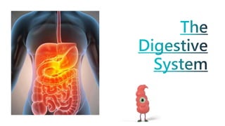
The Digestive System.pptx
1.
2. • Identify the main organs of the digestive system
• Describe the structure and function of each organ in the digestion process
• Explain the mechanical and chemical involvement in digestion
• Explain how nutrients are absorbed and waste is eliminated within the digestive
system.
• Identify the importance of other body systems in digestion.
4. The Alimentary canal:
• Mouth
• Pharynx
• Oesophagus
• Stomach
• Small intestine
• Large intestine Rectum and anal canal
The Accessory Organs;
• Three pairs of salivary glands
• The pancreas
• The liver and biliary tract
5. • Ingestion – Taking in food through the mouth
• Propulsion – Mixes and moves along the alimentary tract
• Digestion – Mechanical breakdown in the stomach, chemical digestion of
food into small molecules by enzymes
• Absorption – Digested food substances pass through the wall of some
organs into blood and lymph capillaries for circulation and use by body
cells
• Elimination – Food eaten but can’t be digested and absorbed is
eliminated
8. AKA the Gastrointestinal Tract and
Digestive Tract
The walls are made up of 4 layers:
• Adventitia or Serosa – Outer covering
• Muscle layer
• Submucosa
• Mucosa – lining
https://youtu.be/R-OhCLIkP5U
9. Specialised cells or glands
release secretions
Saliva – From the Salivary glands
Gastric Juice – From the Gastric Glands
Intestinal Juice – From the Intestinal Glands
Pancreatic Juice – From the Pancreas
Bile – From the Liver
They all contain enzymes that chemically
break down food
Nerve Supply
• The vagus nerve and sacral nerve –
These are both Parasympathetic
which increases muscle activity and
secretions.
• Sympathetic nervous supply – From
the spinal cord, thorax, stomach and
pelvic area. These decrease muscle
activity and decrease secretions
10.
11.
12. • The mouth, also known as the oral cavity, is where food enters
the body and begins its journey through the digestive system.
• The teeth cut and grind food into smaller pieces.
• The salivary glands produce saliva which helps to moisten food
and begin the digestion of carbohydrates.
• The tongue helps to push food pieces into the pharynx.
13. Mechanical Digestion
• Teeth and tongue break food down into
smaller pieces
Chemical Digestion
• Salivary glands produce saliva
• Amylase (enzyme) in saliva begin to
break food down
• Amylase converts starch into glucose.
Your body
produces 1.5L
of saliva each
day!
14. • The oesophagus is a muscular tube connecting the pharynx to the stomach.
• Wave-like peristalsis movements force the food into the stomach.
• At the lower end of the oesophagus is a muscular ring called the oesophageal sphincter which
closes off the end of the oesophagus.
• This traps food in the stomach and stops it re-entering the oesophagus.
15. • The pharynx, or throat, plays a dual role.
• It is a common passageway for air entering the respiratory
system and for food and fluids entering the digestive
system.
• The pharynx contains a flap of tissue known as the
epiglottis that acts as a switch to direct food to the
oesophagus and air to the larynx.
16. • The stomach is divided into four regions: the cardia,
fundus, body, and pylorus.
• It is three muscle layers:
• The longitudinal and circular layers are found throughout
the alimentary canal and move food along using peristaltic
contractions.
• The third, oblique layer in the stomach churns food to
break it down.
17. • The small intestine has three regions: the
duodenum, the jejunum, and the ileum.
• The duodenum is the uppermost part of the small
intestine and only 25-35 cm long.
• During digestion it receives chyme from the
stomach and bile, enzymes, and other digestive
fluids from the liver and the pancreas.
18. • The jejunum is the middle portion of the small intestine (about 2.5
m long and 4 cm wide).
• The ileum is the lower segment and the longest of the three
(about 3.5 m long).
• Finger-like projections called villi line the interior wall throughout
the small intestine.
• The villi absorb most of the nutrients broken down by the
digestive fluids.
19. • Ducts from the pancreas, gallbladder and liver empty
pancreatic juice, bile, and other digestive fluids into the
duodenum.
• The duodenum is the uppermost part of the small intestine, so
it receives chyme from the stomach.
• The duodenum is where most chemical digestion occurs and
where the absorption of vital nutrients, vitamins, & minerals
begins.
20. • Has three sections, it starts with the duodenum, which
has a role in neutralising the low pH of the stomach.
• It then continues into the jejunum, which is where a lot
of absorption of nutrients occurs.
• The final section is the ileum, which connects on to the
large intestine, some fluids are absorbed here.
21. • The regions of the large intestine are the appendix,
cecum, ascending colon, transverse colon,
descending colon, sigmoid colon, rectum, and anal
canal.
• The large intestine absorbs water, electrolytes, and
vitamins that remain after chyme is passed from the
small intestine. It compacts and temporarily stores
faeces for defecation.
22. • The main function of the liver in digestion is the production of bile and its secretion into the
small intestine.
• Bile, a mixture of water, bile salts, cholesterol, and the pigment bilirubin, travels through the
bile ducts and is released into the duodenum where it emulsifies large masses of fat.
• This involves turning these large masses of fat into smaller pieces that are easier for the body
to digest.
23. • The gallbladder is used to store and
recycle excess bile from the small
intestine so that it can be reused for
the digestion of subsequent meals.
24. • The pancreas secretes digestive juices into the small
intestine to complete the chemical digestion of foods.
• This fluid contains enzymes that break down fats,
proteins and carbohydrates.
• It also contains sodium bicarbonate which neutralises
acid in the stomach.
The peritoneum is the largest serous membrane in the body. It is a closed sac with 2 layers:
Parietal peritoneum which lines the abdominal wall
Visceral peritoneum which covers the organs in the abdominopelvic cavity.
There is serous fluid in between the layers to stop friction very much like the lungs.
In men the peritoneal cavity is completely closed. In women the uterine tubes open in to it and the ovaries are the only structure inside.
PERITONTITIS is an inflammation of this lining which can be caused by bacteria entering the space from a rupture such as the appendix or colon. Remember we have bacteria living in out gut and colon which is safe if it stays where it should but is dangerous if it goes to other places.
The muscle consists of 2 layers. The outer layers are arranged long ways and the inner layer encircle the wall of the tub. Between them are blood vessels, lymph vessels and a network of nerves parasympathetic and sympathetic. Contraction and relaxation happens in waves which helps to push the contents onwards this is called peristalsis
The submucosa consists of loose alveolar connective tissue with collegen and elastic fibres which bind the mucosa to the muscle layer. In there are blood and lymph vessels, nerves and lymphoid tissue.
The mucosa is subject to wear and tear. There are stratifies squamous epithelium with mucus secreting glands. This layer lubricates the wall of the tract and provides a physical barrier that protects them from damage from enzymes.
Stomach ulcers are usually caused by Helicobacter pylori (H. pylori) bacteria or non-steroidal anti-inflammatory drugs (NSAIDs). These can break down the stomach's defence against the acid it produces to digest food. The stomach lining then becomes damaged causing an ulcer to form.