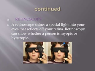
 Refractive error is defined as a problem in eye
due to which light is not correctly focused on
retina that results in blurred image. It mainly
happens due to alterations in shape of eye.
 TYPES:
 • Myopia.
 • Hyperopia.
 • Astigmatism.
 • Presbyopia.
 VISUAL ACUITY TEST:
 A visual acuity test is an exam that checks how will
you see that details of a letter or symbol from a specific
distance. Visual acuity refers to your ability to discern
the shapes and details of things you see. It’s just one
factor in your overall vision.
 PIN HOLE TEST:
 A pin hole occluder is an opaque disc with one or more
small holes through it, used by opthalmologist,
optometrist, orthoptisis to test visual acuity. The ocular
is simple way to focus light, as in a pinhole camera,
temporarily removing the effects of refractive errors
such as myopia.
 RETINOSCOPY:
 A retinoscope shines a special light into your
eyes that reflects off your retina. Retinoscopy
can show whether a person is myopic or
hyperopic.
 VISUAL ACUITY TEST:
 The initial hyperopia diagnosis is by visual acuity
test asking the patient to read letters from an
snells chart, which is keeping at specific distance
 RETINOSCOPY:
 It is useful to shine a light into a patient's eye for
an eye optometrist to examine the reflection off the
retina. The light is moving back and forth across
the pupil. Retinoscope is particularly useful to
assess small kids.
 PHOROPTER:
 It is an instrument used during an eye
examination to measure refractive error. The
patient is requesting to sit behind the
phoropter, and looks through it at an eye chart
hanging at a specific distance. The examiner
then changes lenses and other settings, by
asking the patient for their feedback on which
settings give the best vision, based on this the
doctor assesses the corrective lens value for the
error.
 VISUAL ACUITY TEST:
 As part of the testing, people will ask to read
letters on a distance chart. This test measures
visual acuity, which is writing as a fraction
such as 20/40. The top number is the standard
distance at which testing is performing, twenty
feet. The bottom number is the smallest letter-
size that can read. A person with 20/40 visual
acuity would have to get within 20 feet, for a
letter that should see at 40 feet in order to see it
clearly. Normal distance visual acuity is 20/20.
 CORNEAL TOPOGRAPHY:
 This advanced technology provides the most
detailed information about your cornea’s
shape. The doctor will tell you to look at
something specific. Meanwhile, the device
collects thousands of tiny measurements. A
computer then builds a color map of your
cornea from the data. Your doctor will refer to
it if he plans surgery for astigmatism.
 DYNAMIC RETINOSCOPY:
 Objective technique of assessing
accommodative function.
 Suitable from infancy.
 Non verbal response.
 LAG-UNDER ACCOMMODATION:
 • Lag of approx 0.75D to 4D stimulus is normal.
 LEAD-OVER ACCOMMODATION:
 • Target held at distance X D from subject.
 • Retinoscope reflex observed while retinoscope is also at X D.
 • If this movement observed ret is moved away from until
neutral achieved at distance A D.
 • Target remains at X
 • Target held at 25 cm / 4D from subject.
 • Retinoscope reflex observed while retinoscope is also at 4D
 • With movement is observed
 • Ret moved away from subject
 • Neutral achieved at 40cm / 2.50D
 • Target remains at 4D

assessment of refractive errors

  • 2.
     Refractive erroris defined as a problem in eye due to which light is not correctly focused on retina that results in blurred image. It mainly happens due to alterations in shape of eye.  TYPES:  • Myopia.  • Hyperopia.  • Astigmatism.  • Presbyopia.
  • 3.
     VISUAL ACUITYTEST:  A visual acuity test is an exam that checks how will you see that details of a letter or symbol from a specific distance. Visual acuity refers to your ability to discern the shapes and details of things you see. It’s just one factor in your overall vision.  PIN HOLE TEST:  A pin hole occluder is an opaque disc with one or more small holes through it, used by opthalmologist, optometrist, orthoptisis to test visual acuity. The ocular is simple way to focus light, as in a pinhole camera, temporarily removing the effects of refractive errors such as myopia.
  • 4.
     RETINOSCOPY:  Aretinoscope shines a special light into your eyes that reflects off your retina. Retinoscopy can show whether a person is myopic or hyperopic.
  • 5.
     VISUAL ACUITYTEST:  The initial hyperopia diagnosis is by visual acuity test asking the patient to read letters from an snells chart, which is keeping at specific distance  RETINOSCOPY:  It is useful to shine a light into a patient's eye for an eye optometrist to examine the reflection off the retina. The light is moving back and forth across the pupil. Retinoscope is particularly useful to assess small kids.
  • 6.
     PHOROPTER:  Itis an instrument used during an eye examination to measure refractive error. The patient is requesting to sit behind the phoropter, and looks through it at an eye chart hanging at a specific distance. The examiner then changes lenses and other settings, by asking the patient for their feedback on which settings give the best vision, based on this the doctor assesses the corrective lens value for the error.
  • 7.
     VISUAL ACUITYTEST:  As part of the testing, people will ask to read letters on a distance chart. This test measures visual acuity, which is writing as a fraction such as 20/40. The top number is the standard distance at which testing is performing, twenty feet. The bottom number is the smallest letter- size that can read. A person with 20/40 visual acuity would have to get within 20 feet, for a letter that should see at 40 feet in order to see it clearly. Normal distance visual acuity is 20/20.
  • 8.
     CORNEAL TOPOGRAPHY: This advanced technology provides the most detailed information about your cornea’s shape. The doctor will tell you to look at something specific. Meanwhile, the device collects thousands of tiny measurements. A computer then builds a color map of your cornea from the data. Your doctor will refer to it if he plans surgery for astigmatism.
  • 9.
     DYNAMIC RETINOSCOPY: Objective technique of assessing accommodative function.  Suitable from infancy.  Non verbal response.
  • 10.
     LAG-UNDER ACCOMMODATION: • Lag of approx 0.75D to 4D stimulus is normal.  LEAD-OVER ACCOMMODATION:  • Target held at distance X D from subject.  • Retinoscope reflex observed while retinoscope is also at X D.  • If this movement observed ret is moved away from until neutral achieved at distance A D.  • Target remains at X  • Target held at 25 cm / 4D from subject.  • Retinoscope reflex observed while retinoscope is also at 4D  • With movement is observed  • Ret moved away from subject  • Neutral achieved at 40cm / 2.50D  • Target remains at 4D