SlideShare a Scribd company logo
brazilian journal of microbiology 47S (2016) 3–30
http://www.bjmicrobiol.com.br/
Medical Microbiology
Diarrheagenic Escherichia coli
Tânia A.T. Gomesa,∗
, Waldir P. Eliasb
, Isabel C.A. Scaletskya
, Beatriz E.C. Gutha
,
Juliana F. Rodriguesc
, Roxane M.F. Piazzab
, Luís C.S. Ferreirac
, Marina B. Martinezd
a Universidade Federal de São Paulo, Escola Paulista de Medicina, Departamento de Microbiologia, Imunologia e Parasitologia, São Paulo,
SP, Brazil
b Instituto Butantan, Laboratório de Bacterologia, São Paulo, SP, Brazil
c Universidade de São Paulo, Instituto de Ciências Biomédicas, Departamento de Microbiologia, São Paulo, SP, Brazil
d Universidade de São Paulo, Faculdade de Ciências Farmacêuticas, Departamento de Análises Clínicas e Toxicológicas, São Paulo, SP,
Brazil
a r t i c l e i n f o
Article history:
Received 8 October 2016
Accepted 27 October 2016
Available online 5 November 2016
Keywords:
Escherichia coli
Diarrhea
Pathogenic mechanisms
Virulence factor
Epidemiology
a b s t r a c t
Most Escherichia coli strains live harmlessly in the intestines and rarely cause disease in
healthy individuals. Nonetheless, a number of pathogenic strains can cause diarrhea or
extraintestinal diseases both in healthy and immunocompromised individuals. Diarrheal
illnesses are a severe public health problem and a major cause of morbidity and mortal-
ity in infants and young children, especially in developing countries. E. coli strains that
cause diarrhea have evolved by acquiring, through horizontal gene transfer, a particular set
of characteristics that have successfully persisted in the host. According to the group of
virulence determinants acquired, specific combinations were formed determining the cur-
rently known E. coli pathotypes, which are collectively known as diarrheagenic E. coli. In this
review, we have gathered information on current definitions, serotypes, lineages, virulence
mechanisms, epidemiology, and diagnosis of the major diarrheagenic E. coli pathotypes.
© 2016 Sociedade Brasileira de Microbiologia. Published by Elsevier Editora Ltda. This is
an open access article under the CC BY-NC-ND license (http://creativecommons.org/
licenses/by-nc-nd/4.0/).
The genus Escherichia, which was named after the Ger-
man pediatrician Theodor Escherich, consists of facultative
anaerobic Gram-negative bacilli that belong to the family
Enterobacteriaceae.1 The genus type species Escherichia coli is
widely distributed, where it is the major facultative anaerobe
inhabiting the large intestine of humans and warm-blooded
animals.2 Although most E. coli strains live harmlessly in
the colon and seldom cause disease in healthy individuals,
∗
Corresponding author.
E-mail: tatg.amaral@unifesp.br (T.A. Gomes).
a number of pathogenic strains can cause intestinal and
extraintestinal diseases both in healthy and immunocompro-
mised individuals.3
Diarrheal illnesses are a severe public health problem and
a major cause of morbidity and mortality in infants and young
children.4 Low- and middle-income countries in Africa, Asia
and Latin America are the most affected regions with diarrheal
diseases occurring more often with lethal outcomes mainly
http://dx.doi.org/10.1016/j.bjm.2016.10.015
1517-8382/© 2016 Sociedade Brasileira de Microbiologia. Published by Elsevier Editora Ltda. This is an open access article under the CC
BY-NC-ND license (http://creativecommons.org/licenses/by-nc-nd/4.0/).
4 brazilian journal of microbiology 47S (2016) 3–30
due to poor living conditions (inadequate water supplies,
poor environmental hygiene and sanitation, and insufficient
education).5
E. coli strains involved in diarrheal diseases are one of the
most important of the various etiological agents of diarrhea,
where strains have evolved by the acquisition, through hori-
zontal gene transfer, of a particular set of characteristics that
have successfully persisted in the host.3,5,6 According to the
group of virulence determinants acquired, specific combina-
tions were formed determining the currently known E. coli
pathotypes, which are collectively known as diarrheagenic
E. coli (DEC).6 The DEC pathotypes differ regarding their prefer-
ential host colonization sites, virulence mechanisms, and the
ensuing clinical symptoms and consequences, and are clas-
sified as enteropathogenic E. coli (EPEC), enterohemorrhagic
(Shiga toxin-producing) E. coli (EHEC/STEC), enteroaggregative
E. coli (EAEC), enterotoxigenic E. coli (ETEC), and enteroinvasive
E. coli (EIEC).
Each of these pathotypes represents a group of clones
that share specific virulence factors. Nevertheless, it should
be pointed out that the plasticity of the E. coli genome has
hindered the identification of certain E. coli isolates as a
pathotype, because some isolates combine the main virulence
characteristics of different pathotypes and are thus consid-
ered potentially more virulent hybrid pathogenic strains.5
Another less well-defined pathotype has been described,
that is, the diffusely-adherent E. coli (DAEC) pathotype, which
comprises strains that adhere to epithelial cells in a diffused
distribution.6 Despite their classification as a group distinct
from the other pathotypes, the designation of DAEC as a differ-
ent DEC pathotype requires further epidemiological studies,
which have been hampered by the difficulties in its identifi-
cation and classification.5 Furthermore, certain E. coli strains
that have been classified as the adherent invasive E. coli (AIEC)
pathotype, comprise one of the potential agents for Crohn’s
disease (CD). CD is an inflammatory bowel disease (IBD), which
is thought to be caused by a combination of factors (genetics,
the intestinal microbiota, environmental factors, and enteric
pathogens).7,8
Diarrheal episodes due to DEC infections are an important
public health issue among children and adults in develop-
ing countries, because of their association with morbidity and
mortality of children less than five years of age. It was our aim
with this review to gather information on current definitions,
serotypes, lineages, virulence mechanisms, epidemiology, and
diagnosis of the major DEC pathotypes with emphasis on the
studies conducted in Brazil.
Typical and atypical enteropathogenic E. coli
The term enteropathogenic E. coli (EPEC) was first used in
1995 by Neter et al.,9 to describe a number of E. coli strains
epidemiologically related to a series of outbreaks of infantile
diarrhea in the 1940s and 1950s.10,11 Originally identified by
serotype, EPEC are now defined as those E. coli strains having
the ability to cause diarrhea, to produce a histopathology on
the intestinal epithelium known as the attaching and effac-
ing (AE) lesion, and the inability to produce Shiga toxins and
heat-labile (LT) or heat-stable (ST) enterotoxins.6
Improvements in techniques allowing a better understand-
ing of the genome and virulence mechanisms among EPEC
strains over the years have led to the sub-classification of EPEC
into typical EPEC (tEPEC) and atypical EPEC (aEPEC).3,12 Typi-
cal EPEC strains causing human infectious diarrhea possess
a large virulence plasmid known as the EPEC adherence fac-
tor (EAF) plasmid (pEAF), which encodes the type IV fimbriae
called the bundle-forming pilus (BFP), while aEPEC do not
possess this plasmid.6,12
The majority of tEPEC strains fall into well-recognized O
serotypes. Classical EPEC O serogroups include O55, O86, O111,
O114, O119, O127, and O142. The most common H antigens
associated with EPEC are the H6 and H2 antigens.12–15 A less
common EPEC type is H34, and a number of tEPEC strains
are classified as non-motile (H-) in conventional tests. Typical
EPEC strains belonging to non-classical serotypes have also
been reported.12,16
Based on multilocus enzyme electrophoresis analysis
(MLEE) of allelic differences between housekeeping genes,
tEPEC strains have been subtyped into two major lineages,
previously designated EPEC1 and EPEC2.13,14 EPEC1 includes
widespread serotypes such as O55:H6 and O119:H6, whereas
EPEC2 consists of serotypes with more limited occurrence
such as O111:H2 and O114:H2. Based on a whole-genome
phylogeny and analysis of type III secretion system (T3SS)
effectors, tEPEC strains have been demonstrated to cluster
in three main lineages, designated EPEC1, EPEC2, and EPEC4,
which probably acquired the locus of enterocyte effacement
(LEE) region and pEAF independently.17
In turn, aEPEC belong to a large diversity of classical
and non-classical serotypes.12,16,18 Over 20% of strains of
non-classical EPEC serotypes are O non-typeable and the O-
typeable strains belong to more than 4200 different serotypes,
with many non-motile and H non-typeable strains.12,18 Inter-
estingly, it has been found that 35% of the aEPEC strains also
belong to the tEPEC lineages.17 Thus, it has been hypothe-
sized that at least some aEPEC may have originated from tEPEC
strains that lost pEAF in the host or in the environment.17,19,20
Virulence factors, mechanisms and pathogenesis
Typical EPEC strains adhere to HeLa, HEp-2, and other cell lines
and to organ cultures in vitro in a distinctive pattern of three-
dimensional microcolonies, a so-called localized adherence
(LA) pattern.6,21 A similar adherence pattern has been seen
in tissue biopsies of EPEC-infected humans.22
The LA phenotype is mediated by the BFP,23 which
also contributes to antigenicity, autoaggregation, and biofilm
formation.23–27 An operon of 14 genes contained on the pEAF
is necessary for BFP expression, with bfpA encoding the major
structural subunit (bundlin)28 and being highly conserved
among EPEC1 and EPEC2 strains.
The self-transmissible pEAF pMAR2 is found among
strains of the EPEC1 lineage and contains an intact transfer
region, unlike pB171, which is more common among EPEC2
strains.29,30 Besides the bfp gene cluster, encoding BFP,23 the
pEAF carries the per locus, encoding the transcriptional activa-
tor called plasmid-encoded regulator (Per).29 Between pMAR2
and pB171, the bfp and per loci share 99% sequence similarity,30
and both BFP and PerA have been shown to contribute to
brazilian journal of microbiology 47S (2016) 3–30 5
virulence in human volunteers.24 Recent comparative
genomics of the EAF plasmids from diverse EPEC phyloge-
nomic lineages demonstrated significant plasmid diversity
even among isolates within the same phylogenomic lineage.31
Typical EPEC have the ability to form tight, spherical, bac-
terial autoaggregates when grown in liquid culture.32 Like LA,
autoaggregation requires BFP. Typical EPEC also form biofilms
on abiotic surfaces under static conditions, or in a flow through
continuous culture system, and a model of EPEC biofilm
formation has been proposed.26 Mutagenesis analysis has
identified adhesive structures such as the common type 1
pilus (T1P), antigen 43, BFP and the EspA filament (see below)
as participants in bacterial aggregation during biofilm forma-
tion on abiotic surfaces.26
A hallmark phenotype of both tEPEC and aEPEC is the abil-
ity to produce AE lesions.33 This phenotype is characterized
by effacement of intestinal epithelial-cell microvilli and inti-
mate adherence between the bacterium and the epithelial cell
membrane. Directly beneath the adherent bacterium, marked
cytoskeletal changes are seen in the epithelial cell membrane,
particularly the formation of an actin-rich cup-like pedestal at
the site of bacterial contact. AE lesions are observed in model
EPEC infections with cultured cells and mucosal explants, as
well as in intestinal biopsies from EPEC-infected infants or
animals.6
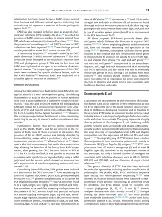
AE lesions are encoded by LEE, which is a ∼35-kb
pathogenicity island (PAI)34 that is organized into five oper-
ons (LEE1 to LEE5).35–37 The LEE1, LEE2, and LEE3 operons
encode components of a T3SS, and the global regulator Ler
(LEE-encoded regulator).38 LEE4 encodes the T3SS-secreted
proteins EspA, EspB, and EspD (EPEC-secreted protein), which
are components of the translocation apparatus by which other
effector proteins are translocated into the cell. LEE5 encodes
the adhesin intimin and its translocated receptor, Tir.39
Intimin is a 94-kDa protein encoded by the eae gene and
required for intimate adherence of EPEC to host cells at the
sites of AE lesions.6 The N-terminus of intimin is highly
conserved, whereas the C-terminus is highly variable.40 Dif-
ferences in the C-terminus of intimin have been used as a
basis for classification into several distinct subtypes, repre-
sented by the Greek letters ␣ (alpha) through ␨ (zeta)41,42; the ␣
subtype is expressed by EPEC1 strains while subtype ␤ is asso-
ciated with human EPEC2 strains. The N-terminus of intimin
anchors the protein in the EPEC outer membrane, whereas
the C-terminus extends from the EPEC surface and binds
to the Tir. Intimin–Tir interaction leads to intimate adher-
ence and pedestal formation beneath adherent bacteria,39 and
inhibits NF-␬B activity through tumor necrosis factor alpha
(TNF-␣) receptor-associated factors.43 In addition to Tir, the
EPEC genome contains six other LEE-encoded effector proteins
that are translocated into the cell (Map, EspF, EspG, EspZ, EspH,
and EspB), which interfere with different aspects of the cell’s
physiology.13,36,37,44
In addition to the LEE effectors, various non-LEE (Nle)-
encoded effector genes (cif, espI/nleA, nleB, nleC, nleD, nleE,
nleH)36,44 have been described, which are located outside the
EPEC LEE region, in at least six chromosomal PAIs, or in
prophage elements.45,46 The Nle proteins have been shown
to disrupt the cytoskeleton and tight junctions of the host
cell, and to modulate or prevent the host inflammatory
response.45–47 Although they are not required for AE lesion
formation, it is understood that they contribute to increased
bacterial virulence.44
Intracellular tEPEC have been observed both in tissue cul-
ture and in small intestinal biopsies from an EPEC-infected
infant.6 Two studies have reported that O111:NM strains con-
tain plasmid sequences that confer invasiveness upon E. coli
K12 strains containing the cloned fragments.48,49 Sequences
homologous to these cloned genes are present in only a minor-
ity of tEPEC strains (Scaletsky et al., unpublished data).
Typical EPEC strains encode a large surface protein,
lymphocyte inhibitory factor (LifA), which inhibits the expres-
sion of multiple lymphokines and inhibits lymphocyte
proliferation.50 Two related genes efa1 and toxB have been
implicated in adhesion to epithelial cells.51,52 There is evi-
dence indicating that Efa1/LifA contributes to epithelial cell
adherence in vitro53 and is required for intestinal colonization
of mice by the related AE pathogen Citrobacter rodentium.54
Some tEPEC strains possess other fimbriae or pili in addi-
tion to BFP. Type 1 fimbriae of EPEC have been found to be
antigenic in volunteer studies; however, they do not have a role
in adherence to epithelial cells in vitro.6 In addition, some EPEC
strains have conserved fimbrial genes encoding homologs of
long polar fimbriae (LPF),55 but a number of polymorphisms
within the lpfA genes have been identified.56 Initial stud-
ies have indicated that LPF is apparently not necessary for
adherence and AE lesion in human biopsies.55 The E. coli com-
mon pilus (ECP) has also been shown to act as an accessory
adherence factor in EPEC, playing a role during cell adher-
ence and/or in bacterium-bacterium interactions.57 However,
the significance of ECP to EPEC pathogenesis has not been
determined. Interestingly, it has been shown that some tEPEC
strains may produce a hybrid adherence phenotype in HeLa
cells, i.e., LA and aggregative (AA)-like pattern concurrently
(LA+/AA-like+).58 Recently, it was shown that at least some of
these LA/AA-like+ strains bear large plasmids, distinct from
the pEAF, that encode a so far unknown adhesin.59 It has
been proposed that the ability of such strains in producing
AE lesions and an AA-associated biofilm concomitantly could
worsen the patient’s clinical condition, leading to persistent
diarrhea.59
Flagella may also be involved in tEPEC adherence to epithe-
lial cells,60 since certain EPEC mutants are markedly impaired
in their ability to adhere and to form microcolonies. Further-
more, in one study, purified EPEC flagella and anti-flagellum
antibodies were both effective in blocking the adherence of
several EPEC serotypes.60 However, another study could not
confirm the role of flagella in EPEC adherence.61
Some tEPEC strains harbor the astA gene, which encodes
the enteroaggregative E. coli heat-stable enterotoxin 1
(EAST1).62,63 A recent study reported that 11 of 70 (16%) tEPEC
strains tested harbored an intact astA gene.64 Typical EPEC
strains of serotype O86:H34 produce cytolethal-distending
toxin (CDT).65 The significance of EAST1 and CDT toxins in
EPEC pathogenesis remains unknown.
Autotransporter (AT) proteins, which have been associ-
ated with bacterial adherence, aggregation, biofilm formation,
invasion, and toxicity66 in Gram-negative bacteria, have also
been described among EPEC strains.67 One such protein, EspC,
which is secreted by the type V secretion system and is
6 brazilian journal of microbiology 47S (2016) 3–30
injected by the T3SS in epithelial cells, has an IgA protease-
like activity and, once in the host cytoplasm, has various
cytopathic effects, including cytoskeletal damage,68 enhanced
lysozyme resistance,70 hemoglobin degradation,69 hydrolysis
of pepsin, factor V, and spectrin,70 and fodrin and focal adhe-
sion protein degradation.71 In addition, oligomerization of
EspC gives rise to rope-like structures that serve as a substra-
tum for adherence and biofilm formation as well as protecting
bacteria from antimicrobial compounds.72
A three-stage model of tEPEC adhesion and pathogen-
esis, consisting of LA, signal transduction, and intimate
attachment with pedestal formation, was proposed.73 Simul-
taneously with intimate attachment, a series of bacterial
effector proteins are injected into host cells, where they sub-
vert actin polymerization and other host cell processes.37,44
In the earliest stage and under correct environmental con-
ditions, tEPEC express BFP, intimin, and the T3SS/translocon
apparatus. Next, EPEC adhere to the surface of the intesti-
nal epithelium via BFP and EspA filaments, and the T3SS
injects the bacterial translocated intimin receptor (Tir) and
effector proteins (EspB, EspD, EspF, EspG, and Map) directly
into the host cell.37 The effectors activate cell-signaling
pathways, causing alterations in the host cell cytoskele-
ton and resulting in the depolymerization of actin and
the loss of microvilli. Finally, bacteria intimately adhere to
host cell by intimin–Tir interactions, causing a cytoskeletal
rearrangement that results in pedestal-like structures. Tir pro-
motes cytoskeletal reorganization through interaction with
neural WASP (Wiskott-Aldrich syndrome protein) (N-WASP)
and subsequent activation of the Arp2/3 complex,45 lead-
ing to the effacement of the microvilli and the production
of pedestals.44,74 The translocated effectors disrupt host cell
processes, resulting in loss of tight-junction integrity and
mitochondrial function, leading to both electrolyte loss and
eventual cell death.45
For actin dynamics subversion, tEPEC usually recruits
Nck to the adhesion site in a Tir phosphorylated Y474-
dependent mechanism. In turn, TirEHEC (enterohemorrhagic
E. coli [EHEC] O157:H7) is devoid of an Y474 equivalent and
employs EspFU/TccP (Tir-cytoskeleton coupling protein), a
T3SS-translocated effector protein that binds N-WASP, leading
to Nck-independent actin polymerization.45
aEPEC are devoid of pEAF and do not produce BFP. It is
important to point out that EPEC strains of serotypes O128:H2
and O119:H2 contain a pEAF with defective bfp operons, which
contain part of the bfpA gene but have the rest of the bfp
gene cluster deleted. Thus, they are classified as aEPEC.12,75
Most aEPEC produce adherence patterns categorized as LA-
like, with loosened microcolonies compared to those of the
tEPEC LA pattern.12,76,77 In addition, some isolates express
the aggregative (AA) or diffuse (DA) patterns of adherence,
which are characteristics of the EAEC and DAEC pathotypes,
respectively,20,78 or adhere in undefined patterns or are non-
adherent.20,78–80 Remarkably, the epithelial cell adherence
phenotype displayed by aEPEC is determined in prolonged
assays (6 h) of bacteria-cell interaction.12,76,77 In addition, it
has been suggested that lack of the pEAF-encoded Per pro-
teins in the regulatory cascade of the aEPEC virulence genes
may promote delayed AE lesion formation, probably making it
difficult for such strains to cause disease.81
The prevalence of intimin subtypes among aEPEC strains
has been reviewed.18,67 Intimins classified as beta1, epsilon1
and theta appear as the most frequent among aEPEC.78,82–85 In
addition, some aEPEC strains bear adhesive-encoding genes
that have been originally described in other DEC pathotypes
and/or in extraintestinal pathogenic E. coli.79,80,82,86–88 This
observation suggests that aEPEC could employ additional
adherence mechanisms besides the Tir-intimin interaction.
The only adhesin first characterized in an aEPEC strain
(serotype O26:H11) is the locus of diffuse adherence (LDA), which
is an afimbrial adhesin that confers the diffuse pattern of
adherence on HEp-2 cells, when cloned in E. coli K-12 strains.89
The T3SS-translocon has been also shown to contribute to the
adherence efficacy of an aEPEC strain in vitro.90 The prevalence
of these different adhesins among aEPEC has been recently
reviewed.18,67,91
Moreover, it has been recently shown that the flagellar
cap protein FliD of an aEPEC strain (serotype O51:H40) binds
to unknown receptors on intestinal Caco-2 cell microvilli.92
Interestingly, an anti-FliD serum and purified FliD reduced
adherence of the aEPEC as well as that of tEPEC, EHEC and ETEC
prototype strains to the same cell line.92 Furthermore, it has
been suggested that adherence of aEPEC of serotype O26:H11
may be mediated by binding of the flagellin protein FliC (the
subunit of the flagella shaft) to cellular fibronectin.93 However,
the role of the flagella in aEPEC in vivo colonization has yet to
be investigated.
Atypical EPEC strains have also been shown to adhere to
abiotic surfaces (polystyrene and glass).94,95 The non-fimbrial
adhesin curli and the T1P have been shown to mediate binding
to these surfaces in some aEPEC at different temperatures.90,96
The LEE region of some aEPEC strains display a genetic
organization similar to that found in the tEPEC prototype
E2348/69 strain.97 Although the T3SS-encoding genes are con-
siderably conserved,97,98 the effector protein-encoding genes
display important differences, and remarkable differences can
be detected at the 5 and 3 flanking regions of aEPEC, suggest-
ing the occurrence of different evolutionary events.99 Atypical
EPEC strains may carry two tccP variants, tccP and/or tccP2, sug-
gesting that some aEPEC strains may use both Tir-Nck and
Tir-TccP pathways to promote actin polymerization.100 Inter-
estingly, Rocha and colleagues101 showed that transformation
of a non-adherent aEPEC strain (serotype O88:HNM) with a
TccP expressing-plasmid, conferred this strain the ability to
adhere to and to induce actin-accumulation in HeLa cells.
The occurrence and prevalence of Nle in aEPEC strains have
been recently reviewed.67 It has been suggested that differ-
ent isolates can employ distinct strategies to promote damage
to the host and cause disease.45 In addition, the Nle effec-
tors Ibe (invasion of endothelial cells) and EspT have been
originally described and characterized in aEPEC strains.102,103
Ibe appears to regulate Tir phosphorylation and to enhance
actin polymerization and pedestal formation,103 while EspT104
modulates actin dynamics, leading to membrane ruffling and
cell invasion, and induces macrophages to produce inter-
leukins IL-8 and IL-1␤ and PGE2.102
Invasion of epithelial cells in vitro in an intimin-dependent
pathway has been described in an aEPEC strain,105 but further
studies pointed out that the invasive phenotype is not a com-
mon characteristic among aEPEC.106 Despite their invasive
brazilian journal of microbiology 47S (2016) 3–30 7
potential in vitro,107 most aEPEC are considered extracellular
pathogens.5
It has been shown that apical infection of cultured human
mucin-secreting intestinal HT29-MTX cells by some aEPEC
strains may induce increased production of secreted MUC2
and MUC5AC mucins and membrane-bound MUC3 and MUC4
mucins.108 This observation suggests that the apically adher-
ing bacteria could exploit large amounts of mucins to grow
more efficiently in the host intestines, characterizing a puta-
tive new virulence mechanism in aEPEC.108
AT proteins have also been shown to be produced by some
aEPEC strains.67 Abreu and collegues109 have shown that the
AT protein encoded by the ehaC gene, which is involved in
biofilm formation in EHEC strains, was the most frequent, with
a significantly higher prevalence than in tEPEC. Although the
prevalence of the AT protein Pic (protein involved in intestinal
colonization), formerly identified in EAEC, is not a common
finding in aEPEC strains, it also appears to mediate coloniza-
tion of mouse intestines, hemagglutination, mucin cleavage,
and complement components degradation.110 More recently,
some aEPEC strains were shown to cause cell damage by
secreting the AT protein Pet (plasmid encoded toxin) to the
extracellular environment.111
Epidemiology
The prevalence of EPEC infections varies between epidemio-
logical studies on the basis of differences in study populations,
age distributions, and methods (serotyping, adherence pat-
terns, and presence of the eae or conserved LEE genes) used
for detection and diagnosis.112 In addition, differences in geo-
graphic regions, periods of time and socioeconomic class may
also contribute to differences in the epidemiology of EPEC-
induced diarrheal disease.113 Lack of discrimination between
tEPEC and aEPEC in some studies also makes such analysis
difficult.
Diarrhea due to tEPEC decreases with age, and infections in
adults are rarely reported. This apparent resistance in adults
and older children has been attributed to the loss of specific
receptors with age or development of immunity.6
For many decades, studies conducted worldwide have
shown that tEPEC serotypes are strongly associated with diar-
rhea in children <1 year of age, mainly in poor children in
urban centers.6,12,15 The association with diarrhea was partic-
ularly strong in infants less than 6 months of age. Studies in
Brazil, Chile, Mexico, and South Africa, showed that 30–40% of
infantile diarrhea cases were due to tEPEC serotypes.15,112,114
However, the epidemiology of EPEC infections has shifted.
In numerous developing countries, where the prevalence of
EPEC infection had been high until the 1990s, recent studies
have not identified a significant association between tEPEC
and infantile diarrhea. In Brazil, 92% of EPEC isolates col-
lected from children between 2001 and 2002 were atypical,115
compared to 38% in a 1998–1999 study.79 However, other stud-
ies still report tEPEC being more prevalent than aEPEC as a
cause of diarrhea.116 In addition, in some less developed areas
(Africa and Asia), tEPEC are still some of the most impor-
tant enteropathogens.117–122 Based on the recently completed
Global Enteric Multicenter Study (GEMS) involving children
less than 5 years of age from seven sites in Africa and Asia,
tEPEC was significantly associated with moderate to severe
diarrhea in children under 2 years of age in Kenya, whereas
aEPEC was not associated with this type of diarrhea.118
Transmission of tEPEC follows a fecal-oral process
through contaminated surfaces, weaning fluids, and human
carriers.123 Although rare, outbreaks among adults seem
to occur through the ingestion of contaminated food and
water; however, no specific environmental reservoir has been
identified.6 The infective dose in adult volunteers is high, at
108 to 1010 organisms,124 while the infective dose that causes
disease in children is unknown. EPEC outbreaks have been
reported to show a seasonal distribution with peaks during
the warm months.6,125 Humans are the only known reservoir
for tEPEC, with symptomatic and asymptomatic children and
asymptomatic adults being the most likely source.6
In contrast to tEPEC, aEPEC have been found in diarrheic
patients of all ages and in adults with HIV-AIDS.82,126–128 Fur-
thermore, the proportion of aEPEC strains has increased, and
aEPEC strains have outnumbered tEPEC strains and have also
been associated with childhood diarrhea in some developing
and developed countries.12,18,67,91,112,129 However, the increase
in prevalence of aEPEC may also reflect the refined discrimi-
nation between tEPEC and aEPEC.12,18,91
The role of aEPEC in diarrhea is not clear because of its
detection at similar rates in both diarrheic and non-diarrheic
patients in various geographical areas.18,91,128 In studies con-
ducted in the last five years, aEPEC have been found at
rates varying from ∼0.05 to ∼12% in diarrheic versus 0 to
∼14% in non-diarrheic patients.67 Some recent studies have
also implicated aEPEC as the cause of persistent and bloody
diarrhea.18,91 Moreover, aEPEC strains have been associated
with diarrheal outbreaks in Finland, United States, Japan,
China18,91,112 and Brazil.85
In contrast to tEPEC, which are seldom found in animals,12
many aEPEC strains have been found in both diarrheic and
healthy animals.18,67 Interestingly, animal aEPEC serogroups
associated with human diarrhea have been identified (e.g.,
O26, O103, O119, O128, O142 and O157).18,130,131 Serotyping
and molecular methods such as multilocus sequence typing
(MSLT) and pulsed field gel electrophoresis (PFGE) have con-
tributed to demonstrating that domestic and wild animals and
the environment are potential sources of aEPEC for human
infections in several regions.18,67,91,131 Therefore, although no
direct transmission from animals to humans has been shown
so far, it is reasonable to suggest that some aEPEC strains are
potentially zoonotic pathogens, with a large variety of ani-
mal species serving as important reservoirs.67,91 In addition,
foods including raw meat, pasteurized milk and vegetables
and water have also been implicated as vehicles of aEPEC in
human infections.67
aEPEC strains comprise a very assorted group with various
additional virulence mechanisms that altogether can modu-
late the disease outcome or their occurrence in asymptomatic
persons. There have been continuous advances in our knowl-
edge of the genetic background and pathogenicity of aEPEC
as well as in the information gathered from epidemiological
studies, and may contribute to the discrimination between
strains that cause diarrhea and those that cause asymp-
tomatic infections.
8 brazilian journal of microbiology 47S (2016) 3–30
Detection and diagnosis
EPEC can be detected by DNA probes or PCR assays using
primers targeting the eae and stx genes.132,133 All eae-positive
and stx-negative E. coli strains are further tested by PCR for
the presence of the bfpA gene encoding bundlin6 and/or the
EAF plasmid to differentiate tEPEC from aEPEC.134,135 How-
ever, this may fail to identify all bfpA-positive EPEC strains,
since multiple alleles of bfpA have been identified,136 suggest-
ing that some current PCR methods may fail to identify all
bfpA-positive EPEC strains.
However, in routine microbiology laboratories, all E. coli
colonies obtained from primary isolation plates are tradition-
ally screened by slide agglutination assays using sera against
the classical EPEC serogroups O26, O55, O86, O111, O114, O119,
O125, O126, O127, O128, O142, and O158.137 This method is
practical and easy to perform, the main advantage of which is
the commercial availability of the sera. However, the disadvan-
tage of this method is the heterogeneity of EPEC serogroups
that can comprise categories other than EPEC, the inability to
distinguish tEPEC from aEPEC within these serogroups, and
the occurrence of EPEC strains belonging to serogroups other
than the classical EPEC serogroups.12,18,138,139
Since EPEC strains are defined based on their virulence
properties, a set of proteins, including intimin, BFP and T3SS
secreted proteins can be considered targets for diagnosis. BFP
expression has been considered the phenotypic marker of
tEPEC.18,78,140 Immunofluorescence and immunoblotting tests
using monoclonal or polyclonal antibodies against BFP have
been employed.141,142 These cited authors detected the pro-
duction of BFP on different media, in which they reported
that 91% of the tEPEC strains tested produced BFP in Dul-
becco’s Modified Eagle Medium (DMEM), 89% in MacConkey,
and 83% in EMB agars. These results are particularly inter-
esting, since MacConkey and EMB agars are routinely used
for the identification of lactose-fermenting E. coli isolated
from diarrheal stools. A colony immunoblot assay for tEPEC
detection based on BFP expression was also standardized
using a rabbit tEPEC anti-BFP polyclonal serum. Standardiza-
tion was done after growing the bacterial isolates on DMEM
agar containing fetal bovine serum or tryptic soy agar con-
taining 5% washed sheep blood (TSAB). This test showed a
positivity of 92 and 83% and specificity of 96 and 97%, respec-
tively, when the culture was done in DMEM and TSAB. This
method combines the simplicity of an immunoserological
assay with the high efficiency of testing a large number of EPEC
colonies.140
Concerning intimin detection, a rabbit polyclonal sera
raised against the conserved region of intimin (Int388-667)143
was employed in order to detect tEPEC isolates expressing ␣,
␤, ␥, ␦ and ␧ intimin reported an application of immunoblot-
ting with 100% specificity and 97% sensitivity in the detection
of eae positive E. coli strains.144–146 These authors clearly
demonstrated that polyclonal rabbit antisera is suitable for
immunoblotting as a diagnostic tool, and showed that protein
denaturation and linearization is a critical step for anti-
intimin antibody accessibility. Indeed, even employing the
recombinant antibody such as single chain fragment variable
(scFv-intimin),147,148 merely by immunofluorescence the scFv-
intimin was able to detect tEPEC, aEPEC, and EHEC isolates,
showing that intimin can be a target for EPEC and EHEC diag-
nosis after bacterial permeabilization.148
Regarding secreted proteins, Lu et al.149 developed a new
practical method to identify EPEC by detecting the E. coli
secreted protein B (EspB) in the culture supernatant by
reversed passive latex agglutination (RPLA), after the strains
have been cultivated in DMEM. In addition, Nakasone et al.,150
established a rapid immunochromatographic (IC) test to iden-
tify the presence of EspB in EPEC and EHEC isolates. The
detection limit of the test has been reported to be 4 ng/mL, and
the results showed 96.9% sensitivity and 100% specificity. The
IC test for the detection of EspB may be a practical method
to define EPEC or EHEC both in clinical laboratories and the
field.150
In addition, a rapid agglutination test using latex beads
coated with anti-EspB mAb was standardized, showing 97%
sensitivity, 98% specificity and 97% efficiency, which is
required for the diagnosis of enteropathogenic diseases and
can be employed in developing countries with poorly equipped
laboratories.151
Enterohemorrhagic (Shiga toxin-producing)
E. coli (EHEC/STEC)
EHEC/STEC represent a well-known group of foodborne
pathogens distributed worldwide. The ability to produce one
or more of the Shiga toxin (Stx) family cytotoxins152 con-
stitutes the main virulence attribute of this pathogroup of
E. coli. A wide array of infections from mild and almost
unapparent diarrhea to more serious manifestations such
as hemorrhagic colitis (HC) and the development of a life-
threatening syndrome known as hemolytic uremic syndrome
(HUS) are caused by EHEC/STEC. Infants and children are the
main affected patients, and although the incidence of infec-
tion varies in different regions, the impact and importance
of EHEC/STEC infections in public health is immense, being
the main cause of acute renal failure in children in many
countries. The perspective of EHEC/STEC infections has been
previously described,153,154 but a considerable amount of infor-
mation has been obtained in more recent years related to the
epidemiology, ecology and virulence properties of these bacte-
ria.
E. coli O157:H7 serotype was the first to be linked to HC
and HUS cases in the early 1980s, and has been since then
responsible for numerous outbreaks and sporadic cases of
severe diseases all over the world, therefore considered to be
the prototype of this pathogenic group of bacteria.155 It is well
known that hundreds of other E. coli serotypes can harbor the
stx genes, but epidemiological studies carried out worldwide
have proven that only some of them have been responsible
for causing human diseases. Some serogroups including O26,
O45, O103, O111, O121 and O145 can be highlighted among
those most commonly related to human infections.156 More-
over, in recent years the emergence of some particular clones
such as the hybrid O104:H4 enteroaggregative E. coli carrying
Stx2 genes, responsible for a severe outbreak of HUS start-
ing in Germany in 2011,157 the spread of a new O26:H11 clone
in Europe,158 and some other hybrid clones,159 suggests that
brazilian journal of microbiology 47S (2016) 3–30 9
the mobility of genes and certainly the host background are
important features implicated in their pathogenic potential.
Virulence factors, mechanisms and pathogenesis
The common feature among EHEC/STEC isolates is the abil-
ity to produce Stx. This family of toxins has a conserved
AB5 subunit structure, composed of one active A subunit
linked to a pentameric B subunit responsible for the bind-
ing of the toxin to specific glycolipid receptors on the surface
of target cells. The stx operon is usually found within the
sequence for an inducible, lysogenic, lambda-like bacterio-
phage. Stxs inhibit protein synthesis by removing an adenine
residue from the 28S rRNA of the 60S ribosome.152 However,
besides this activity, studies have described that Stx also acts
on cell signal transduction and immune modulation causing
proinflammatory and pro-apoptotic responses.159 Two major
families, Stx1 and Stx2, have been recognized, and on the
basis of sequence diversity, each is composed of several vari-
ants. The Stx1 family is more homogenous and includes Stx1a,
Stx1c and Stx1d; while the heterogenous Stx2 group is com-
posed of Stx2a, Stx2b, Stx2c, Stx2d, Stx2e, Stx2f, and Stx2g.160
It should be mentioned that the association of some vari-
ants such as Stx2a, Stx2c or Stx2d with HC and HUS has
been highlighted compared to some others that seemed to
be more related to uncomplicated cases of diarrhea such as
Stx1variants or even Stx2e, Stx2f and Stx2g, which are uncom-
monly found causing human infections so far.161,162 Indeed,
the higher association of Stx2 with severe diseases has been
extensively studied by using Vero and endothelial cell lines
as well as some animal models.159 Moreover, knowledge of
stx phage characteristics and behavior has helped our under-
standing of how differences in expression of Stx between
EHEC/STEC isolates may contribute to pathogenesis and
disease.163
The ability to adhere to intestinal epithelial cells is
another key event in EHEC/STEC pathogenesis. The pres-
ence of the chromosomal pathogenicity island LEE,164 also
present in isolates belonging to the EPEC pathotype, is
common. Although LEE has been described in the major
EHEC/STEC serotypes responsible for a high proportion of
HC and HUS cases in several countries, its presence is not a
required condition for the occurrence of more serious infec-
tions as initially thought, because some LEE-negative strains
are also capable of causing outbreaks and sporadic cases of
HUS.165,166
Therefore, it is clear that EHEC/STEC pathogenesis is a mul-
tistep process, and besides the production of Stx toxins and
the AE lesion, other factors including different types of toxins
and adhesins have been described and found to be involved in
virulence.159
One should also consider that as a pathogen of the
human gastrointestinal tract the ability of EHEC/STEC
to monitor nutrients in the gut milieu, and translate
this information to sense the host physiological state in
order to program the expression of its virulence mark-
ers has a pivotal role on the development of infection.167
In addition, it has been shown that EHEC/STEC can
also cross-communicate with the host by exploiting the
autoinducer-3 (AI-3)/epinephrine/norepinephrine signaling
system to express two important virulence traits, motility and
A/E lesion, required at different time points during intestinal
colonization.168
The ability to adhere, colonize and form biofilm on food
and several types of surfaces may be a way to be an impor-
tant source and/or vehicle of transmission of EHEC/STEC. In
addition, biofilm may also act as bacterial protection against
adverse environmental conditions. A study conducted by Bis-
cola et al.,169 evaluated the capacity of biofilm formation
in EHEC/STEC strains isolated from different reservoirs and
serotypes. The authors observed that the ability to adhere
to abiotic surfaces forming biofilms, under defined culture
conditions, occurred in an array of wild-type O157 and non-
O157 strains. Biofilm production was identified in several
non-O157 STEC serotypes of human, animal, and food ori-
gin. On the other hand, among the O157 strains, only those
isolated from the animal reservoir and from a water sample
produced biofilm. A close correlation between biofilm for-
mation and expression of curli fimbriae and cellulose was
observed among O157 strains. However, in addition to curli,
the presence of other factors such as type 1 fimbriae and AT
proteins may be associated with the ability to form biofilm in
non-O157 strains. Matheus-Guimarães et al.,170 studied O157
and non-O157 EHEC/STEC strains isolated from bovine hides
and carcasses and showed that different sets of genes were
involved in the interactions of the bacteria with biotic and
abiotic surfaces. Moreover, the detection of an O157 strain
that was able to form biofilm on both glass and polystyrene
and that adhered to and invaded human cells, suggests an
important ability of this isolate to persist in the environment
and interact with the host. In fact, cell invasion and sur-
vival of some EHEC/STEC strains in cultured human intestinal
epithelial cells has been previously described.171 It should be
mentioned that this invasive characteristic has been identified
in some EHEC/STEC serotypes, many of which are responsi-
ble for human infections.170–173 Therefore, it is conceivable
that this virulence strategy may help bacteria to overcome
host defense mechanisms and certainly contributes to their
persistence in the zoonotic reservoir, ensuring efficient envi-
ronmental and food transmission.
Another topic of interest has been the analysis and com-
parison of the virulence profile of EHEC/STEC strains isolated
from the animal reservoir and environment with strains recov-
ered from human infections. In general, these studies have
shown that despite serotype diversity, the stx subtypes and
the virulence profile identified among isolates from the animal
reservoir and environment are similar to the isolates recov-
ered from patients.173,178–180 There has been particular interest
in some STEC serotypes that have been responsible for causing
severe human infections, such as O113:H21, but unlike others,
they do not produce adhesins encoded by LEE. By using a PCR
microarray, 41 virulence or genetic markers were tested in a
panel of 65 O113:H21 strains isolated from clinical infections,
environment and food from various countries.174 The results
obtained showed no clear differences in these genetic mark-
ers between the pathogens recovered from HUS cases and the
environmental strains. Moreover, only stx subtypes associated
with human infections were identified in all isolates, therefore
suggesting that the environmental isolates have the potential
to cause human diseases.
10 brazilian journal of microbiology 47S (2016) 3–30
Epidemiology
The incidence of HUS cases in Brazil is low,175 and although
some hypothesis has been proposed to explain this fact, there
are limited data on the immune response against Stx. In an
attempt to overcome this gap, prevalence of anti-Stx2 anti-
bodies in sera of children diagnosed with HUS and of healthy
children was recently determined.176 The percentage of indi-
viduals showing antibodies against Stx2 was higher among
HUS patients than controls, and the results also confirmed
that STEC strains are circulating in our settings despite the
low number of identified HUS cases.
Among the several serotypes associated with human
infections, O157:H7 is responsible for more severe cases. Epi-
demiological investigations of diarrheal outbreaks conducted
in four Brazilian states showed that O157:H7 strains were iso-
lated from two hospitalized patients, one with HUS and the
other with bloody diarrhea.177 Besides, O157:H7, EHEC/STEC
strains belonging to the top six most important non-O157
serogroups such as O26, O103, O111 and O145 were identi-
fied, all of which were recovered from ambulatory patients.
In addition, some uncommon serogroups including O1, O24
and O77 among others were also detected, but they were
all associated with acute diarrhea. It is interesting to note
that the majority of patients from whom STEC was isolated
were female (57%), and that patients’ ages ranged from 8
months to 80 years, with most being less than five years old
(54%).177
The distribution of EHEC/STEC in the gastrointestinal tract
of a wide variety of animals indicates the zoonotic charac-
ter of its infections. The role of different animal species as
asymptomatic carriers of EHEC/STEC has been extensively
studied in the last years in Brazil. Besides cattle, which
are their most common natural reservoir,173,178 the pres-
ence of these pathogens has been identified in the feces
of dairy buffaloes,179 sheep,180,181 pigs,182,183 birds,184 and
fishes.185 It is noteworthy that some relevant serotypes linked
to human infections such as O103:H2 and O157:H7 have
been recovered from the feces of sheep186 and cattle173
respectively. Additionally, the high prevalence of O157:H7
EHEC/STEC strains identified in hides of cattle sent to
slaughter in a Brazilian processing plant178 certainly rep-
resents a relevant issue that should be considered when
thinking about interventions targeting EHEC/STEC related to
animal handling, from farm to slaughter, as well as the
implementation of food safety throughout production and
processing.
The presence of EHEC/STEC in the environment is another
issue of concern, since they can survive in the soil, manure,
pastures and water, which thus represent important vehicles
of transmission. The isolation of STEC strains from drink-
ing water supplies, collected in different municipalities in
northern Paraná State, has been recently described, highlight-
ing the importance of drinking water, especially that from
untreated water supplies, as a source of STEC strains poten-
tially pathogenic for humans.187 Taking into account that
chicken litter is very useful as an organic soil fertilizer for the
production of fruits and vegetables in our settings, the detec-
tion of STEC in organic chicken fertilizer used on farms188 also
represents a significant public health safety hazard.
Although data on the detection of EHEC/STEC in foods
in Brazil are still scarce, the isolation and identification of
O157:H7 serotype from a ground beef sample was described for
the first time,189 while O125:H19 and O149:H8 STEC serotypes
were found in refrigerated raw kibbe collected from retail
establishments.190 On the other hand, EHEC/STEC has not
been detected in pasteurized cow’s milk samples collected in
dairies in northwestern Paraná State191 or in raw milk, pas-
teurized milk, Minas Frescal cheese and ground beef samples
collected in Minas Gerais.192 One should be aware that despite
difficulties in the detection and isolation of EHEC/STEC from
foods, the implementation of the most sensitive methods in
most laboratories should be the main goal in the near future
to help in the analysis of the risk posed by foods as vehicles of
STEC transmission to humans.
Detection and diagnosis
An important concern is how to detect Shiga toxin-producing
strains either in stools of infected patients or contaminated
food, since selective enrichment is necessary.193,194 For rou-
tine diagnosis, some protocols have already been described.139
However, the gold standard for Stx detection is still the eval-
uation of the cytotoxicity of bacterial culture supernatants
to eukaryotic cells.195,196 Thus, multiplex PCR including stx
gene and other virulence genes could be useful in screening
for STEC using bacterial confluent growth zones or sor-
bitol fermenting and non-fermenting colonies taken from
SMAC.197
Numerous assays for the diagnosis of STEC have been
developed on the basis of the detection of Stx1 and/or
Stx2, which represents the major virulence factors of this
E. coli category.198 Sensitivities and specificities vary according
to the test format and the manufacturer.199–205 Neverthe-
less, the standard by which each manufacturer evaluates its
tests also varies; therefore, a direct comparison of perfor-
mance characteristics of various immunoassays has not been
performed.198,206,207 Moreover, these commercially available
tests are not affordable for developing countries. Thus, to out-
line this, previous works have established different formats of
immunoassays, employing either a mixture of rabbit anti-Stx1
and anti-Stx2 sera by indirect ELISA or polyclonal and mono-
clonal antibodies in a capture ELISA assay for the detection of
STEC.207–209 The standardized methods are reproducible, fast,
easy to perform, showing high sensitivity in detecting Stx by
capture ELISA, even in low-producing isolates. These assays
have not yet been evaluated in terms of industrial quality con-
trol and commercial availability, but the estimated cost of the
assay is around US$70 per 96 detections, which is realistically
inexpensive for developing countries.
These monoclonal antibodies were rebuilt resulting in sin-
gle chain fragment variable (scFv) fragments. Stx2-scFv was
obtained from a bacteria-induced culture and showed diag-
nostic ability; the scFv fragment was able to recognize the
majority of Stx2-producing strains, with 79.3% sensitivity
(confidence interval of 60.3 to 92%), and no reactivity was
observed with the non-producing strains, indicating as high
as 100% specificity (confidence interval of 86.8–100%).210 It is
worth mentioning that none of the commercially available
brazilian journal of microbiology 47S (2016) 3–30 11
immunoenzymatic tests for Stx1/2 toxin detection employ
recombinant antibodies produced in bacteria, which indeed
will reduce the costs of the diagnostic assays.198
Enteroaggregative E. coli
EAEC is the diarrheagenic E. coli pathotype defined by showing
the characteristic AA pattern on epithelial cells in culture.211
The AA pattern was defined in 1987 when Nataro et al.,212 dis-
tinguished the previously described “diffuse adherence” as the
truly diffuse adherence (DA) and the AA pattern. The standard
AA was characterized by adherent bacteria in a stacked-brick
arrangement on the surface of epithelial cells and also on
the coverslip between cells. Strains displaying the AA pat-
tern were then categorized as “enteroadherent-aggregative
E. coli” but afterwards the category was called enteroaggrega-
tive E. coli or EAEC, the current nomenclature. The detection
of AA in vitro is still the gold standard test to define EAEC;
however, as described before, the AA pattern may be found
in strains of other DEC pathotypes, such as aEPEC. Therefore,
an up-to-date definition of EAEC is the diarrheagenic E. coli
that produce AA in cultured epithelial cells but lack the main
genetic markers that define other DEC pathotypes (EPEC, ETEC,
EHEC, EIEC). An exception for that is the hybrid EAEC/STEC
strain responsible for a massive outbreak of diarrhea and HUS
in 2011 in Europe.213 This strain consists of an EAEC strain
that acquired the Stx2-encoding phage. Therefore, this specific
O104:H4 strain is a Stx-producing EAEC.
Diarrhea caused by EAEC is watery, often with the pres-
ence of mucus, with or without blood and abdominal pain,
vomiting and low fever. Acute self-limiting diarrhea is the
usual pathology, but some patients may develop protracted
diarrhea, i.e., lasting more than 14 days.214 Prolonged diar-
rhea occurs depending on the host’s immunity, nutritional
status and genetic susceptibility.215 Genetic susceptibilities
associated with EAEC diarrhea were identified in North Amer-
ican travelers to Mexico. Single nucleotide polymorphisms
(SNP) in the IL-8 gene promoter and the promotor regions
of the genes encoding lactoferrin, CD14 and osteoprotegerin
as well were recognized as indicators for symptomatic EAEC
infection.216–219
A well-described characteristic of EAEC strains is their
heterogeneous nature when serotypes, genetic markers of
virulence and phylogenetic groups are analyzed.220–225 This
indicates that only EAEC strains carrying specific virulence
factors are able to cause diarrhea. While these factors are
unknown, some studies have demonstrated the association
of specific virulence genes with diarrhea, such as pet or aafA
in Brazil226 and sepA in Mali.223
Virulence factors, mechanisms and pathogenesis
Most of our knowledge about EAEC pathogenesis is based on
data accumulated from studies with EAEC strain 042, since
its association with human diarrhea in a volunteer study.227
These putative virulence factors include adhesins, toxins and
secreted proteins. However, none of these factors are found in
all EAEC strains.
The majority of these virulence factors are plasmid borne,
including those mediating AA. Consequently, these high-
molecular-weight plasmids are called pAA.220 Baudry et al.,228
developed a genetic probe (CVD432) for EAEC diagnosis on the
basis of a fragment from pAA1 present in EAEC strain 17-2.
In EAEC 042, many putative virulence factors are present in
pAA2.220
Recently, a division of EAEC strains into typical or atypi-
cal subgroups was proposed. This classification is based on
the presence or absence of aggR, a gene that encodes a global
regulator of EAEC virulence genes.229 Therefore, it has been
proposed that typical EAEC have more pathogenic potential
by the presence of the AggR regulon and, consequently, pAA
virulence factors.230 However, at least two outbreaks of diar-
rhea were caused by atypical EAEC,231,232 and atypical EAEC
are commonly isolated from children with diarrhea, in some
cases more frequently than typical strains.233,234
Numerous adhesins, cytotoxins, enterotoxins and secreted
proteins have been characterized in EAEC strains since this
pathotype definition.211,214
The most studied adhesins are the aggregative adherence
fimbria (AAF/I-AAF/V) family, which includes five types.235–239
They mediate the AA pattern and biofilm formation. Afimbrial
adhesins have also been characterized in EAEC strains, includ-
ing outer membrane proteins between 30 and 58 kDa.240–242
However, it has been shown that these structures are
present in low frequencies in EAEC collections from different
settings.221,226,243–245
Located in pAA2 of EAEC 042 is the aap gene, encoding
an antiaggregation protein called dispersin.246 This protein
is secreted and linked to lipopolysaccharide, neutralizing
the negative charge of the bacterial surface leading to AAF
projection and consequent dispersion along the intestinal
mucosa.247 Although immunogenic, dispersin is found in
other E. coli pathotypes and in commensal E. coli.248
Various toxins have been described in EAEC in association
with the cytotoxic or enterotoxic effects of culture super-
natants in vitro. The heat-stable toxin enteroaggregative E. coli
heat-stable enterotoxin 1 (EAST-1) was the first toxin charac-
terized in the EAEC pathotype.249 EAST-1 activates adenylate
cyclase inducing increased cyclic GMP levels, effects observed
in a Ussing chamber with rabbit ileum.250 ShET1 is an A:B type
toxin that causes accumulation of fluid in rabbit ileal loops and
has secretory response in Ussing chamber assays.251,252
The two AT proteins characterized in EAEC 042, Pet and
Pic,253–254 are members of the serine protease autotrans-
porters of Enterobacteriaceae, or SPATE.255 Pet is a cytotoxin
that modifies the cytoskeleton of enterocytes, leading to
rounding and cell detachment. The cytotoxic mechanism of
Pet arises from the degradation of ␣-fodrin, a membrane pro-
tein of the enterocytes.256 Pic is a multitask protein that
mediates hemagglutination, mucus cleavage and hypersecre-
tion, intestinal colonization in mice, cleavage of surface
glycoproteins involved in leukocyte trafficking and cleavage of
key complement molecules.257,258 The phenotypes identified
for Pic suggest its role in promoting colonization of the intes-
tine and immune system evasion. SPATEs are immunogenic
proteins, as evidenced by the presence of serum antibod-
ies against Pet and Pic in children recovering from diarrhea
caused by EAEC.259
12 brazilian journal of microbiology 47S (2016) 3–30
In the years that followed the definition of EAEC as
a pathotype, research in the field was dedicated to prove
the pathogenic capacity of EAEC using different animal
models260–262 and human volunteers receiving oral inoculum
of different EAEC strains.227,235,263 Not all volunteers devel-
oped diarrhea after ingestion of different EAEC strains, the
first evidence that strains of this pathotype are heterogeneous.
Among the strains tested, EAEC 042 (serotype O44:H18) caused
diarrhea in three out of five volunteers.227 Since then, strain
042 has been considered the prototype EAEC strain and is cer-
tainly the most studied strain of the pathotype.264 EAEC 042
was isolated from a case of acute infantile diarrhea in Peru.265
The clinical data obtained from the volunteers who developed
diarrhea suggested that EAEC 042 caused secretory diarrhea,
with abundant presence of mucus and absence of blood in the
stool.
Studies employing different EAEC strains interacting with
intestinal cells from animals or humans have been performed
to elucidate the pathogenesis of this pathotype. Data from
these in vitro, in vivo and ex vivo experiments strongly indi-
cate that EAEC can bind to jejunal, ileal and colonic epithelium
in the characteristic aggregative pattern, forming a strong
biofilm in a mucus layer, followed by cytotoxic and proinflam-
matory effects.260,266–270 Fragments from terminal ileum and
colon excised from pediatric and adult patients were incu-
bated with EAEC strains that were capable to colonize the ileal
and colonic mucosa in the typical stacked-brick pattern over
an augmented mucus layer.270
All these lines of evidence in combination with the iden-
tification of several putative virulence factors in prototype
EAEC strains allowed the proposal of a three-stage model of
EAEC pathogenesis: (a) abundant adherence to the intestinal
mucosa, (b) production of cytotoxins and enterotoxins, and
(c) induction of mucosal inflammation.211 In the first stage,
the contribution of fimbrial and afimbrial adhesins as well
as other adhesive structures is essential. Several colonization
factors have been identified in EAEC strains.271 In this stage, a
characteristic increased secretion of mucus on the intestinal
mucosa leads to the formation of a strong biofilm where EAEC
are embedded.234,266,272 In the following step, EAEC produce
cytotoxic effects on the intestinal mucosa due to the secretion
of toxins, inducing microvillus vesiculation, enlarged crypt
openings, and increased epithelial cell extrusion.266,273 EAEC-
induced inflammation results from the strong colonization
of the intestinal mucosa; however, all bacterial factors that
contribute to this condition have not been identified. Inflam-
matory markers such as IL-8, IL-1␤, interferon (INF)-␥ and
lactoferrin have been detected in stools of children and adults
colonized by EAEC.274–276 Although this model summarizes
the data so far obtained using in vivo, in vitro and ex vivo
approaches it may not be valid for all strains.
A large foodborne outbreak of bloody diarrhea and HUS
occurred in 2011 in Europe, affecting more than 4000 patients,
most of them from Germany. This outbreak was caused
by a Stx2-producing E. coli strain belonging to the serotype
O104:H4. The genome of that strain was rapidly sequenced,
revealing a unique hybrid combination of EAEC and STEC,
i.e. the EAEC strain Ec55989 harboring the Shiga toxin 2-
encoding prophage.166,213 Several virulence factors of typical
EAEC are present in that strain, including AggR, dispersin, Pic
and ShET-1. Also expressed are two Shigella autotransporter
proteins called SigA and SepA, implicated in mucosal dam-
age and colonization.277,278 Interestingly, the EAEC Ec55989
is the prototype strain for AAF/III.237 Conversely, the out-
break hybrid strain produces AAF/I, showing that the outbreak
EAEC/STEC acquired an AAF/I-encoding plasmid.166,279 It has
been proposed that the presence of these virulence factors
combined is responsible for the highly virulent attributes of
that strain.166,213
Epidemiology
EAEC is an emerging pathogen affecting children and adults
worldwide, responsible for cases of acute and persistent diar-
rhea. Nevertheless, the most important impact in terms of
morbidity is among children younger than 5 years living in
developing countries.214 A meta-analysis study of the lit-
erature on the epidemiology of diarrhea that included the
search of EAEC showed a statistical association of EAEC with
acute and persistent diarrhea in developed and developing
countries, with diarrhea in HIV-infected patients in developing
countries, and adult traveler’s diarrhea.280 In another meta-
analysis study EAEC was associated with acute diarrhea in
children living in South Asian countries.281
It is important to mention that data on the epidemi-
ology of EAEC infection are somewhat inconsistent due to
large variation in terms of method of detection, geographical
location and patient age and socioeconomic status. Nonethe-
less, EAEC has been systematically identified as an emerging
enteropathogen, strongly associated with acute and persis-
tent diarrhea in children of developing countries. Moreover, in
developed countries, EAEC have been frequently isolated from
cases of diarrhea in children and adults in the last years.282,283
In addition, several foodborne outbreaks of diarrhea caused
by EAEC have been reported in Europe, Japan, Mexico and
India.231,232,284–286 One of them affected 2697 school children
in Japan, after consumption of school lunches.232
Several studies have implicated EAEC as the predominant
agent of persistent diarrhea in children.287–289 EAEC-mediated
persistent diarrhea has been linked to malnutrition and
decrease in physical and intellectual development in several
studies from Brazil.274,288,290 Notably, asymptomatic patients
infected with EAEC also exhibit growth retardation.274 Since
its definition as a pathotype, high rates of asymptomatic
young children carrying EAEC have been reported in sev-
eral studies, involving subjects with low socioeconomic status
in developing countries.214 The persistence of EAEC may
induce chronic intestinal inflammation, even in the absence
of diarrhea, reducing its absorptive function and leading
to malnutrition.274,291 Growth impairment has also been
observed in a mouse model of EAEC oral infection.292 Consid-
ering the high number of asymptomatic EAEC-colonized
children in low-income countries, this pathotype has an
important impact on public health as one cause of impaired
physical and cognitive development.
EAEC is transmitted by the fecal-oral route by food or con-
taminated water.232,285,286 EAEC were detected in milk samples
from infant feeding bottles that were handled by mothers
with low socioeconomic status.293 Also, viable EAEC were
isolated in tabletop sauces from Mexican restaurants.294 No
brazilian journal of microbiology 47S (2016) 3–30 13
relationship has been found between EAEC strains isolated
from humans and different animal species, indicating that
animals may not represent a reservoir of human pathogenic
typical EAEC.295
EAEC has also emerged in the last years as an agent of uri-
nary tract infections (UTI). Initially, Abe et al.,296 described the
presence of EAEC virulence markers in strains isolated from
UTI, which was subsequently observed by others.297–300 Also,
the presence of uropathogenic E. coli (UPEC) markers in EAEC
collections has been reported.301,302 These findings pointed
out the potential for some EAEC strains to cause UTI.
A community acquired UTI outbreak caused by an EAEC
strain of serotype O78:H10, occurred in Denmark.303 This mul-
tiresistant strain belonged to the multilocus sequence type
ST10 and phylogenetic group A. This was the first time that
EAEC was implicated as an agent of an outbreak of extrain-
testinal disease. The uropathogenic properties of this EAEC
strain were conferred by specific virulence factors, such as
the AAF/I fimbriae.304 Recently, EAEC was implicated as a
causative agent of one case of urosepsis.300
Detection and diagnosis
Among the DEC pathotypes, EAEC is the most difficult to cat-
egorize, since it is a very heterogeneous group. The defining
characteristic of EAEC is the AA pattern in human epithelial
cells or on a glass substrate in a distinctive stacked-brick for-
mation. Thus, the gold standard method for distinguishing
EAEC is to culture five E. coli colonies per patient in static Luria-
broth at 37 ◦C, and then to infect semi-confluent HEp-2 cells
for 3 or 6 h, looking for the typical AA pattern.212,305 However,
this test requires specialized facilities and is time-consuming,
restricting its use only to research and certain reference labo-
ratories.
Furthermore, despite that several protein components
such as Pic, ShET1, EAST-1, and Pet are involved in the vir-
ulence of EAEC, none of them is present in all isolates. The
presence of Pet in EAEC isolates was initially detected by
immunoblotting assays after a preliminary step of culture
supernatant concentration.256 Vilhena-Costa et al.306 devel-
oped a slot blot immunoassay that avoids the concentration
step, allowing the detection of Pet directly from EAEC super-
natant, after growing the EAEC bacterial isolate in TSB at
37 ◦C for 4 h. In this method, it was possible to evaluate Pet
expression with specificity and reproducibility, using a rabbit
polyclonal anti-Pet serum, which showed no cross-reaction
with supernatants of non-Pet-expressing isolates and com-
mensal E. coli.
Considering these difficulties, DNA probes were included
as a valuable tool for EAEC detection.307 After sequencing the
EcoRI-PstI fragment of pCVD432 (AA or EAEC probe) developed
by Baudry et al.,228 primers complementary to this probe for
PCR amplification were designed.308 This PCR assay was found
to be a rapid, simple, and highly sensitive method, and there-
fore considered to be useful for screening stool specimens for
the presence of EAEC strains. Rapid and practical multiplex
PCR assays targeting more genes (aggR, aap and aatA, encod-
ing the AggR regulator, dispersin and an ABC secretion system
outer membrane protein, respectively) or aggR, pic and astA,
encoding AggR, Pic and an EAST-1) have also been employed to
detect EAEC strains.309–311 Monteiro et al.248 used PCR to evalu-
ate aggR, aatA and aap in a collection of E. coli strains and found
that aggR and aatA were more specific to EAEC than aap, sug-
gesting that the simultaneous detection of aggR, aatA, and aaiA
(a type VI secretion system protein) could be an improvement
in the PCR detection of EAEC.
All these proposed PCR-based protocols detect plas-
mid genes, which disfavors the detection of atypical EAEC
strains.305,309,312 Others, employing plasmid and chromosome
loci, have not reported sensitivity and specificity of the
assay.226,313,314 However, a multiplex PCR based on two genes
encoded in the plasmid and two chromosome-borne genes
is recommended to increase the ability to detect both typi-
cal and atypical EAEC strains. The aggR and aatA genes309,313
and aaiA and aaiG genes315 incorporated in the assay detec-
ting aaiA, aaiG, aggR and aatA demonstrated 94.8% sensitivity
and 94.3% specificity, and the assay was able to effectively
detect both groups of EAEC among E. coli isolated from stool
cultures.316 This method should improve EAEC detection,
since this pathotype is responsible for acute and persistent
diarrhea in children and adults and is also associated with
foodborne diarrheal outbreaks.
Enterotoxigenic E. coli
ETEC strains are characterized by the production of coloniza-
tion factors (CFs) and at least one of two enterotoxins: LT and
ST. ETEC represents one of the most common causes of diar-
rhea in children in developing countries and in travelers to
these regions. ETEC is also an economic burden to farmers and
industry, where it is an important pathogen for broilers, swine,
cattle and other farm animals. The group represents a highly
diverse pathovar of diarrheiogenic E. coli, harboring mobile
genetic elements such as plasmids and phages. ETEC hetero-
geneity was first demonstrated by phenotypic traits including
the large diversity of lipopolysshacaride (LPS) and flagelin
composition and the expression of different CFs and toxin
types.317,318 Serological typing of ETEC strains have relied on
the composition of outer membrane proteins and, mainly, in
the somatic LPS (O) and flagellar (H) antigens.318–320 ETEC com-
prise more than 100 somatic serogroups (O) and at least 34
flagellar types (H), combined in an unpredicted number of
O:H serotypes, but only a limited number of serotypes are
associated with infectious diseases, such as O8:H9, O6:H16,
O78:H12 and O25:H42, and are therefore of major clinical
relevance.318,321
The genetic diversity of ETEC has also been evaluated
by molecular approaches including random amplification of
polymorphic DNA (RAPD), MLEE, PFGE, multilocus sequence
type (MLST) and whole-genome sequencing.322–330 More
recently, 362 human-derived strains were subjected to next-
generation whole-genome sequencing; 21 genotypes could
be identified, and ETEC strains could be classified into
5 major phylogroups (A, B1, B2, D and E).330 Genetic
analyses demonstrated that clonally related ETEC lineages
sharing the same serotypes and CF and toxin profiles
have worldwide distribution.327,328,330–332 On the other hand,
genetically distinct ETEC strains, frequently found among
asymptomatic subjects show high antigen heterogeneity with
14 brazilian journal of microbiology 47S (2016) 3–30
regard to virulence traits and serotypes.333 Apparently, these
strains have recently acquired the genes encoding virulence-
associated traits, and their maintenance is driven by selective
pressure.328,330
Virulence factors, mechanisms and pathogenesis
Following the initial discovery of the association of ETEC with
diarrheic disease in humans in the 1950s, there was an intense
effort to identify ETEC virulence-associated traits that could
help to understand the physiology of the pathological process
and lead to the development of specific diagnostic meth-
ods. ETEC strains characteristically produce adhesins, or CFs,
proteinaceus complex that may take the shape of fimbrial,
fibrillar or nonfimbrial structures on the bacterial surface. The
adhesins expressed by ETEC strains facilitate the adherence
of the bacteria to the intestinal mucosa and confer host speci-
ficity to the different strains.317,321
Approximately 30 antigenically distinct CFs have been
identified in clinically relevant ETEC strains, but only a few
are usually found among samples collected from diarrheic
patients.317,330 Besides differences regarding biogenesis and
structural organization, ETEC CFs show specific antigenic,
genetic and biochemical features, which are currently used to
cluster them into three main groups: the colonization factor
antigen I (CFA/I)-like group, the coli surface antigen 5 (CS5)-like
group and the class 1b group.317,334,335 The CFA/I-like group
harbors the first described CF (CFA/I) and some of the most
clinically prevalent CFs, including CS1, CS2, CS4, CS14, CS17,
CS19 and putative colonization factor O71(PCFO71), while the
CS5-like group comprises only CS5 and CS7. The class 1b group
includes CS12, CS18, CS20 and the recently described CS26-
28 and CS30 types.317,334,335 Additionally, genetic relationships
are also observed between strains expressing CS8 and CS21,
CS13 and CS23, as well as between strains expressing CS15
and CS22.336–338 Other previously characterized CFs, such as
CS3, CS6, CS10 and CS11, are not classified into the known CS
families.317 Some CFs, such as CS18 and CS20, are related to
swine-derived ETEC fimbriae, which show a lower heterogene-
ity than those found in strains isolated from humans.317,321,339
Strains expressing CFA/I, CFA-II (CS1/CS3, CS2/CS3 or CS3),
CFA-IV (CS4/CS6, CS5/CS6 or CS6), CS17 and/or CS21 are the
most prevalent CFs found in epidemiological studies, whereas
other CFs are found in ETEC strains not clearly linked to diar-
rheal disease.317,318,331
After adherence to the intestinal mucosa, ETEC strains
produce enterotoxins, which are recognized as the second
component associated with diarrheal disease. Two major cat-
egories of enterotoxins have been identified among ETEC
strains, isolated either from humans or other animal hosts: LT
and ST. Both toxin types mediate deregulation of membrane
ion channels in the epithelial membrane, leading to the loss of
ions and massive amounts of water, the major characteristic
of watery diarrhea caused by these bacterial strains.340
LT are composed of five identical monomers (11.5 kDa)
arranged in a ring shape to form a pentameric B subunit, and
a 28-kDa A subunit linked to the B subunit by the helical A2
domain. The B subunit binds to cell surface receptors, par-
ticularly to gangliosides, promoting toxin internalization and
retrograte transport up to the endoplasmic reticulum, where
the A1 domain is cleaved from the A2 domain and released
to the cytoplasm. The A1 domain transfers the ADP-ribose
moiety from the NAD+ cofactor to stimulatory G protein,
which becomes active and capable of stimulating adenylate
cyclase, leading to an intracellular increase in cyclic adenosine
monophosphate (cAMP). Higher cAMP levels in the cell induce
protein kinase A activation, which in turn leads to phos-
phorylation of ion channels, resulting in Cl− release as well
as decrease in Na+ uptake and, consequently, massive water
release to intestinal lumen, the major characteristic of secre-
tory diarrhea caused by these pathogens.6,341 ST, a monomeric
protein of about 5 kDa, may also induce osmotic deregula-
tion, activating directly the guanylate cyclase C located at the
apical membrane of the intestinal cells to produce intracel-
lular cyclic guanosine monophosphate and consequently to
generate secretion of Cl− ions and water from the intestinal
epithelium. However, an ST variant first isolated from pigs
shows distinct physiological activity, characterized by the loss
of villus epithelial cells and net bicarbonate secretion.6 The
toxins LT and ST, separately or in combination, are able to
induce cellular water-electrolyte imbalance, which surely con-
tributes to ETEC pathogenesis.
A hallmark in ETEC biology is the expression of enterotox-
ins, which also display a significant antigenic heterogeneity.
Approximately one-third of the strains isolated from diar-
rheic patients express only LT or only ST, while another
third express both toxin types. In addition, two unrelated
ST groups, with different functional and structural features,
have been identified: (i) STa, comprising two variants (STh
and STp) associated with human disease, and (ii) STb, which
is generally found among swine-derived ETEC strains. Simi-
larly, LT are divided into two antigenically distinct groups: LT-I
and LT-II.6 Initially, two LT-I variants, isolated from human
or swine-derived ETECs (LTh and LTp, respectively), were
described and shown to have high amino acid sequence iden-
tity and similar but not equal antigenicity and biochemical
and receptor-binding properties.342,343 The related LT-II vari-
ants (LT-IIa,-IIb,-IIc) have been isolated from human beings
or other hosts and contaminated food and bind to different
receptors.344–347 The LT-IIa, LT-IIb and LT-IIc share 51, 52 and
49% or 15, 16 and 7% identity with LT-Ih regarding the A and B
subunits, respectively.347,348
More recently, a pioneer study carried out with ETEC strains
isolated in Brazil demonstrated a rather high intraspecific
LTh variability between LT-producing ETEC strains.333,349,350
In a collection of 51 ETEC strains expressing LT and/or ST,
50 genetic polymorphic sites were found in the LT-encoding
genes, which revealed 16 natural LT variants according to
differences in amino acid sequences. Among these variants,
named LT1 to LT16, two (LT1 and LT2) were associated with
a limited number of serotypes with a global distribution and
mainly isolated from diarrheic patients.333 In contrast, most of
the detected LT variants were observed among LT-producing
ETEC strains isolated from asymptomatic subjects.333 More
recently, 12 additional LT types were identified in a larger col-
lection of ETEC strains isolated from different regions of the
world.351 Interestingly, a much reduced genetic variability was
found in the LT-encoding genes among ETEC strains isolated
from pigs (LTp) and ST-encoding genes.352,353
brazilian journal of microbiology 47S (2016) 3–30 15
The natural diversity of LT types found among ETEC strains
isolated from symptomatic and asymptomatic humans sug-
gests that some LT types can show higher toxicity to
eukaryotic cells and can be expressed at different levels com-
pared to other toxin types. Indeed, previous observations
indicated that some LT types are endowed with different tox-
icity, under in vitro and in vivo conditions.333,349,350 A natural LT
variant, similar to the LT expressed by swine-derived strains,
showed reduced toxicity due to an amino acid replacement at
a key polymorphic site in the A subunit.333,350 This amino acid
change provided a less flexible A subunit structure, impairing
appropriate contact with the cofactor (NAD+) at the catalytic
site.350 Other authors observed that natural polymorphisms
in the B subunit resulted in decreased receptor binding and
therefore reduced toxicity to eukaryotic cells.352 These results
suggest that the presence of ETEC strains expressing differ-
ent LT variants may correlate with the incidence of symptoms
among infected subjects, particularly among infected infants
not previously exposed to ETEC infections.
Variable LT expression may also impact the severity
of ETEC-associated disease. Previous observations demon-
strated that the amounts of LT produced and/or secreted
by ETEC are dramatically different among strains and clin-
ical isolates.332,351,354–356 The presence of single nucleotide
changes in the etx operon regulatory region may be found
and, at least for some of them, are associated with dif-
ferent transcriptional and translational activity among wild
ETEC strains.332,unpublished data Nonetheless, further studies are
required to demonstrate a clear link between transcriptional
and post-transcriptional events and the severity of the symp-
toms associated with ETEC infection.
Epidemiology
Annually, infections with different ETEC strains cause an
astonishing number of diarrheal episodes, greatly exceeding
200 million cases and causing approximately 75,000 deaths,
mainly among babies and young children in tropical areas
with poor sanitary conditions.118,357 In Brazil, epidemiological
data harvested at different times between 1978 and 2007 have
demonstrated that the incidence of ETEC-induced diarrhea
ranges from 3.5 to 20.45%.115,358–361
Detection and diagnosis
This pathotype is mainly characterized by the enterotoxins
it produces, and diagnosis depends upon identifying either
LT and/or ST. One or both toxins may be expressed by ETEC
strains.340,362–364 The diagnosis of ETEC strains should include,
in addition to LT and ST detection, complementary PCR assays
for the detection of virulence genes such as clyA, eatA, tia,
tibC, leoA, and east-1.340 A sensitive and specific PCR assay
with primers targeting the genes lt and st was reported by
Stacy-Phipps et al.,365 and later by Youmans et al.,366 using
quantitative real-time PCR. Moreover, several multiplex PCR
assays were also developed using these two genes.367–369
Phenotypical detection of ETEC was initially performed
using supernatants obtained from single E. coli colonies and
by laborious procedures such as rabbit ileal loop test,370 suck-
ling mouse assay371 or cytopathic effect studies on CHO or Y1
adrenal cell monolayers, in which the presence of LT in super-
natants was indicated by rounding of Y1 cells or elongation of
CHO cells after 24 h of incubation.372,373
A number of immunoassays have been developed for ST
detection, including radioimmunoassay and enzyme-linked
immunosorbent assay (ELISA). Both tests correlate well with
results obtained with the suckling-mouse assay and require
substantially less expertise.374,375 ELISA assays were then
developed using the GM1 receptor to bind LT obtained from
filtered culture supernatants or employing a competitive test
for LT, which replaced former procedures.376
Immunological assays for LT detection includes the tradi-
tional Biken test, latex agglutination, and reliable and easy
to perform commercially available tests, such as the reversed
passive latex agglutination and the staphylococcal coagglu-
tination test.321 Several immunological assays where LT is
captured either by ganglioside GM1 (its receptor in the host
cell) or by antibodies have been described.139,321,377,378 Assays
for ST by indirect ELISA using IgG1 ST-mAb and for LT by
capture ELISA employing IgG enriched fraction of a rabbit poly-
clonal as a capture antibody and IgG2b LT-mAb as a second
antibody have been employed as tools for diagnosis. The pres-
ence of bile salts and the use of certain antibiotics improved
ETEC toxin production/release. Triton X-100, as chemical treat-
ment, proved to be an alternative method for toxin release.
Consequently, a common protocol that can increase the pro-
duction and release of LT and ST could facilitate and enhance
the sensitivity of diagnostic tests for ETEC.355 Afterwards,
those monoclonal antibodies were rebuilt resulting in sin-
gle chain fragment variable (scFv) fragments. The developed
recombinant scFvs against LT and ST constitute a promising
starting point for simple and cost-effective ETEC diagnosis.379
Enteroinvasive E. coli
Enteroinvasive E. coli (EIEC) is a causative agent of dysentery in
humans, especially in developing countries.380 It causes ker-
atoconjunctivitis in experimental guinea pigs381 and invades
human colon cells, causing an infection similar to that caused
by Shigella sp.382,383 The first description of EIEC was per-
formed by EWING and GRAWATTI in 1947.384 The first works
emphasizing the particular biochemical characteristics of EIEC
samples were presented in 1967 by Trabulsi et al.,385 in Brazil
and by Sakazaki et al.,386 in Japan. All isolates studied were
Serény test positive (guinea pig keratoconjunctivitis) and the
strains were lysine decarboxylase negative, late fermenting
lactose and generally non-motile, except for samples of the
O124 serogroup. The study of the biochemical behavior of
97 samples of EIEC381 corroborated the results obtained pre-
viously. It has been shown that this group of diarrheagenic
E. coli belonged to well-defined bioserotypes, O28ac:H-, O29:H-,
O112ac:H-, O121:H-, O124:H-, O124:H30, O135:H-, O136:H-,
O143:H, O144:H-, O152:H-, O159:H-, O164:H-, O167:H- and
O173:H-.381,387–389 In 1964, it was demonstrated that samples
of the O32 and O42 serogroups of E. coli also had the abil-
ity to cause keratoconjunctivitis in guinea pigs.389 However,
the existence of enteroinvasive bioserotypes in O42 serogroup
was not confirmed, and O32 bioserotype is actually an aero-
genic variant of S. boydii 14, as shown by Toledo et al.390 There
16 brazilian journal of microbiology 47S (2016) 3–30
are reports of isolation of EIEC samples belonging to other
mobile serotypes, O144H25391; however, these are sporadic
cases. Recently, the serotype of E. coli O96:H19 was described as
enteroinvasive E. coli in two large outbreaks occurring in Italy
and United Kingdom.392,393 It is worth mentioning that EIEC
serotypes considered to be nonmotile produce an unusually
large (77 kDa) flagellin that is assembled into functional fla-
gellum filaments that allow the bacteria to swim in modified
motility agar (0.2%).394 Analysis of the fliC gene showed that
11 different EIEC serotypes have six molecular profiles of fliC.
The major EIEC serotypes showed low fliC diversity. The den-
drogram showed two major clusters, suggesting two different
origins for the flagellin gene among these strains. In addition,
the presence of the same pattern among strains of the same
serotype suggests the existence of a common clone.395
Virulence factors, mechanisms and pathogenesis
Diarrhea due to EIEC and Shigella is caused by the invasion
and penetration of bacteria in the enterocytes, leading to their
destruction. These bacteria bind specifically to the mucosa
of the large intestine and invade cells by endocytosis.396,397
Shigella flexneri strains are used as template for most studies
of invasion.
The complex process in colonization and EIEC survival in
the gastrointestinal barrier depends on the presence of a large
plasmid of about 220 kb (pInv), very similar to that found in
Shigella.397–400 In this process, multiple bacterial genes are
involved, both chromosomal and plasmidial. Bacteria with-
out the virulence plasmid do not cause keratoconjunctivitis
in guinea pigs, being considered non-virulent.397,401
Most of these functions are related to proteins encoded
by a 31-kb fragment from pInv, containing 38 genes. In this
fragment are genes responsible for bacterial invasion and
escape, by cell spreading, inhibition of autophagy, regula-
tion of immune response of the host apparatus and type III
secretion system (TTSS). Once injected into the host cell, the
virulence or effector factors induce or inhibit cell signaling
pathways. The changes in host cells induced by bacteria allow
intracellular survival of these microorganisms.402–404
Due to the great similarity between Shigella and EIEC, it can
be assumed that the two would share the same ancestor and
that at a given moment in evolution there was a division. Why
has EIEC retained some E. coli properties that have been lost in
multiple lineages of Shigella? Data obtained by different groups
lead to the speculation that EIEC strains are in an intermediate
stage and are a potential pre-cursor of “full-blown” Shigella
strains.405–409
Despite the similarities invasion mechanism and symp-
toms of the disease (dysentery), the infectious dose of EIEC
is much higher than that of Shigella.410 Furthermore, the dis-
ease caused by EIEC appears to be a milder and self-limiting
form.
In the Serény test, it was observed that EIEC induces a
milder form of the disease (mild/moderate inflammation),
while Shigella leads to an exacerbation of proinflammatory
response (severe inflammation). Furthermore, keratoconjunc-
tivitis develops more rapidly in guinea pigs inoculated with
Shigella (two days) than in guinea pigs inoculated with EIEC
(4–5 days).411
Samples from different serotypes of EIEC have shown poly-
morphism in some regions of genes involved in invasion.
However, the data reveal that there are no changes in genes
of the invasion plasmid antigens that could explain the differ-
ences in pathogenicity between Shigella and EIEC.400 Moreover,
recent studies from our group showed that the genes respon-
sible for cell spreading (icsA and icsB) and regulation of the
immune response of the host (osp) did not indicate changes
that could explain the difference in pathogenicity between
Shigella and EIEC (data not shown).
Another important aspect of bacterial colonization is the
uptake of iron (Fe) under conditions limited in the host. Iron
is an essential element for all living organisms, it is estimated
that the microorganisms require iron at concentrations from
10 to 10−6 M to meet their metabolic needs. It was shown that
EIEC has a high adaptability, using, if necessary, the iron cap-
ture system that consumes less energy. The ability to capture
Fe from different sources can facilitate the development of
infectious processes by this bacterium.412,413
EIEC, like other enteric pathogens, target M cells (micro-
fold cells) present in the intestinal mucosa as a route of
entry to deeper tissues of the host.403,414 Reaching the lam-
ina through the M cells, the bacterial cells are phagocytized by
macrophages and dendritic cells. These cells are the first step
in the production of the inflammatory response against bac-
terial invasion. After escape from macrophages and dendritic
cells, EIEC are able to invade enterocyte cells from the baso-
lateral side, escaping from the phagosome and replicating in
the cytoplasm.403,414
Our group first described the phenotypic and genotypic
characteristics explaining the lower capacity of EIEC to cause
disease when compared with the species of Shigella. To this
end, use has been made of experimental models that mimick
the intestinal microenvironment of the host, such as cul-
tures of intestinal epithelial cells, macrophages and dendritic
cells.411,415,416 Our results showed that the initial ability to
invade the intestinal cell is similar between EIEC and Shigella,
but that the expression of virulence genes (ipaABCD, icsA, icsB,
virF, virB), capacity to escape from the phagosome, intracel-
lular proliferation and dissemination of EIEC, as well as the
ability to cause cell damage during the infection, are much
lower than with S. flexneri.411 A significantly greater number
of EIEC are seen inside macrophages compared to Shigella
after phagocytosis. Furthermore, Shigella shows greater capac-
ity to escape from macrophages as compared to EIEC. The
expression of virulence genes, production of proinflammatory
cytokines and cell death was found to be less in macrophages
infected by EIEC when compared to Shigella. It should be noted
that the production of antiinflammatory cytokine IL-10 by
macrophages is greater in infection by EIEC than Shigella.415
EIEC interaction with dendritic cells has been evaluated.
The data suggest that EIEC induces the production of IL-10,
IL-12 and TNF-␣ by infected dendritic cells, while S. flexneri
induce TNF-␣ production. Unlike Shigella, infection with EIEC
increases the expression of TLR-4 and TLR-5 receptors on
dendritic cells and decreases the expression of costimula-
tory molecules that may cooperate to induce the proliferation
of T-lymphocytes, and in addition, there is a greater prolif-
eration of lymphocytes challenged with S. flexneri than with
EIEC.416
brazilian journal of microbiology 47S (2016) 3–30 17
Epidemiology
The EIEC strains have similar biochemical, genetic and
pathogenic characteristics as Shigella species, which can
often make the correct identification of this pathotype
difficult.381,385,386,417 Epidemiological data may be underesti-
mated due to the difficulty in differentiating between Shigella
and EIEC.
EIEC was responsible for several outbreaks, but there are
few reports on routes of transmission and distribution of
this bacterium in nature. Water and cheese were described
as potential sources,418–421 as well as the direct transmis-
sion through person-to-person contact.422 In the 1970s, a
major outbreak of diarrhea was reported in the United States,
which affected 387 patients. The transmission vehicle was an
imported cheese, contaminated by O124 serogroup.419 Accord-
ing to the food and drug control agency of the United States
(Food and Drug Administration-FDA), outbreaks caused by EIEC
have been associated with milk and milk products and beef;
however, any food or water contaminated with human feces of
an individual patient can cause disease in other individuals.423
In Brazil, there is a report of three samples isolated from
water.421 Outbreaks involving two EIEC were recently reported
in Europe, one in Italy in 2012 involving 109 cases and another
in the United Kingdom in 2014 involving 50 cases.392,393 In
both, vegetables were to blame.
In Calcutta, the prevalence of EIEC in a group of 263 patients
hospitalized with diarrhea was high, 16.3% of cases.424 How-
ever, there are reports of a prevalence of 2%.425 In Thailand,
China and other Asian countries, a prevalence of 4 to 7%
has been seen.426–429 In Bolivia, the reports showed a 2%
prevalence.430 Some studies have shown that in Nigeria, Iran
and Thailand, the distribution of EIEC is below (less than
0.1%) the rates found in developed countries; in Spain, for
example, a prevalence of 0.2% was found.431–434 The low inci-
dence can be due to difficulties in differentiating EIEC from
Shigella.
The isolation of EIEC in Brazil has ranged from 0.5 to 15%,
depending on the population investigated.435–440 The data sug-
gest that the presence of EIEC is related to socioeconomic
conditions. Toledo and Trabulsi439 investigated the presence
of this microorganism from children under five years of age
and non-slum-dwelling children from different areas of the
city of São Paulo. This bacterium has been found in 17 of
107 slum-dwelling children with diarrhea (15.9%) and in 16 of
701 non-slum-dwelling children with diarrhea (2.3%). In the
first group, EIEC was the enteropathogen most frequently iso-
lated from children over 2 years of age. In non-slum-dwelling
children of the same age, it was the fourth most common
agent, being more frequent than EPEC, Salmonella, Rotavirus,
and Yersinia enterocolitica. Studies performed outside the city
of São Paulo showed a low prevalence of these bacteria,
0.5–2.5%.435,440
Detection and diagnosis
Samples of EIEC grow well in culture medium routinely used
for isolation of Enterobacteriaceae, such as MacConkey agar,
xylose-lysine-deoxycholate (XLD) agar and Hektoen enteric
(HE). Highly selective media such as Salmonella Shigella agar
(SS) or bismuth sulfite agar may not be as effective for some
serotypes.381
The identification of E. coli species may be carried out
using conventional biochemical tests, such as production
of indole, fermentation of glucose, sucrose and lactose, gas
production from glucose fermentation, pathway glucose fer-
mentation, using citrate as sole carbon source, motility, lysine,
arginine and ornithine decarboxylation.1,441 Fermentation of
lactose varies according to the strain; EIEC samples can fer-
ment lactose slowly (72 h), making it difficult to differentiate
from Shigella.381 Complementary to the physiological and bio-
chemical characteristics, serotyping may be required for the
differentiation, since some serotypes of S. flexneri produce
indole. In such cases, O antisera of EIEC and Shigella should
be used.1,441 Bacterial colonies with this characteristic can
be screened for the classical EIEC serogroups O28ac, O29,
O112, O124, O136, O143, O144, O152, O159, O164, O169, and
O173.1,441,443 EIEC invasive capacity can be evaluated using
the Sereny guinea pig eye test444 and tissue culture assays,445
which are more markedly limited to reference laboratories.
To characterize the EIEC pathotype, it is necessary to search
for plasmid virulence genes. Currently, the investigation of
the ipaH gene, a multi-copy gene (4–10) present in EIEC and
Shigella, by PCR is recommended,442,446 or studies of other
DNA sequences are needed, such as the invasion-associated
locus gene (ial).447 The presence of the iudA and lacY genes
can differentiate EIEC from S. flexneri.446 A simple and rapid
stool test based on apyrase (ATP-diphosphohydrolase) activ-
ity was described for EIEC detection.448 This is an essential
periplasmic enzyme required for unipolar localization of IcsA,
which is involved in the pathogen’s intracellular and inter-
cellular spread, and is only expressed by EIEC and Shigella.449
The enzyme activity is measured by a colorimetric reaction.
The method is robust, requires widely available equipment
and affordable reagents, and can be applied for routine use in
laboratories with limited resources.448
Conclusions
The genomic plasticity of E. coli strains is noteworthy, as can
be seen by the variety of strains ranging from commensal
residents of the gastrointestinal tract to assorted pathogens
that are able to promote intestinal or extraintestinal illnesses
with different clinical consequences. It is thus important to
note that the continuous evolution of the E. coli genome has
hindered the classification of certain E. coli isolates into a
pathotype, because some isolates combine the main virulence
characteristics of different pathotypes and are thus consid-
ered hybrid pathotypes (reviewed in 5) with the potential of
allowing the rise of new and more virulent pathogenic E. coli
hybrids.
Whole-genome sequencing has provided a great amount
of useful information on the genome of pathogenic E. coli,
which will help improve diagnosis, typing, disease manage-
ment, epidemiology and outbreak investigations as well as
helping to monitor the spread of pathogens.5 Despite the
recent advances in our knowledge of the genetic background
and pathogenicity of strains of different DEC pathotypes, var-
ious novel genes encoding unknown functions are yet to be
Diarrheagenic escherichia coli

More Related Content

What's hot

Laboratory Virology
Laboratory VirologyLaboratory Virology
Laboratory Virology
raj kumar
 
Carbapenem-resistant Enterobacteriaceae ppt 8.18.16.NOWICKI
Carbapenem-resistant Enterobacteriaceae ppt 8.18.16.NOWICKICarbapenem-resistant Enterobacteriaceae ppt 8.18.16.NOWICKI
Carbapenem-resistant Enterobacteriaceae ppt 8.18.16.NOWICKI
Diana Nicole Nowicki, CPhT
 

What's hot (20)

Laboratory Virology
Laboratory VirologyLaboratory Virology
Laboratory Virology
 
esbl
esblesbl
esbl
 
Drug resistant gram - ve bacteria
Drug resistant gram -  ve bacteriaDrug resistant gram -  ve bacteria
Drug resistant gram - ve bacteria
 
MRSA
MRSAMRSA
MRSA
 
Superficial mycoses
Superficial mycosesSuperficial mycoses
Superficial mycoses
 
Automation in microbiology, changing concept and defeating challenges
Automation in microbiology, changing concept and defeating challengesAutomation in microbiology, changing concept and defeating challenges
Automation in microbiology, changing concept and defeating challenges
 
Serological tests in mycology
Serological tests in mycologySerological tests in mycology
Serological tests in mycology
 
Automated methods in the microbiology lab
Automated methods in the microbiology labAutomated methods in the microbiology lab
Automated methods in the microbiology lab
 
Recent advances in fungal infections
Recent advances in fungal infections Recent advances in fungal infections
Recent advances in fungal infections
 
MRSA (Methicillin resistant staphylococcus aureus)
MRSA (Methicillin resistant staphylococcus aureus)MRSA (Methicillin resistant staphylococcus aureus)
MRSA (Methicillin resistant staphylococcus aureus)
 
Medical Microbiology - Bacteriology
Medical Microbiology - BacteriologyMedical Microbiology - Bacteriology
Medical Microbiology - Bacteriology
 
Antibiogram
AntibiogramAntibiogram
Antibiogram
 
beta lactamases
beta lactamasesbeta lactamases
beta lactamases
 
Carbapenem-resistant Enterobacteriaceae ppt 8.18.16.NOWICKI
Carbapenem-resistant Enterobacteriaceae ppt 8.18.16.NOWICKICarbapenem-resistant Enterobacteriaceae ppt 8.18.16.NOWICKI
Carbapenem-resistant Enterobacteriaceae ppt 8.18.16.NOWICKI
 
MRSA Detection
MRSA DetectionMRSA Detection
MRSA Detection
 
CLINICOPATHOLOGICAL CASE DISCUSSION _ ON ESBL producing bacteria
CLINICOPATHOLOGICAL CASE DISCUSSION _ ON ESBL producing bacteriaCLINICOPATHOLOGICAL CASE DISCUSSION _ ON ESBL producing bacteria
CLINICOPATHOLOGICAL CASE DISCUSSION _ ON ESBL producing bacteria
 
Antibiotic resistance
Antibiotic resistanceAntibiotic resistance
Antibiotic resistance
 
beta lactamases : structure , classification and investigations
beta lactamases : structure , classification and investigationsbeta lactamases : structure , classification and investigations
beta lactamases : structure , classification and investigations
 
Amp C
Amp CAmp C
Amp C
 
Antimicrobial susceptibility testing- Microbiology
Antimicrobial susceptibility testing- MicrobiologyAntimicrobial susceptibility testing- Microbiology
Antimicrobial susceptibility testing- Microbiology
 

Similar to Diarrheagenic escherichia coli

sinh bệnh học escherichia coli
sinh bệnh học escherichia colisinh bệnh học escherichia coli
sinh bệnh học escherichia coli
SoM
 
Respiration of e. coli in the mouse intestine
Respiration of e. coli in the mouse intestineRespiration of e. coli in the mouse intestine
Respiration of e. coli in the mouse intestine
Andrew Fabich
 
A new pathogenic e coli francisco muñoz maestre
A new pathogenic e coli francisco muñoz maestreA new pathogenic e coli francisco muñoz maestre
A new pathogenic e coli francisco muñoz maestre
Francisco Maestre
 
Alan Moran_Thesis submission (1)
Alan Moran_Thesis submission (1)Alan Moran_Thesis submission (1)
Alan Moran_Thesis submission (1)
Alan Moran
 
E coli lectur revised alpana
E coli lectur revised alpanaE coli lectur revised alpana
E coli lectur revised alpana
Bruno Mmassy
 
LINDA TORKUMA POWER POINT SEMINAR PRESENTATION
LINDA TORKUMA POWER POINT SEMINAR PRESENTATIONLINDA TORKUMA POWER POINT SEMINAR PRESENTATION
LINDA TORKUMA POWER POINT SEMINAR PRESENTATION
amee terdue
 
The molecular characterisation of Escherichia coli K1 isolated from neonatal ...
The molecular characterisation of Escherichia coli K1 isolated from neonatal ...The molecular characterisation of Escherichia coli K1 isolated from neonatal ...
The molecular characterisation of Escherichia coli K1 isolated from neonatal ...
Pauline Ogrodzki
 

Similar to Diarrheagenic escherichia coli (20)

Understanding Colloquial laboratory workhorse: Escherichia coli
Understanding Colloquial laboratory workhorse:  Escherichia coliUnderstanding Colloquial laboratory workhorse:  Escherichia coli
Understanding Colloquial laboratory workhorse: Escherichia coli
 
sinh bệnh học escherichia coli
sinh bệnh học escherichia colisinh bệnh học escherichia coli
sinh bệnh học escherichia coli
 
ESBL.Pdf
ESBL.PdfESBL.Pdf
ESBL.Pdf
 
Respiration of e. coli in the mouse intestine
Respiration of e. coli in the mouse intestineRespiration of e. coli in the mouse intestine
Respiration of e. coli in the mouse intestine
 
Enterobacteria Microbiology
Enterobacteria MicrobiologyEnterobacteria Microbiology
Enterobacteria Microbiology
 
A new pathogenic e coli francisco muñoz maestre
A new pathogenic e coli francisco muñoz maestreA new pathogenic e coli francisco muñoz maestre
A new pathogenic e coli francisco muñoz maestre
 
Alan Moran_Thesis submission (1)
Alan Moran_Thesis submission (1)Alan Moran_Thesis submission (1)
Alan Moran_Thesis submission (1)
 
E coli lectur revised alpana
E coli lectur revised alpanaE coli lectur revised alpana
E coli lectur revised alpana
 
LINDA TORKUMA POWER POINT SEMINAR PRESENTATION
LINDA TORKUMA POWER POINT SEMINAR PRESENTATIONLINDA TORKUMA POWER POINT SEMINAR PRESENTATION
LINDA TORKUMA POWER POINT SEMINAR PRESENTATION
 
Bacteriophage therapy of infections diseases.
Bacteriophage therapy of infections diseases.Bacteriophage therapy of infections diseases.
Bacteriophage therapy of infections diseases.
 
5. diarheal diseases of E.coli Dr. Mahadi H Abdallah
5. diarheal diseases of E.coli  Dr. Mahadi  H  Abdallah5. diarheal diseases of E.coli  Dr. Mahadi  H  Abdallah
5. diarheal diseases of E.coli Dr. Mahadi H Abdallah
 
Molecular Biology - Seminary
Molecular Biology - SeminaryMolecular Biology - Seminary
Molecular Biology - Seminary
 
The molecular characterisation of Escherichia coli K1 isolated from neonatal ...
The molecular characterisation of Escherichia coli K1 isolated from neonatal ...The molecular characterisation of Escherichia coli K1 isolated from neonatal ...
The molecular characterisation of Escherichia coli K1 isolated from neonatal ...
 
4-4
4-44-4
4-4
 
Characterization of shiga toxin in escherichia coli during
Characterization of shiga toxin in escherichia coli duringCharacterization of shiga toxin in escherichia coli during
Characterization of shiga toxin in escherichia coli during
 
Escherichia coli
Escherichia coliEscherichia coli
Escherichia coli
 
Study of E. coli on basis of Morphological, Cultural, Biochemical, clinical a...
Study of E. coli on basis of Morphological, Cultural, Biochemical, clinical a...Study of E. coli on basis of Morphological, Cultural, Biochemical, clinical a...
Study of E. coli on basis of Morphological, Cultural, Biochemical, clinical a...
 
AmpC
AmpCAmpC
AmpC
 
Classification of Bacteria.pptx
Classification of Bacteria.pptxClassification of Bacteria.pptx
Classification of Bacteria.pptx
 
2_5193183418749290508.pptx
2_5193183418749290508.pptx2_5193183418749290508.pptx
2_5193183418749290508.pptx
 

Recently uploaded

Pests of Green Manures_Bionomics_IPM_Dr.UPR.pdf
Pests of Green Manures_Bionomics_IPM_Dr.UPR.pdfPests of Green Manures_Bionomics_IPM_Dr.UPR.pdf
Pests of Green Manures_Bionomics_IPM_Dr.UPR.pdf
PirithiRaju
 
RNA INTERFERENCE: UNRAVELING GENETIC SILENCING
RNA INTERFERENCE: UNRAVELING GENETIC SILENCINGRNA INTERFERENCE: UNRAVELING GENETIC SILENCING
RNA INTERFERENCE: UNRAVELING GENETIC SILENCING
AADYARAJPANDEY1
 
Circulatory system_ Laplace law. Ohms law.reynaults law,baro-chemo-receptors-...
Circulatory system_ Laplace law. Ohms law.reynaults law,baro-chemo-receptors-...Circulatory system_ Laplace law. Ohms law.reynaults law,baro-chemo-receptors-...
Circulatory system_ Laplace law. Ohms law.reynaults law,baro-chemo-receptors-...
muralinath2
 
Detectability of Solar Panels as a Technosignature
Detectability of Solar Panels as a TechnosignatureDetectability of Solar Panels as a Technosignature
Detectability of Solar Panels as a Technosignature
Sérgio Sacani
 
FAIR & AI Ready KGs for Explainable Predictions
FAIR & AI Ready KGs for Explainable PredictionsFAIR & AI Ready KGs for Explainable Predictions
FAIR & AI Ready KGs for Explainable Predictions
Michel Dumontier
 

Recently uploaded (20)

Pests of Green Manures_Bionomics_IPM_Dr.UPR.pdf
Pests of Green Manures_Bionomics_IPM_Dr.UPR.pdfPests of Green Manures_Bionomics_IPM_Dr.UPR.pdf
Pests of Green Manures_Bionomics_IPM_Dr.UPR.pdf
 
SAMPLING.pptx for analystical chemistry sample techniques
SAMPLING.pptx for analystical chemistry sample techniquesSAMPLING.pptx for analystical chemistry sample techniques
SAMPLING.pptx for analystical chemistry sample techniques
 
Hemoglobin metabolism: C Kalyan & E. Muralinath
Hemoglobin metabolism: C Kalyan & E. MuralinathHemoglobin metabolism: C Kalyan & E. Muralinath
Hemoglobin metabolism: C Kalyan & E. Muralinath
 
RNA INTERFERENCE: UNRAVELING GENETIC SILENCING
RNA INTERFERENCE: UNRAVELING GENETIC SILENCINGRNA INTERFERENCE: UNRAVELING GENETIC SILENCING
RNA INTERFERENCE: UNRAVELING GENETIC SILENCING
 
A Giant Impact Origin for the First Subduction on Earth
A Giant Impact Origin for the First Subduction on EarthA Giant Impact Origin for the First Subduction on Earth
A Giant Impact Origin for the First Subduction on Earth
 
THE IMPORTANCE OF MARTIAN ATMOSPHERE SAMPLE RETURN.
THE IMPORTANCE OF MARTIAN ATMOSPHERE SAMPLE RETURN.THE IMPORTANCE OF MARTIAN ATMOSPHERE SAMPLE RETURN.
THE IMPORTANCE OF MARTIAN ATMOSPHERE SAMPLE RETURN.
 
GEOLOGICAL FIELD REPORT On Kaptai Rangamati Road-Cut Section.pdf
GEOLOGICAL FIELD REPORT  On  Kaptai Rangamati Road-Cut Section.pdfGEOLOGICAL FIELD REPORT  On  Kaptai Rangamati Road-Cut Section.pdf
GEOLOGICAL FIELD REPORT On Kaptai Rangamati Road-Cut Section.pdf
 
EY - Supply Chain Services 2018_template.pptx
EY - Supply Chain Services 2018_template.pptxEY - Supply Chain Services 2018_template.pptx
EY - Supply Chain Services 2018_template.pptx
 
SCHIZOPHRENIA Disorder/ Brain Disorder.pdf
SCHIZOPHRENIA Disorder/ Brain Disorder.pdfSCHIZOPHRENIA Disorder/ Brain Disorder.pdf
SCHIZOPHRENIA Disorder/ Brain Disorder.pdf
 
Circulatory system_ Laplace law. Ohms law.reynaults law,baro-chemo-receptors-...
Circulatory system_ Laplace law. Ohms law.reynaults law,baro-chemo-receptors-...Circulatory system_ Laplace law. Ohms law.reynaults law,baro-chemo-receptors-...
Circulatory system_ Laplace law. Ohms law.reynaults law,baro-chemo-receptors-...
 
biotech-regenration of plants, pharmaceutical applications.pptx
biotech-regenration of plants, pharmaceutical applications.pptxbiotech-regenration of plants, pharmaceutical applications.pptx
biotech-regenration of plants, pharmaceutical applications.pptx
 
word2vec, node2vec, graph2vec, X2vec: Towards a Theory of Vector Embeddings o...
word2vec, node2vec, graph2vec, X2vec: Towards a Theory of Vector Embeddings o...word2vec, node2vec, graph2vec, X2vec: Towards a Theory of Vector Embeddings o...
word2vec, node2vec, graph2vec, X2vec: Towards a Theory of Vector Embeddings o...
 
The ASGCT Annual Meeting was packed with exciting progress in the field advan...
The ASGCT Annual Meeting was packed with exciting progress in the field advan...The ASGCT Annual Meeting was packed with exciting progress in the field advan...
The ASGCT Annual Meeting was packed with exciting progress in the field advan...
 
Topography and sediments of the floor of the Bay of Bengal
Topography and sediments of the floor of the Bay of BengalTopography and sediments of the floor of the Bay of Bengal
Topography and sediments of the floor of the Bay of Bengal
 
Detectability of Solar Panels as a Technosignature
Detectability of Solar Panels as a TechnosignatureDetectability of Solar Panels as a Technosignature
Detectability of Solar Panels as a Technosignature
 
Microbial Type Culture Collection (MTCC)
Microbial Type Culture Collection (MTCC)Microbial Type Culture Collection (MTCC)
Microbial Type Culture Collection (MTCC)
 
Astronomy Update- Curiosity’s exploration of Mars _ Local Briefs _ leadertele...
Astronomy Update- Curiosity’s exploration of Mars _ Local Briefs _ leadertele...Astronomy Update- Curiosity’s exploration of Mars _ Local Briefs _ leadertele...
Astronomy Update- Curiosity’s exploration of Mars _ Local Briefs _ leadertele...
 
FAIR & AI Ready KGs for Explainable Predictions
FAIR & AI Ready KGs for Explainable PredictionsFAIR & AI Ready KGs for Explainable Predictions
FAIR & AI Ready KGs for Explainable Predictions
 
INSIGHT Partner Profile: Tampere University
INSIGHT Partner Profile: Tampere UniversityINSIGHT Partner Profile: Tampere University
INSIGHT Partner Profile: Tampere University
 
Shuaib Y-basedComprehensive mahmudj.pptx
Shuaib Y-basedComprehensive mahmudj.pptxShuaib Y-basedComprehensive mahmudj.pptx
Shuaib Y-basedComprehensive mahmudj.pptx
 

Diarrheagenic escherichia coli

  • 1. brazilian journal of microbiology 47S (2016) 3–30 http://www.bjmicrobiol.com.br/ Medical Microbiology Diarrheagenic Escherichia coli Tânia A.T. Gomesa,∗ , Waldir P. Eliasb , Isabel C.A. Scaletskya , Beatriz E.C. Gutha , Juliana F. Rodriguesc , Roxane M.F. Piazzab , Luís C.S. Ferreirac , Marina B. Martinezd a Universidade Federal de São Paulo, Escola Paulista de Medicina, Departamento de Microbiologia, Imunologia e Parasitologia, São Paulo, SP, Brazil b Instituto Butantan, Laboratório de Bacterologia, São Paulo, SP, Brazil c Universidade de São Paulo, Instituto de Ciências Biomédicas, Departamento de Microbiologia, São Paulo, SP, Brazil d Universidade de São Paulo, Faculdade de Ciências Farmacêuticas, Departamento de Análises Clínicas e Toxicológicas, São Paulo, SP, Brazil a r t i c l e i n f o Article history: Received 8 October 2016 Accepted 27 October 2016 Available online 5 November 2016 Keywords: Escherichia coli Diarrhea Pathogenic mechanisms Virulence factor Epidemiology a b s t r a c t Most Escherichia coli strains live harmlessly in the intestines and rarely cause disease in healthy individuals. Nonetheless, a number of pathogenic strains can cause diarrhea or extraintestinal diseases both in healthy and immunocompromised individuals. Diarrheal illnesses are a severe public health problem and a major cause of morbidity and mortal- ity in infants and young children, especially in developing countries. E. coli strains that cause diarrhea have evolved by acquiring, through horizontal gene transfer, a particular set of characteristics that have successfully persisted in the host. According to the group of virulence determinants acquired, specific combinations were formed determining the cur- rently known E. coli pathotypes, which are collectively known as diarrheagenic E. coli. In this review, we have gathered information on current definitions, serotypes, lineages, virulence mechanisms, epidemiology, and diagnosis of the major diarrheagenic E. coli pathotypes. © 2016 Sociedade Brasileira de Microbiologia. Published by Elsevier Editora Ltda. This is an open access article under the CC BY-NC-ND license (http://creativecommons.org/ licenses/by-nc-nd/4.0/). The genus Escherichia, which was named after the Ger- man pediatrician Theodor Escherich, consists of facultative anaerobic Gram-negative bacilli that belong to the family Enterobacteriaceae.1 The genus type species Escherichia coli is widely distributed, where it is the major facultative anaerobe inhabiting the large intestine of humans and warm-blooded animals.2 Although most E. coli strains live harmlessly in the colon and seldom cause disease in healthy individuals, ∗ Corresponding author. E-mail: tatg.amaral@unifesp.br (T.A. Gomes). a number of pathogenic strains can cause intestinal and extraintestinal diseases both in healthy and immunocompro- mised individuals.3 Diarrheal illnesses are a severe public health problem and a major cause of morbidity and mortality in infants and young children.4 Low- and middle-income countries in Africa, Asia and Latin America are the most affected regions with diarrheal diseases occurring more often with lethal outcomes mainly http://dx.doi.org/10.1016/j.bjm.2016.10.015 1517-8382/© 2016 Sociedade Brasileira de Microbiologia. Published by Elsevier Editora Ltda. This is an open access article under the CC BY-NC-ND license (http://creativecommons.org/licenses/by-nc-nd/4.0/).
  • 2. 4 brazilian journal of microbiology 47S (2016) 3–30 due to poor living conditions (inadequate water supplies, poor environmental hygiene and sanitation, and insufficient education).5 E. coli strains involved in diarrheal diseases are one of the most important of the various etiological agents of diarrhea, where strains have evolved by the acquisition, through hori- zontal gene transfer, of a particular set of characteristics that have successfully persisted in the host.3,5,6 According to the group of virulence determinants acquired, specific combina- tions were formed determining the currently known E. coli pathotypes, which are collectively known as diarrheagenic E. coli (DEC).6 The DEC pathotypes differ regarding their prefer- ential host colonization sites, virulence mechanisms, and the ensuing clinical symptoms and consequences, and are clas- sified as enteropathogenic E. coli (EPEC), enterohemorrhagic (Shiga toxin-producing) E. coli (EHEC/STEC), enteroaggregative E. coli (EAEC), enterotoxigenic E. coli (ETEC), and enteroinvasive E. coli (EIEC). Each of these pathotypes represents a group of clones that share specific virulence factors. Nevertheless, it should be pointed out that the plasticity of the E. coli genome has hindered the identification of certain E. coli isolates as a pathotype, because some isolates combine the main virulence characteristics of different pathotypes and are thus consid- ered potentially more virulent hybrid pathogenic strains.5 Another less well-defined pathotype has been described, that is, the diffusely-adherent E. coli (DAEC) pathotype, which comprises strains that adhere to epithelial cells in a diffused distribution.6 Despite their classification as a group distinct from the other pathotypes, the designation of DAEC as a differ- ent DEC pathotype requires further epidemiological studies, which have been hampered by the difficulties in its identifi- cation and classification.5 Furthermore, certain E. coli strains that have been classified as the adherent invasive E. coli (AIEC) pathotype, comprise one of the potential agents for Crohn’s disease (CD). CD is an inflammatory bowel disease (IBD), which is thought to be caused by a combination of factors (genetics, the intestinal microbiota, environmental factors, and enteric pathogens).7,8 Diarrheal episodes due to DEC infections are an important public health issue among children and adults in develop- ing countries, because of their association with morbidity and mortality of children less than five years of age. It was our aim with this review to gather information on current definitions, serotypes, lineages, virulence mechanisms, epidemiology, and diagnosis of the major DEC pathotypes with emphasis on the studies conducted in Brazil. Typical and atypical enteropathogenic E. coli The term enteropathogenic E. coli (EPEC) was first used in 1995 by Neter et al.,9 to describe a number of E. coli strains epidemiologically related to a series of outbreaks of infantile diarrhea in the 1940s and 1950s.10,11 Originally identified by serotype, EPEC are now defined as those E. coli strains having the ability to cause diarrhea, to produce a histopathology on the intestinal epithelium known as the attaching and effac- ing (AE) lesion, and the inability to produce Shiga toxins and heat-labile (LT) or heat-stable (ST) enterotoxins.6 Improvements in techniques allowing a better understand- ing of the genome and virulence mechanisms among EPEC strains over the years have led to the sub-classification of EPEC into typical EPEC (tEPEC) and atypical EPEC (aEPEC).3,12 Typi- cal EPEC strains causing human infectious diarrhea possess a large virulence plasmid known as the EPEC adherence fac- tor (EAF) plasmid (pEAF), which encodes the type IV fimbriae called the bundle-forming pilus (BFP), while aEPEC do not possess this plasmid.6,12 The majority of tEPEC strains fall into well-recognized O serotypes. Classical EPEC O serogroups include O55, O86, O111, O114, O119, O127, and O142. The most common H antigens associated with EPEC are the H6 and H2 antigens.12–15 A less common EPEC type is H34, and a number of tEPEC strains are classified as non-motile (H-) in conventional tests. Typical EPEC strains belonging to non-classical serotypes have also been reported.12,16 Based on multilocus enzyme electrophoresis analysis (MLEE) of allelic differences between housekeeping genes, tEPEC strains have been subtyped into two major lineages, previously designated EPEC1 and EPEC2.13,14 EPEC1 includes widespread serotypes such as O55:H6 and O119:H6, whereas EPEC2 consists of serotypes with more limited occurrence such as O111:H2 and O114:H2. Based on a whole-genome phylogeny and analysis of type III secretion system (T3SS) effectors, tEPEC strains have been demonstrated to cluster in three main lineages, designated EPEC1, EPEC2, and EPEC4, which probably acquired the locus of enterocyte effacement (LEE) region and pEAF independently.17 In turn, aEPEC belong to a large diversity of classical and non-classical serotypes.12,16,18 Over 20% of strains of non-classical EPEC serotypes are O non-typeable and the O- typeable strains belong to more than 4200 different serotypes, with many non-motile and H non-typeable strains.12,18 Inter- estingly, it has been found that 35% of the aEPEC strains also belong to the tEPEC lineages.17 Thus, it has been hypothe- sized that at least some aEPEC may have originated from tEPEC strains that lost pEAF in the host or in the environment.17,19,20 Virulence factors, mechanisms and pathogenesis Typical EPEC strains adhere to HeLa, HEp-2, and other cell lines and to organ cultures in vitro in a distinctive pattern of three- dimensional microcolonies, a so-called localized adherence (LA) pattern.6,21 A similar adherence pattern has been seen in tissue biopsies of EPEC-infected humans.22 The LA phenotype is mediated by the BFP,23 which also contributes to antigenicity, autoaggregation, and biofilm formation.23–27 An operon of 14 genes contained on the pEAF is necessary for BFP expression, with bfpA encoding the major structural subunit (bundlin)28 and being highly conserved among EPEC1 and EPEC2 strains. The self-transmissible pEAF pMAR2 is found among strains of the EPEC1 lineage and contains an intact transfer region, unlike pB171, which is more common among EPEC2 strains.29,30 Besides the bfp gene cluster, encoding BFP,23 the pEAF carries the per locus, encoding the transcriptional activa- tor called plasmid-encoded regulator (Per).29 Between pMAR2 and pB171, the bfp and per loci share 99% sequence similarity,30 and both BFP and PerA have been shown to contribute to
  • 3. brazilian journal of microbiology 47S (2016) 3–30 5 virulence in human volunteers.24 Recent comparative genomics of the EAF plasmids from diverse EPEC phyloge- nomic lineages demonstrated significant plasmid diversity even among isolates within the same phylogenomic lineage.31 Typical EPEC have the ability to form tight, spherical, bac- terial autoaggregates when grown in liquid culture.32 Like LA, autoaggregation requires BFP. Typical EPEC also form biofilms on abiotic surfaces under static conditions, or in a flow through continuous culture system, and a model of EPEC biofilm formation has been proposed.26 Mutagenesis analysis has identified adhesive structures such as the common type 1 pilus (T1P), antigen 43, BFP and the EspA filament (see below) as participants in bacterial aggregation during biofilm forma- tion on abiotic surfaces.26 A hallmark phenotype of both tEPEC and aEPEC is the abil- ity to produce AE lesions.33 This phenotype is characterized by effacement of intestinal epithelial-cell microvilli and inti- mate adherence between the bacterium and the epithelial cell membrane. Directly beneath the adherent bacterium, marked cytoskeletal changes are seen in the epithelial cell membrane, particularly the formation of an actin-rich cup-like pedestal at the site of bacterial contact. AE lesions are observed in model EPEC infections with cultured cells and mucosal explants, as well as in intestinal biopsies from EPEC-infected infants or animals.6 AE lesions are encoded by LEE, which is a ∼35-kb pathogenicity island (PAI)34 that is organized into five oper- ons (LEE1 to LEE5).35–37 The LEE1, LEE2, and LEE3 operons encode components of a T3SS, and the global regulator Ler (LEE-encoded regulator).38 LEE4 encodes the T3SS-secreted proteins EspA, EspB, and EspD (EPEC-secreted protein), which are components of the translocation apparatus by which other effector proteins are translocated into the cell. LEE5 encodes the adhesin intimin and its translocated receptor, Tir.39 Intimin is a 94-kDa protein encoded by the eae gene and required for intimate adherence of EPEC to host cells at the sites of AE lesions.6 The N-terminus of intimin is highly conserved, whereas the C-terminus is highly variable.40 Dif- ferences in the C-terminus of intimin have been used as a basis for classification into several distinct subtypes, repre- sented by the Greek letters ␣ (alpha) through ␨ (zeta)41,42; the ␣ subtype is expressed by EPEC1 strains while subtype ␤ is asso- ciated with human EPEC2 strains. The N-terminus of intimin anchors the protein in the EPEC outer membrane, whereas the C-terminus extends from the EPEC surface and binds to the Tir. Intimin–Tir interaction leads to intimate adher- ence and pedestal formation beneath adherent bacteria,39 and inhibits NF-␬B activity through tumor necrosis factor alpha (TNF-␣) receptor-associated factors.43 In addition to Tir, the EPEC genome contains six other LEE-encoded effector proteins that are translocated into the cell (Map, EspF, EspG, EspZ, EspH, and EspB), which interfere with different aspects of the cell’s physiology.13,36,37,44 In addition to the LEE effectors, various non-LEE (Nle)- encoded effector genes (cif, espI/nleA, nleB, nleC, nleD, nleE, nleH)36,44 have been described, which are located outside the EPEC LEE region, in at least six chromosomal PAIs, or in prophage elements.45,46 The Nle proteins have been shown to disrupt the cytoskeleton and tight junctions of the host cell, and to modulate or prevent the host inflammatory response.45–47 Although they are not required for AE lesion formation, it is understood that they contribute to increased bacterial virulence.44 Intracellular tEPEC have been observed both in tissue cul- ture and in small intestinal biopsies from an EPEC-infected infant.6 Two studies have reported that O111:NM strains con- tain plasmid sequences that confer invasiveness upon E. coli K12 strains containing the cloned fragments.48,49 Sequences homologous to these cloned genes are present in only a minor- ity of tEPEC strains (Scaletsky et al., unpublished data). Typical EPEC strains encode a large surface protein, lymphocyte inhibitory factor (LifA), which inhibits the expres- sion of multiple lymphokines and inhibits lymphocyte proliferation.50 Two related genes efa1 and toxB have been implicated in adhesion to epithelial cells.51,52 There is evi- dence indicating that Efa1/LifA contributes to epithelial cell adherence in vitro53 and is required for intestinal colonization of mice by the related AE pathogen Citrobacter rodentium.54 Some tEPEC strains possess other fimbriae or pili in addi- tion to BFP. Type 1 fimbriae of EPEC have been found to be antigenic in volunteer studies; however, they do not have a role in adherence to epithelial cells in vitro.6 In addition, some EPEC strains have conserved fimbrial genes encoding homologs of long polar fimbriae (LPF),55 but a number of polymorphisms within the lpfA genes have been identified.56 Initial stud- ies have indicated that LPF is apparently not necessary for adherence and AE lesion in human biopsies.55 The E. coli com- mon pilus (ECP) has also been shown to act as an accessory adherence factor in EPEC, playing a role during cell adher- ence and/or in bacterium-bacterium interactions.57 However, the significance of ECP to EPEC pathogenesis has not been determined. Interestingly, it has been shown that some tEPEC strains may produce a hybrid adherence phenotype in HeLa cells, i.e., LA and aggregative (AA)-like pattern concurrently (LA+/AA-like+).58 Recently, it was shown that at least some of these LA/AA-like+ strains bear large plasmids, distinct from the pEAF, that encode a so far unknown adhesin.59 It has been proposed that the ability of such strains in producing AE lesions and an AA-associated biofilm concomitantly could worsen the patient’s clinical condition, leading to persistent diarrhea.59 Flagella may also be involved in tEPEC adherence to epithe- lial cells,60 since certain EPEC mutants are markedly impaired in their ability to adhere and to form microcolonies. Further- more, in one study, purified EPEC flagella and anti-flagellum antibodies were both effective in blocking the adherence of several EPEC serotypes.60 However, another study could not confirm the role of flagella in EPEC adherence.61 Some tEPEC strains harbor the astA gene, which encodes the enteroaggregative E. coli heat-stable enterotoxin 1 (EAST1).62,63 A recent study reported that 11 of 70 (16%) tEPEC strains tested harbored an intact astA gene.64 Typical EPEC strains of serotype O86:H34 produce cytolethal-distending toxin (CDT).65 The significance of EAST1 and CDT toxins in EPEC pathogenesis remains unknown. Autotransporter (AT) proteins, which have been associ- ated with bacterial adherence, aggregation, biofilm formation, invasion, and toxicity66 in Gram-negative bacteria, have also been described among EPEC strains.67 One such protein, EspC, which is secreted by the type V secretion system and is
  • 4. 6 brazilian journal of microbiology 47S (2016) 3–30 injected by the T3SS in epithelial cells, has an IgA protease- like activity and, once in the host cytoplasm, has various cytopathic effects, including cytoskeletal damage,68 enhanced lysozyme resistance,70 hemoglobin degradation,69 hydrolysis of pepsin, factor V, and spectrin,70 and fodrin and focal adhe- sion protein degradation.71 In addition, oligomerization of EspC gives rise to rope-like structures that serve as a substra- tum for adherence and biofilm formation as well as protecting bacteria from antimicrobial compounds.72 A three-stage model of tEPEC adhesion and pathogen- esis, consisting of LA, signal transduction, and intimate attachment with pedestal formation, was proposed.73 Simul- taneously with intimate attachment, a series of bacterial effector proteins are injected into host cells, where they sub- vert actin polymerization and other host cell processes.37,44 In the earliest stage and under correct environmental con- ditions, tEPEC express BFP, intimin, and the T3SS/translocon apparatus. Next, EPEC adhere to the surface of the intesti- nal epithelium via BFP and EspA filaments, and the T3SS injects the bacterial translocated intimin receptor (Tir) and effector proteins (EspB, EspD, EspF, EspG, and Map) directly into the host cell.37 The effectors activate cell-signaling pathways, causing alterations in the host cell cytoskele- ton and resulting in the depolymerization of actin and the loss of microvilli. Finally, bacteria intimately adhere to host cell by intimin–Tir interactions, causing a cytoskeletal rearrangement that results in pedestal-like structures. Tir pro- motes cytoskeletal reorganization through interaction with neural WASP (Wiskott-Aldrich syndrome protein) (N-WASP) and subsequent activation of the Arp2/3 complex,45 lead- ing to the effacement of the microvilli and the production of pedestals.44,74 The translocated effectors disrupt host cell processes, resulting in loss of tight-junction integrity and mitochondrial function, leading to both electrolyte loss and eventual cell death.45 For actin dynamics subversion, tEPEC usually recruits Nck to the adhesion site in a Tir phosphorylated Y474- dependent mechanism. In turn, TirEHEC (enterohemorrhagic E. coli [EHEC] O157:H7) is devoid of an Y474 equivalent and employs EspFU/TccP (Tir-cytoskeleton coupling protein), a T3SS-translocated effector protein that binds N-WASP, leading to Nck-independent actin polymerization.45 aEPEC are devoid of pEAF and do not produce BFP. It is important to point out that EPEC strains of serotypes O128:H2 and O119:H2 contain a pEAF with defective bfp operons, which contain part of the bfpA gene but have the rest of the bfp gene cluster deleted. Thus, they are classified as aEPEC.12,75 Most aEPEC produce adherence patterns categorized as LA- like, with loosened microcolonies compared to those of the tEPEC LA pattern.12,76,77 In addition, some isolates express the aggregative (AA) or diffuse (DA) patterns of adherence, which are characteristics of the EAEC and DAEC pathotypes, respectively,20,78 or adhere in undefined patterns or are non- adherent.20,78–80 Remarkably, the epithelial cell adherence phenotype displayed by aEPEC is determined in prolonged assays (6 h) of bacteria-cell interaction.12,76,77 In addition, it has been suggested that lack of the pEAF-encoded Per pro- teins in the regulatory cascade of the aEPEC virulence genes may promote delayed AE lesion formation, probably making it difficult for such strains to cause disease.81 The prevalence of intimin subtypes among aEPEC strains has been reviewed.18,67 Intimins classified as beta1, epsilon1 and theta appear as the most frequent among aEPEC.78,82–85 In addition, some aEPEC strains bear adhesive-encoding genes that have been originally described in other DEC pathotypes and/or in extraintestinal pathogenic E. coli.79,80,82,86–88 This observation suggests that aEPEC could employ additional adherence mechanisms besides the Tir-intimin interaction. The only adhesin first characterized in an aEPEC strain (serotype O26:H11) is the locus of diffuse adherence (LDA), which is an afimbrial adhesin that confers the diffuse pattern of adherence on HEp-2 cells, when cloned in E. coli K-12 strains.89 The T3SS-translocon has been also shown to contribute to the adherence efficacy of an aEPEC strain in vitro.90 The prevalence of these different adhesins among aEPEC has been recently reviewed.18,67,91 Moreover, it has been recently shown that the flagellar cap protein FliD of an aEPEC strain (serotype O51:H40) binds to unknown receptors on intestinal Caco-2 cell microvilli.92 Interestingly, an anti-FliD serum and purified FliD reduced adherence of the aEPEC as well as that of tEPEC, EHEC and ETEC prototype strains to the same cell line.92 Furthermore, it has been suggested that adherence of aEPEC of serotype O26:H11 may be mediated by binding of the flagellin protein FliC (the subunit of the flagella shaft) to cellular fibronectin.93 However, the role of the flagella in aEPEC in vivo colonization has yet to be investigated. Atypical EPEC strains have also been shown to adhere to abiotic surfaces (polystyrene and glass).94,95 The non-fimbrial adhesin curli and the T1P have been shown to mediate binding to these surfaces in some aEPEC at different temperatures.90,96 The LEE region of some aEPEC strains display a genetic organization similar to that found in the tEPEC prototype E2348/69 strain.97 Although the T3SS-encoding genes are con- siderably conserved,97,98 the effector protein-encoding genes display important differences, and remarkable differences can be detected at the 5 and 3 flanking regions of aEPEC, suggest- ing the occurrence of different evolutionary events.99 Atypical EPEC strains may carry two tccP variants, tccP and/or tccP2, sug- gesting that some aEPEC strains may use both Tir-Nck and Tir-TccP pathways to promote actin polymerization.100 Inter- estingly, Rocha and colleagues101 showed that transformation of a non-adherent aEPEC strain (serotype O88:HNM) with a TccP expressing-plasmid, conferred this strain the ability to adhere to and to induce actin-accumulation in HeLa cells. The occurrence and prevalence of Nle in aEPEC strains have been recently reviewed.67 It has been suggested that differ- ent isolates can employ distinct strategies to promote damage to the host and cause disease.45 In addition, the Nle effec- tors Ibe (invasion of endothelial cells) and EspT have been originally described and characterized in aEPEC strains.102,103 Ibe appears to regulate Tir phosphorylation and to enhance actin polymerization and pedestal formation,103 while EspT104 modulates actin dynamics, leading to membrane ruffling and cell invasion, and induces macrophages to produce inter- leukins IL-8 and IL-1␤ and PGE2.102 Invasion of epithelial cells in vitro in an intimin-dependent pathway has been described in an aEPEC strain,105 but further studies pointed out that the invasive phenotype is not a com- mon characteristic among aEPEC.106 Despite their invasive
  • 5. brazilian journal of microbiology 47S (2016) 3–30 7 potential in vitro,107 most aEPEC are considered extracellular pathogens.5 It has been shown that apical infection of cultured human mucin-secreting intestinal HT29-MTX cells by some aEPEC strains may induce increased production of secreted MUC2 and MUC5AC mucins and membrane-bound MUC3 and MUC4 mucins.108 This observation suggests that the apically adher- ing bacteria could exploit large amounts of mucins to grow more efficiently in the host intestines, characterizing a puta- tive new virulence mechanism in aEPEC.108 AT proteins have also been shown to be produced by some aEPEC strains.67 Abreu and collegues109 have shown that the AT protein encoded by the ehaC gene, which is involved in biofilm formation in EHEC strains, was the most frequent, with a significantly higher prevalence than in tEPEC. Although the prevalence of the AT protein Pic (protein involved in intestinal colonization), formerly identified in EAEC, is not a common finding in aEPEC strains, it also appears to mediate coloniza- tion of mouse intestines, hemagglutination, mucin cleavage, and complement components degradation.110 More recently, some aEPEC strains were shown to cause cell damage by secreting the AT protein Pet (plasmid encoded toxin) to the extracellular environment.111 Epidemiology The prevalence of EPEC infections varies between epidemio- logical studies on the basis of differences in study populations, age distributions, and methods (serotyping, adherence pat- terns, and presence of the eae or conserved LEE genes) used for detection and diagnosis.112 In addition, differences in geo- graphic regions, periods of time and socioeconomic class may also contribute to differences in the epidemiology of EPEC- induced diarrheal disease.113 Lack of discrimination between tEPEC and aEPEC in some studies also makes such analysis difficult. Diarrhea due to tEPEC decreases with age, and infections in adults are rarely reported. This apparent resistance in adults and older children has been attributed to the loss of specific receptors with age or development of immunity.6 For many decades, studies conducted worldwide have shown that tEPEC serotypes are strongly associated with diar- rhea in children <1 year of age, mainly in poor children in urban centers.6,12,15 The association with diarrhea was partic- ularly strong in infants less than 6 months of age. Studies in Brazil, Chile, Mexico, and South Africa, showed that 30–40% of infantile diarrhea cases were due to tEPEC serotypes.15,112,114 However, the epidemiology of EPEC infections has shifted. In numerous developing countries, where the prevalence of EPEC infection had been high until the 1990s, recent studies have not identified a significant association between tEPEC and infantile diarrhea. In Brazil, 92% of EPEC isolates col- lected from children between 2001 and 2002 were atypical,115 compared to 38% in a 1998–1999 study.79 However, other stud- ies still report tEPEC being more prevalent than aEPEC as a cause of diarrhea.116 In addition, in some less developed areas (Africa and Asia), tEPEC are still some of the most impor- tant enteropathogens.117–122 Based on the recently completed Global Enteric Multicenter Study (GEMS) involving children less than 5 years of age from seven sites in Africa and Asia, tEPEC was significantly associated with moderate to severe diarrhea in children under 2 years of age in Kenya, whereas aEPEC was not associated with this type of diarrhea.118 Transmission of tEPEC follows a fecal-oral process through contaminated surfaces, weaning fluids, and human carriers.123 Although rare, outbreaks among adults seem to occur through the ingestion of contaminated food and water; however, no specific environmental reservoir has been identified.6 The infective dose in adult volunteers is high, at 108 to 1010 organisms,124 while the infective dose that causes disease in children is unknown. EPEC outbreaks have been reported to show a seasonal distribution with peaks during the warm months.6,125 Humans are the only known reservoir for tEPEC, with symptomatic and asymptomatic children and asymptomatic adults being the most likely source.6 In contrast to tEPEC, aEPEC have been found in diarrheic patients of all ages and in adults with HIV-AIDS.82,126–128 Fur- thermore, the proportion of aEPEC strains has increased, and aEPEC strains have outnumbered tEPEC strains and have also been associated with childhood diarrhea in some developing and developed countries.12,18,67,91,112,129 However, the increase in prevalence of aEPEC may also reflect the refined discrimi- nation between tEPEC and aEPEC.12,18,91 The role of aEPEC in diarrhea is not clear because of its detection at similar rates in both diarrheic and non-diarrheic patients in various geographical areas.18,91,128 In studies con- ducted in the last five years, aEPEC have been found at rates varying from ∼0.05 to ∼12% in diarrheic versus 0 to ∼14% in non-diarrheic patients.67 Some recent studies have also implicated aEPEC as the cause of persistent and bloody diarrhea.18,91 Moreover, aEPEC strains have been associated with diarrheal outbreaks in Finland, United States, Japan, China18,91,112 and Brazil.85 In contrast to tEPEC, which are seldom found in animals,12 many aEPEC strains have been found in both diarrheic and healthy animals.18,67 Interestingly, animal aEPEC serogroups associated with human diarrhea have been identified (e.g., O26, O103, O119, O128, O142 and O157).18,130,131 Serotyping and molecular methods such as multilocus sequence typing (MSLT) and pulsed field gel electrophoresis (PFGE) have con- tributed to demonstrating that domestic and wild animals and the environment are potential sources of aEPEC for human infections in several regions.18,67,91,131 Therefore, although no direct transmission from animals to humans has been shown so far, it is reasonable to suggest that some aEPEC strains are potentially zoonotic pathogens, with a large variety of ani- mal species serving as important reservoirs.67,91 In addition, foods including raw meat, pasteurized milk and vegetables and water have also been implicated as vehicles of aEPEC in human infections.67 aEPEC strains comprise a very assorted group with various additional virulence mechanisms that altogether can modu- late the disease outcome or their occurrence in asymptomatic persons. There have been continuous advances in our knowl- edge of the genetic background and pathogenicity of aEPEC as well as in the information gathered from epidemiological studies, and may contribute to the discrimination between strains that cause diarrhea and those that cause asymp- tomatic infections.
  • 6. 8 brazilian journal of microbiology 47S (2016) 3–30 Detection and diagnosis EPEC can be detected by DNA probes or PCR assays using primers targeting the eae and stx genes.132,133 All eae-positive and stx-negative E. coli strains are further tested by PCR for the presence of the bfpA gene encoding bundlin6 and/or the EAF plasmid to differentiate tEPEC from aEPEC.134,135 How- ever, this may fail to identify all bfpA-positive EPEC strains, since multiple alleles of bfpA have been identified,136 suggest- ing that some current PCR methods may fail to identify all bfpA-positive EPEC strains. However, in routine microbiology laboratories, all E. coli colonies obtained from primary isolation plates are tradition- ally screened by slide agglutination assays using sera against the classical EPEC serogroups O26, O55, O86, O111, O114, O119, O125, O126, O127, O128, O142, and O158.137 This method is practical and easy to perform, the main advantage of which is the commercial availability of the sera. However, the disadvan- tage of this method is the heterogeneity of EPEC serogroups that can comprise categories other than EPEC, the inability to distinguish tEPEC from aEPEC within these serogroups, and the occurrence of EPEC strains belonging to serogroups other than the classical EPEC serogroups.12,18,138,139 Since EPEC strains are defined based on their virulence properties, a set of proteins, including intimin, BFP and T3SS secreted proteins can be considered targets for diagnosis. BFP expression has been considered the phenotypic marker of tEPEC.18,78,140 Immunofluorescence and immunoblotting tests using monoclonal or polyclonal antibodies against BFP have been employed.141,142 These cited authors detected the pro- duction of BFP on different media, in which they reported that 91% of the tEPEC strains tested produced BFP in Dul- becco’s Modified Eagle Medium (DMEM), 89% in MacConkey, and 83% in EMB agars. These results are particularly inter- esting, since MacConkey and EMB agars are routinely used for the identification of lactose-fermenting E. coli isolated from diarrheal stools. A colony immunoblot assay for tEPEC detection based on BFP expression was also standardized using a rabbit tEPEC anti-BFP polyclonal serum. Standardiza- tion was done after growing the bacterial isolates on DMEM agar containing fetal bovine serum or tryptic soy agar con- taining 5% washed sheep blood (TSAB). This test showed a positivity of 92 and 83% and specificity of 96 and 97%, respec- tively, when the culture was done in DMEM and TSAB. This method combines the simplicity of an immunoserological assay with the high efficiency of testing a large number of EPEC colonies.140 Concerning intimin detection, a rabbit polyclonal sera raised against the conserved region of intimin (Int388-667)143 was employed in order to detect tEPEC isolates expressing ␣, ␤, ␥, ␦ and ␧ intimin reported an application of immunoblot- ting with 100% specificity and 97% sensitivity in the detection of eae positive E. coli strains.144–146 These authors clearly demonstrated that polyclonal rabbit antisera is suitable for immunoblotting as a diagnostic tool, and showed that protein denaturation and linearization is a critical step for anti- intimin antibody accessibility. Indeed, even employing the recombinant antibody such as single chain fragment variable (scFv-intimin),147,148 merely by immunofluorescence the scFv- intimin was able to detect tEPEC, aEPEC, and EHEC isolates, showing that intimin can be a target for EPEC and EHEC diag- nosis after bacterial permeabilization.148 Regarding secreted proteins, Lu et al.149 developed a new practical method to identify EPEC by detecting the E. coli secreted protein B (EspB) in the culture supernatant by reversed passive latex agglutination (RPLA), after the strains have been cultivated in DMEM. In addition, Nakasone et al.,150 established a rapid immunochromatographic (IC) test to iden- tify the presence of EspB in EPEC and EHEC isolates. The detection limit of the test has been reported to be 4 ng/mL, and the results showed 96.9% sensitivity and 100% specificity. The IC test for the detection of EspB may be a practical method to define EPEC or EHEC both in clinical laboratories and the field.150 In addition, a rapid agglutination test using latex beads coated with anti-EspB mAb was standardized, showing 97% sensitivity, 98% specificity and 97% efficiency, which is required for the diagnosis of enteropathogenic diseases and can be employed in developing countries with poorly equipped laboratories.151 Enterohemorrhagic (Shiga toxin-producing) E. coli (EHEC/STEC) EHEC/STEC represent a well-known group of foodborne pathogens distributed worldwide. The ability to produce one or more of the Shiga toxin (Stx) family cytotoxins152 con- stitutes the main virulence attribute of this pathogroup of E. coli. A wide array of infections from mild and almost unapparent diarrhea to more serious manifestations such as hemorrhagic colitis (HC) and the development of a life- threatening syndrome known as hemolytic uremic syndrome (HUS) are caused by EHEC/STEC. Infants and children are the main affected patients, and although the incidence of infec- tion varies in different regions, the impact and importance of EHEC/STEC infections in public health is immense, being the main cause of acute renal failure in children in many countries. The perspective of EHEC/STEC infections has been previously described,153,154 but a considerable amount of infor- mation has been obtained in more recent years related to the epidemiology, ecology and virulence properties of these bacte- ria. E. coli O157:H7 serotype was the first to be linked to HC and HUS cases in the early 1980s, and has been since then responsible for numerous outbreaks and sporadic cases of severe diseases all over the world, therefore considered to be the prototype of this pathogenic group of bacteria.155 It is well known that hundreds of other E. coli serotypes can harbor the stx genes, but epidemiological studies carried out worldwide have proven that only some of them have been responsible for causing human diseases. Some serogroups including O26, O45, O103, O111, O121 and O145 can be highlighted among those most commonly related to human infections.156 More- over, in recent years the emergence of some particular clones such as the hybrid O104:H4 enteroaggregative E. coli carrying Stx2 genes, responsible for a severe outbreak of HUS start- ing in Germany in 2011,157 the spread of a new O26:H11 clone in Europe,158 and some other hybrid clones,159 suggests that
  • 7. brazilian journal of microbiology 47S (2016) 3–30 9 the mobility of genes and certainly the host background are important features implicated in their pathogenic potential. Virulence factors, mechanisms and pathogenesis The common feature among EHEC/STEC isolates is the abil- ity to produce Stx. This family of toxins has a conserved AB5 subunit structure, composed of one active A subunit linked to a pentameric B subunit responsible for the bind- ing of the toxin to specific glycolipid receptors on the surface of target cells. The stx operon is usually found within the sequence for an inducible, lysogenic, lambda-like bacterio- phage. Stxs inhibit protein synthesis by removing an adenine residue from the 28S rRNA of the 60S ribosome.152 However, besides this activity, studies have described that Stx also acts on cell signal transduction and immune modulation causing proinflammatory and pro-apoptotic responses.159 Two major families, Stx1 and Stx2, have been recognized, and on the basis of sequence diversity, each is composed of several vari- ants. The Stx1 family is more homogenous and includes Stx1a, Stx1c and Stx1d; while the heterogenous Stx2 group is com- posed of Stx2a, Stx2b, Stx2c, Stx2d, Stx2e, Stx2f, and Stx2g.160 It should be mentioned that the association of some vari- ants such as Stx2a, Stx2c or Stx2d with HC and HUS has been highlighted compared to some others that seemed to be more related to uncomplicated cases of diarrhea such as Stx1variants or even Stx2e, Stx2f and Stx2g, which are uncom- monly found causing human infections so far.161,162 Indeed, the higher association of Stx2 with severe diseases has been extensively studied by using Vero and endothelial cell lines as well as some animal models.159 Moreover, knowledge of stx phage characteristics and behavior has helped our under- standing of how differences in expression of Stx between EHEC/STEC isolates may contribute to pathogenesis and disease.163 The ability to adhere to intestinal epithelial cells is another key event in EHEC/STEC pathogenesis. The pres- ence of the chromosomal pathogenicity island LEE,164 also present in isolates belonging to the EPEC pathotype, is common. Although LEE has been described in the major EHEC/STEC serotypes responsible for a high proportion of HC and HUS cases in several countries, its presence is not a required condition for the occurrence of more serious infec- tions as initially thought, because some LEE-negative strains are also capable of causing outbreaks and sporadic cases of HUS.165,166 Therefore, it is clear that EHEC/STEC pathogenesis is a mul- tistep process, and besides the production of Stx toxins and the AE lesion, other factors including different types of toxins and adhesins have been described and found to be involved in virulence.159 One should also consider that as a pathogen of the human gastrointestinal tract the ability of EHEC/STEC to monitor nutrients in the gut milieu, and translate this information to sense the host physiological state in order to program the expression of its virulence mark- ers has a pivotal role on the development of infection.167 In addition, it has been shown that EHEC/STEC can also cross-communicate with the host by exploiting the autoinducer-3 (AI-3)/epinephrine/norepinephrine signaling system to express two important virulence traits, motility and A/E lesion, required at different time points during intestinal colonization.168 The ability to adhere, colonize and form biofilm on food and several types of surfaces may be a way to be an impor- tant source and/or vehicle of transmission of EHEC/STEC. In addition, biofilm may also act as bacterial protection against adverse environmental conditions. A study conducted by Bis- cola et al.,169 evaluated the capacity of biofilm formation in EHEC/STEC strains isolated from different reservoirs and serotypes. The authors observed that the ability to adhere to abiotic surfaces forming biofilms, under defined culture conditions, occurred in an array of wild-type O157 and non- O157 strains. Biofilm production was identified in several non-O157 STEC serotypes of human, animal, and food ori- gin. On the other hand, among the O157 strains, only those isolated from the animal reservoir and from a water sample produced biofilm. A close correlation between biofilm for- mation and expression of curli fimbriae and cellulose was observed among O157 strains. However, in addition to curli, the presence of other factors such as type 1 fimbriae and AT proteins may be associated with the ability to form biofilm in non-O157 strains. Matheus-Guimarães et al.,170 studied O157 and non-O157 EHEC/STEC strains isolated from bovine hides and carcasses and showed that different sets of genes were involved in the interactions of the bacteria with biotic and abiotic surfaces. Moreover, the detection of an O157 strain that was able to form biofilm on both glass and polystyrene and that adhered to and invaded human cells, suggests an important ability of this isolate to persist in the environment and interact with the host. In fact, cell invasion and sur- vival of some EHEC/STEC strains in cultured human intestinal epithelial cells has been previously described.171 It should be mentioned that this invasive characteristic has been identified in some EHEC/STEC serotypes, many of which are responsi- ble for human infections.170–173 Therefore, it is conceivable that this virulence strategy may help bacteria to overcome host defense mechanisms and certainly contributes to their persistence in the zoonotic reservoir, ensuring efficient envi- ronmental and food transmission. Another topic of interest has been the analysis and com- parison of the virulence profile of EHEC/STEC strains isolated from the animal reservoir and environment with strains recov- ered from human infections. In general, these studies have shown that despite serotype diversity, the stx subtypes and the virulence profile identified among isolates from the animal reservoir and environment are similar to the isolates recov- ered from patients.173,178–180 There has been particular interest in some STEC serotypes that have been responsible for causing severe human infections, such as O113:H21, but unlike others, they do not produce adhesins encoded by LEE. By using a PCR microarray, 41 virulence or genetic markers were tested in a panel of 65 O113:H21 strains isolated from clinical infections, environment and food from various countries.174 The results obtained showed no clear differences in these genetic mark- ers between the pathogens recovered from HUS cases and the environmental strains. Moreover, only stx subtypes associated with human infections were identified in all isolates, therefore suggesting that the environmental isolates have the potential to cause human diseases.
  • 8. 10 brazilian journal of microbiology 47S (2016) 3–30 Epidemiology The incidence of HUS cases in Brazil is low,175 and although some hypothesis has been proposed to explain this fact, there are limited data on the immune response against Stx. In an attempt to overcome this gap, prevalence of anti-Stx2 anti- bodies in sera of children diagnosed with HUS and of healthy children was recently determined.176 The percentage of indi- viduals showing antibodies against Stx2 was higher among HUS patients than controls, and the results also confirmed that STEC strains are circulating in our settings despite the low number of identified HUS cases. Among the several serotypes associated with human infections, O157:H7 is responsible for more severe cases. Epi- demiological investigations of diarrheal outbreaks conducted in four Brazilian states showed that O157:H7 strains were iso- lated from two hospitalized patients, one with HUS and the other with bloody diarrhea.177 Besides, O157:H7, EHEC/STEC strains belonging to the top six most important non-O157 serogroups such as O26, O103, O111 and O145 were identi- fied, all of which were recovered from ambulatory patients. In addition, some uncommon serogroups including O1, O24 and O77 among others were also detected, but they were all associated with acute diarrhea. It is interesting to note that the majority of patients from whom STEC was isolated were female (57%), and that patients’ ages ranged from 8 months to 80 years, with most being less than five years old (54%).177 The distribution of EHEC/STEC in the gastrointestinal tract of a wide variety of animals indicates the zoonotic charac- ter of its infections. The role of different animal species as asymptomatic carriers of EHEC/STEC has been extensively studied in the last years in Brazil. Besides cattle, which are their most common natural reservoir,173,178 the pres- ence of these pathogens has been identified in the feces of dairy buffaloes,179 sheep,180,181 pigs,182,183 birds,184 and fishes.185 It is noteworthy that some relevant serotypes linked to human infections such as O103:H2 and O157:H7 have been recovered from the feces of sheep186 and cattle173 respectively. Additionally, the high prevalence of O157:H7 EHEC/STEC strains identified in hides of cattle sent to slaughter in a Brazilian processing plant178 certainly rep- resents a relevant issue that should be considered when thinking about interventions targeting EHEC/STEC related to animal handling, from farm to slaughter, as well as the implementation of food safety throughout production and processing. The presence of EHEC/STEC in the environment is another issue of concern, since they can survive in the soil, manure, pastures and water, which thus represent important vehicles of transmission. The isolation of STEC strains from drink- ing water supplies, collected in different municipalities in northern Paraná State, has been recently described, highlight- ing the importance of drinking water, especially that from untreated water supplies, as a source of STEC strains poten- tially pathogenic for humans.187 Taking into account that chicken litter is very useful as an organic soil fertilizer for the production of fruits and vegetables in our settings, the detec- tion of STEC in organic chicken fertilizer used on farms188 also represents a significant public health safety hazard. Although data on the detection of EHEC/STEC in foods in Brazil are still scarce, the isolation and identification of O157:H7 serotype from a ground beef sample was described for the first time,189 while O125:H19 and O149:H8 STEC serotypes were found in refrigerated raw kibbe collected from retail establishments.190 On the other hand, EHEC/STEC has not been detected in pasteurized cow’s milk samples collected in dairies in northwestern Paraná State191 or in raw milk, pas- teurized milk, Minas Frescal cheese and ground beef samples collected in Minas Gerais.192 One should be aware that despite difficulties in the detection and isolation of EHEC/STEC from foods, the implementation of the most sensitive methods in most laboratories should be the main goal in the near future to help in the analysis of the risk posed by foods as vehicles of STEC transmission to humans. Detection and diagnosis An important concern is how to detect Shiga toxin-producing strains either in stools of infected patients or contaminated food, since selective enrichment is necessary.193,194 For rou- tine diagnosis, some protocols have already been described.139 However, the gold standard for Stx detection is still the eval- uation of the cytotoxicity of bacterial culture supernatants to eukaryotic cells.195,196 Thus, multiplex PCR including stx gene and other virulence genes could be useful in screening for STEC using bacterial confluent growth zones or sor- bitol fermenting and non-fermenting colonies taken from SMAC.197 Numerous assays for the diagnosis of STEC have been developed on the basis of the detection of Stx1 and/or Stx2, which represents the major virulence factors of this E. coli category.198 Sensitivities and specificities vary according to the test format and the manufacturer.199–205 Neverthe- less, the standard by which each manufacturer evaluates its tests also varies; therefore, a direct comparison of perfor- mance characteristics of various immunoassays has not been performed.198,206,207 Moreover, these commercially available tests are not affordable for developing countries. Thus, to out- line this, previous works have established different formats of immunoassays, employing either a mixture of rabbit anti-Stx1 and anti-Stx2 sera by indirect ELISA or polyclonal and mono- clonal antibodies in a capture ELISA assay for the detection of STEC.207–209 The standardized methods are reproducible, fast, easy to perform, showing high sensitivity in detecting Stx by capture ELISA, even in low-producing isolates. These assays have not yet been evaluated in terms of industrial quality con- trol and commercial availability, but the estimated cost of the assay is around US$70 per 96 detections, which is realistically inexpensive for developing countries. These monoclonal antibodies were rebuilt resulting in sin- gle chain fragment variable (scFv) fragments. Stx2-scFv was obtained from a bacteria-induced culture and showed diag- nostic ability; the scFv fragment was able to recognize the majority of Stx2-producing strains, with 79.3% sensitivity (confidence interval of 60.3 to 92%), and no reactivity was observed with the non-producing strains, indicating as high as 100% specificity (confidence interval of 86.8–100%).210 It is worth mentioning that none of the commercially available
  • 9. brazilian journal of microbiology 47S (2016) 3–30 11 immunoenzymatic tests for Stx1/2 toxin detection employ recombinant antibodies produced in bacteria, which indeed will reduce the costs of the diagnostic assays.198 Enteroaggregative E. coli EAEC is the diarrheagenic E. coli pathotype defined by showing the characteristic AA pattern on epithelial cells in culture.211 The AA pattern was defined in 1987 when Nataro et al.,212 dis- tinguished the previously described “diffuse adherence” as the truly diffuse adherence (DA) and the AA pattern. The standard AA was characterized by adherent bacteria in a stacked-brick arrangement on the surface of epithelial cells and also on the coverslip between cells. Strains displaying the AA pat- tern were then categorized as “enteroadherent-aggregative E. coli” but afterwards the category was called enteroaggrega- tive E. coli or EAEC, the current nomenclature. The detection of AA in vitro is still the gold standard test to define EAEC; however, as described before, the AA pattern may be found in strains of other DEC pathotypes, such as aEPEC. Therefore, an up-to-date definition of EAEC is the diarrheagenic E. coli that produce AA in cultured epithelial cells but lack the main genetic markers that define other DEC pathotypes (EPEC, ETEC, EHEC, EIEC). An exception for that is the hybrid EAEC/STEC strain responsible for a massive outbreak of diarrhea and HUS in 2011 in Europe.213 This strain consists of an EAEC strain that acquired the Stx2-encoding phage. Therefore, this specific O104:H4 strain is a Stx-producing EAEC. Diarrhea caused by EAEC is watery, often with the pres- ence of mucus, with or without blood and abdominal pain, vomiting and low fever. Acute self-limiting diarrhea is the usual pathology, but some patients may develop protracted diarrhea, i.e., lasting more than 14 days.214 Prolonged diar- rhea occurs depending on the host’s immunity, nutritional status and genetic susceptibility.215 Genetic susceptibilities associated with EAEC diarrhea were identified in North Amer- ican travelers to Mexico. Single nucleotide polymorphisms (SNP) in the IL-8 gene promoter and the promotor regions of the genes encoding lactoferrin, CD14 and osteoprotegerin as well were recognized as indicators for symptomatic EAEC infection.216–219 A well-described characteristic of EAEC strains is their heterogeneous nature when serotypes, genetic markers of virulence and phylogenetic groups are analyzed.220–225 This indicates that only EAEC strains carrying specific virulence factors are able to cause diarrhea. While these factors are unknown, some studies have demonstrated the association of specific virulence genes with diarrhea, such as pet or aafA in Brazil226 and sepA in Mali.223 Virulence factors, mechanisms and pathogenesis Most of our knowledge about EAEC pathogenesis is based on data accumulated from studies with EAEC strain 042, since its association with human diarrhea in a volunteer study.227 These putative virulence factors include adhesins, toxins and secreted proteins. However, none of these factors are found in all EAEC strains. The majority of these virulence factors are plasmid borne, including those mediating AA. Consequently, these high- molecular-weight plasmids are called pAA.220 Baudry et al.,228 developed a genetic probe (CVD432) for EAEC diagnosis on the basis of a fragment from pAA1 present in EAEC strain 17-2. In EAEC 042, many putative virulence factors are present in pAA2.220 Recently, a division of EAEC strains into typical or atypi- cal subgroups was proposed. This classification is based on the presence or absence of aggR, a gene that encodes a global regulator of EAEC virulence genes.229 Therefore, it has been proposed that typical EAEC have more pathogenic potential by the presence of the AggR regulon and, consequently, pAA virulence factors.230 However, at least two outbreaks of diar- rhea were caused by atypical EAEC,231,232 and atypical EAEC are commonly isolated from children with diarrhea, in some cases more frequently than typical strains.233,234 Numerous adhesins, cytotoxins, enterotoxins and secreted proteins have been characterized in EAEC strains since this pathotype definition.211,214 The most studied adhesins are the aggregative adherence fimbria (AAF/I-AAF/V) family, which includes five types.235–239 They mediate the AA pattern and biofilm formation. Afimbrial adhesins have also been characterized in EAEC strains, includ- ing outer membrane proteins between 30 and 58 kDa.240–242 However, it has been shown that these structures are present in low frequencies in EAEC collections from different settings.221,226,243–245 Located in pAA2 of EAEC 042 is the aap gene, encoding an antiaggregation protein called dispersin.246 This protein is secreted and linked to lipopolysaccharide, neutralizing the negative charge of the bacterial surface leading to AAF projection and consequent dispersion along the intestinal mucosa.247 Although immunogenic, dispersin is found in other E. coli pathotypes and in commensal E. coli.248 Various toxins have been described in EAEC in association with the cytotoxic or enterotoxic effects of culture super- natants in vitro. The heat-stable toxin enteroaggregative E. coli heat-stable enterotoxin 1 (EAST-1) was the first toxin charac- terized in the EAEC pathotype.249 EAST-1 activates adenylate cyclase inducing increased cyclic GMP levels, effects observed in a Ussing chamber with rabbit ileum.250 ShET1 is an A:B type toxin that causes accumulation of fluid in rabbit ileal loops and has secretory response in Ussing chamber assays.251,252 The two AT proteins characterized in EAEC 042, Pet and Pic,253–254 are members of the serine protease autotrans- porters of Enterobacteriaceae, or SPATE.255 Pet is a cytotoxin that modifies the cytoskeleton of enterocytes, leading to rounding and cell detachment. The cytotoxic mechanism of Pet arises from the degradation of ␣-fodrin, a membrane pro- tein of the enterocytes.256 Pic is a multitask protein that mediates hemagglutination, mucus cleavage and hypersecre- tion, intestinal colonization in mice, cleavage of surface glycoproteins involved in leukocyte trafficking and cleavage of key complement molecules.257,258 The phenotypes identified for Pic suggest its role in promoting colonization of the intes- tine and immune system evasion. SPATEs are immunogenic proteins, as evidenced by the presence of serum antibod- ies against Pet and Pic in children recovering from diarrhea caused by EAEC.259
  • 10. 12 brazilian journal of microbiology 47S (2016) 3–30 In the years that followed the definition of EAEC as a pathotype, research in the field was dedicated to prove the pathogenic capacity of EAEC using different animal models260–262 and human volunteers receiving oral inoculum of different EAEC strains.227,235,263 Not all volunteers devel- oped diarrhea after ingestion of different EAEC strains, the first evidence that strains of this pathotype are heterogeneous. Among the strains tested, EAEC 042 (serotype O44:H18) caused diarrhea in three out of five volunteers.227 Since then, strain 042 has been considered the prototype EAEC strain and is cer- tainly the most studied strain of the pathotype.264 EAEC 042 was isolated from a case of acute infantile diarrhea in Peru.265 The clinical data obtained from the volunteers who developed diarrhea suggested that EAEC 042 caused secretory diarrhea, with abundant presence of mucus and absence of blood in the stool. Studies employing different EAEC strains interacting with intestinal cells from animals or humans have been performed to elucidate the pathogenesis of this pathotype. Data from these in vitro, in vivo and ex vivo experiments strongly indi- cate that EAEC can bind to jejunal, ileal and colonic epithelium in the characteristic aggregative pattern, forming a strong biofilm in a mucus layer, followed by cytotoxic and proinflam- matory effects.260,266–270 Fragments from terminal ileum and colon excised from pediatric and adult patients were incu- bated with EAEC strains that were capable to colonize the ileal and colonic mucosa in the typical stacked-brick pattern over an augmented mucus layer.270 All these lines of evidence in combination with the iden- tification of several putative virulence factors in prototype EAEC strains allowed the proposal of a three-stage model of EAEC pathogenesis: (a) abundant adherence to the intestinal mucosa, (b) production of cytotoxins and enterotoxins, and (c) induction of mucosal inflammation.211 In the first stage, the contribution of fimbrial and afimbrial adhesins as well as other adhesive structures is essential. Several colonization factors have been identified in EAEC strains.271 In this stage, a characteristic increased secretion of mucus on the intestinal mucosa leads to the formation of a strong biofilm where EAEC are embedded.234,266,272 In the following step, EAEC produce cytotoxic effects on the intestinal mucosa due to the secretion of toxins, inducing microvillus vesiculation, enlarged crypt openings, and increased epithelial cell extrusion.266,273 EAEC- induced inflammation results from the strong colonization of the intestinal mucosa; however, all bacterial factors that contribute to this condition have not been identified. Inflam- matory markers such as IL-8, IL-1␤, interferon (INF)-␥ and lactoferrin have been detected in stools of children and adults colonized by EAEC.274–276 Although this model summarizes the data so far obtained using in vivo, in vitro and ex vivo approaches it may not be valid for all strains. A large foodborne outbreak of bloody diarrhea and HUS occurred in 2011 in Europe, affecting more than 4000 patients, most of them from Germany. This outbreak was caused by a Stx2-producing E. coli strain belonging to the serotype O104:H4. The genome of that strain was rapidly sequenced, revealing a unique hybrid combination of EAEC and STEC, i.e. the EAEC strain Ec55989 harboring the Shiga toxin 2- encoding prophage.166,213 Several virulence factors of typical EAEC are present in that strain, including AggR, dispersin, Pic and ShET-1. Also expressed are two Shigella autotransporter proteins called SigA and SepA, implicated in mucosal dam- age and colonization.277,278 Interestingly, the EAEC Ec55989 is the prototype strain for AAF/III.237 Conversely, the out- break hybrid strain produces AAF/I, showing that the outbreak EAEC/STEC acquired an AAF/I-encoding plasmid.166,279 It has been proposed that the presence of these virulence factors combined is responsible for the highly virulent attributes of that strain.166,213 Epidemiology EAEC is an emerging pathogen affecting children and adults worldwide, responsible for cases of acute and persistent diar- rhea. Nevertheless, the most important impact in terms of morbidity is among children younger than 5 years living in developing countries.214 A meta-analysis study of the lit- erature on the epidemiology of diarrhea that included the search of EAEC showed a statistical association of EAEC with acute and persistent diarrhea in developed and developing countries, with diarrhea in HIV-infected patients in developing countries, and adult traveler’s diarrhea.280 In another meta- analysis study EAEC was associated with acute diarrhea in children living in South Asian countries.281 It is important to mention that data on the epidemi- ology of EAEC infection are somewhat inconsistent due to large variation in terms of method of detection, geographical location and patient age and socioeconomic status. Nonethe- less, EAEC has been systematically identified as an emerging enteropathogen, strongly associated with acute and persis- tent diarrhea in children of developing countries. Moreover, in developed countries, EAEC have been frequently isolated from cases of diarrhea in children and adults in the last years.282,283 In addition, several foodborne outbreaks of diarrhea caused by EAEC have been reported in Europe, Japan, Mexico and India.231,232,284–286 One of them affected 2697 school children in Japan, after consumption of school lunches.232 Several studies have implicated EAEC as the predominant agent of persistent diarrhea in children.287–289 EAEC-mediated persistent diarrhea has been linked to malnutrition and decrease in physical and intellectual development in several studies from Brazil.274,288,290 Notably, asymptomatic patients infected with EAEC also exhibit growth retardation.274 Since its definition as a pathotype, high rates of asymptomatic young children carrying EAEC have been reported in sev- eral studies, involving subjects with low socioeconomic status in developing countries.214 The persistence of EAEC may induce chronic intestinal inflammation, even in the absence of diarrhea, reducing its absorptive function and leading to malnutrition.274,291 Growth impairment has also been observed in a mouse model of EAEC oral infection.292 Consid- ering the high number of asymptomatic EAEC-colonized children in low-income countries, this pathotype has an important impact on public health as one cause of impaired physical and cognitive development. EAEC is transmitted by the fecal-oral route by food or con- taminated water.232,285,286 EAEC were detected in milk samples from infant feeding bottles that were handled by mothers with low socioeconomic status.293 Also, viable EAEC were isolated in tabletop sauces from Mexican restaurants.294 No
  • 11. brazilian journal of microbiology 47S (2016) 3–30 13 relationship has been found between EAEC strains isolated from humans and different animal species, indicating that animals may not represent a reservoir of human pathogenic typical EAEC.295 EAEC has also emerged in the last years as an agent of uri- nary tract infections (UTI). Initially, Abe et al.,296 described the presence of EAEC virulence markers in strains isolated from UTI, which was subsequently observed by others.297–300 Also, the presence of uropathogenic E. coli (UPEC) markers in EAEC collections has been reported.301,302 These findings pointed out the potential for some EAEC strains to cause UTI. A community acquired UTI outbreak caused by an EAEC strain of serotype O78:H10, occurred in Denmark.303 This mul- tiresistant strain belonged to the multilocus sequence type ST10 and phylogenetic group A. This was the first time that EAEC was implicated as an agent of an outbreak of extrain- testinal disease. The uropathogenic properties of this EAEC strain were conferred by specific virulence factors, such as the AAF/I fimbriae.304 Recently, EAEC was implicated as a causative agent of one case of urosepsis.300 Detection and diagnosis Among the DEC pathotypes, EAEC is the most difficult to cat- egorize, since it is a very heterogeneous group. The defining characteristic of EAEC is the AA pattern in human epithelial cells or on a glass substrate in a distinctive stacked-brick for- mation. Thus, the gold standard method for distinguishing EAEC is to culture five E. coli colonies per patient in static Luria- broth at 37 ◦C, and then to infect semi-confluent HEp-2 cells for 3 or 6 h, looking for the typical AA pattern.212,305 However, this test requires specialized facilities and is time-consuming, restricting its use only to research and certain reference labo- ratories. Furthermore, despite that several protein components such as Pic, ShET1, EAST-1, and Pet are involved in the vir- ulence of EAEC, none of them is present in all isolates. The presence of Pet in EAEC isolates was initially detected by immunoblotting assays after a preliminary step of culture supernatant concentration.256 Vilhena-Costa et al.306 devel- oped a slot blot immunoassay that avoids the concentration step, allowing the detection of Pet directly from EAEC super- natant, after growing the EAEC bacterial isolate in TSB at 37 ◦C for 4 h. In this method, it was possible to evaluate Pet expression with specificity and reproducibility, using a rabbit polyclonal anti-Pet serum, which showed no cross-reaction with supernatants of non-Pet-expressing isolates and com- mensal E. coli. Considering these difficulties, DNA probes were included as a valuable tool for EAEC detection.307 After sequencing the EcoRI-PstI fragment of pCVD432 (AA or EAEC probe) developed by Baudry et al.,228 primers complementary to this probe for PCR amplification were designed.308 This PCR assay was found to be a rapid, simple, and highly sensitive method, and there- fore considered to be useful for screening stool specimens for the presence of EAEC strains. Rapid and practical multiplex PCR assays targeting more genes (aggR, aap and aatA, encod- ing the AggR regulator, dispersin and an ABC secretion system outer membrane protein, respectively) or aggR, pic and astA, encoding AggR, Pic and an EAST-1) have also been employed to detect EAEC strains.309–311 Monteiro et al.248 used PCR to evalu- ate aggR, aatA and aap in a collection of E. coli strains and found that aggR and aatA were more specific to EAEC than aap, sug- gesting that the simultaneous detection of aggR, aatA, and aaiA (a type VI secretion system protein) could be an improvement in the PCR detection of EAEC. All these proposed PCR-based protocols detect plas- mid genes, which disfavors the detection of atypical EAEC strains.305,309,312 Others, employing plasmid and chromosome loci, have not reported sensitivity and specificity of the assay.226,313,314 However, a multiplex PCR based on two genes encoded in the plasmid and two chromosome-borne genes is recommended to increase the ability to detect both typi- cal and atypical EAEC strains. The aggR and aatA genes309,313 and aaiA and aaiG genes315 incorporated in the assay detec- ting aaiA, aaiG, aggR and aatA demonstrated 94.8% sensitivity and 94.3% specificity, and the assay was able to effectively detect both groups of EAEC among E. coli isolated from stool cultures.316 This method should improve EAEC detection, since this pathotype is responsible for acute and persistent diarrhea in children and adults and is also associated with foodborne diarrheal outbreaks. Enterotoxigenic E. coli ETEC strains are characterized by the production of coloniza- tion factors (CFs) and at least one of two enterotoxins: LT and ST. ETEC represents one of the most common causes of diar- rhea in children in developing countries and in travelers to these regions. ETEC is also an economic burden to farmers and industry, where it is an important pathogen for broilers, swine, cattle and other farm animals. The group represents a highly diverse pathovar of diarrheiogenic E. coli, harboring mobile genetic elements such as plasmids and phages. ETEC hetero- geneity was first demonstrated by phenotypic traits including the large diversity of lipopolysshacaride (LPS) and flagelin composition and the expression of different CFs and toxin types.317,318 Serological typing of ETEC strains have relied on the composition of outer membrane proteins and, mainly, in the somatic LPS (O) and flagellar (H) antigens.318–320 ETEC com- prise more than 100 somatic serogroups (O) and at least 34 flagellar types (H), combined in an unpredicted number of O:H serotypes, but only a limited number of serotypes are associated with infectious diseases, such as O8:H9, O6:H16, O78:H12 and O25:H42, and are therefore of major clinical relevance.318,321 The genetic diversity of ETEC has also been evaluated by molecular approaches including random amplification of polymorphic DNA (RAPD), MLEE, PFGE, multilocus sequence type (MLST) and whole-genome sequencing.322–330 More recently, 362 human-derived strains were subjected to next- generation whole-genome sequencing; 21 genotypes could be identified, and ETEC strains could be classified into 5 major phylogroups (A, B1, B2, D and E).330 Genetic analyses demonstrated that clonally related ETEC lineages sharing the same serotypes and CF and toxin profiles have worldwide distribution.327,328,330–332 On the other hand, genetically distinct ETEC strains, frequently found among asymptomatic subjects show high antigen heterogeneity with
  • 12. 14 brazilian journal of microbiology 47S (2016) 3–30 regard to virulence traits and serotypes.333 Apparently, these strains have recently acquired the genes encoding virulence- associated traits, and their maintenance is driven by selective pressure.328,330 Virulence factors, mechanisms and pathogenesis Following the initial discovery of the association of ETEC with diarrheic disease in humans in the 1950s, there was an intense effort to identify ETEC virulence-associated traits that could help to understand the physiology of the pathological process and lead to the development of specific diagnostic meth- ods. ETEC strains characteristically produce adhesins, or CFs, proteinaceus complex that may take the shape of fimbrial, fibrillar or nonfimbrial structures on the bacterial surface. The adhesins expressed by ETEC strains facilitate the adherence of the bacteria to the intestinal mucosa and confer host speci- ficity to the different strains.317,321 Approximately 30 antigenically distinct CFs have been identified in clinically relevant ETEC strains, but only a few are usually found among samples collected from diarrheic patients.317,330 Besides differences regarding biogenesis and structural organization, ETEC CFs show specific antigenic, genetic and biochemical features, which are currently used to cluster them into three main groups: the colonization factor antigen I (CFA/I)-like group, the coli surface antigen 5 (CS5)-like group and the class 1b group.317,334,335 The CFA/I-like group harbors the first described CF (CFA/I) and some of the most clinically prevalent CFs, including CS1, CS2, CS4, CS14, CS17, CS19 and putative colonization factor O71(PCFO71), while the CS5-like group comprises only CS5 and CS7. The class 1b group includes CS12, CS18, CS20 and the recently described CS26- 28 and CS30 types.317,334,335 Additionally, genetic relationships are also observed between strains expressing CS8 and CS21, CS13 and CS23, as well as between strains expressing CS15 and CS22.336–338 Other previously characterized CFs, such as CS3, CS6, CS10 and CS11, are not classified into the known CS families.317 Some CFs, such as CS18 and CS20, are related to swine-derived ETEC fimbriae, which show a lower heterogene- ity than those found in strains isolated from humans.317,321,339 Strains expressing CFA/I, CFA-II (CS1/CS3, CS2/CS3 or CS3), CFA-IV (CS4/CS6, CS5/CS6 or CS6), CS17 and/or CS21 are the most prevalent CFs found in epidemiological studies, whereas other CFs are found in ETEC strains not clearly linked to diar- rheal disease.317,318,331 After adherence to the intestinal mucosa, ETEC strains produce enterotoxins, which are recognized as the second component associated with diarrheal disease. Two major cat- egories of enterotoxins have been identified among ETEC strains, isolated either from humans or other animal hosts: LT and ST. Both toxin types mediate deregulation of membrane ion channels in the epithelial membrane, leading to the loss of ions and massive amounts of water, the major characteristic of watery diarrhea caused by these bacterial strains.340 LT are composed of five identical monomers (11.5 kDa) arranged in a ring shape to form a pentameric B subunit, and a 28-kDa A subunit linked to the B subunit by the helical A2 domain. The B subunit binds to cell surface receptors, par- ticularly to gangliosides, promoting toxin internalization and retrograte transport up to the endoplasmic reticulum, where the A1 domain is cleaved from the A2 domain and released to the cytoplasm. The A1 domain transfers the ADP-ribose moiety from the NAD+ cofactor to stimulatory G protein, which becomes active and capable of stimulating adenylate cyclase, leading to an intracellular increase in cyclic adenosine monophosphate (cAMP). Higher cAMP levels in the cell induce protein kinase A activation, which in turn leads to phos- phorylation of ion channels, resulting in Cl− release as well as decrease in Na+ uptake and, consequently, massive water release to intestinal lumen, the major characteristic of secre- tory diarrhea caused by these pathogens.6,341 ST, a monomeric protein of about 5 kDa, may also induce osmotic deregula- tion, activating directly the guanylate cyclase C located at the apical membrane of the intestinal cells to produce intracel- lular cyclic guanosine monophosphate and consequently to generate secretion of Cl− ions and water from the intestinal epithelium. However, an ST variant first isolated from pigs shows distinct physiological activity, characterized by the loss of villus epithelial cells and net bicarbonate secretion.6 The toxins LT and ST, separately or in combination, are able to induce cellular water-electrolyte imbalance, which surely con- tributes to ETEC pathogenesis. A hallmark in ETEC biology is the expression of enterotox- ins, which also display a significant antigenic heterogeneity. Approximately one-third of the strains isolated from diar- rheic patients express only LT or only ST, while another third express both toxin types. In addition, two unrelated ST groups, with different functional and structural features, have been identified: (i) STa, comprising two variants (STh and STp) associated with human disease, and (ii) STb, which is generally found among swine-derived ETEC strains. Simi- larly, LT are divided into two antigenically distinct groups: LT-I and LT-II.6 Initially, two LT-I variants, isolated from human or swine-derived ETECs (LTh and LTp, respectively), were described and shown to have high amino acid sequence iden- tity and similar but not equal antigenicity and biochemical and receptor-binding properties.342,343 The related LT-II vari- ants (LT-IIa,-IIb,-IIc) have been isolated from human beings or other hosts and contaminated food and bind to different receptors.344–347 The LT-IIa, LT-IIb and LT-IIc share 51, 52 and 49% or 15, 16 and 7% identity with LT-Ih regarding the A and B subunits, respectively.347,348 More recently, a pioneer study carried out with ETEC strains isolated in Brazil demonstrated a rather high intraspecific LTh variability between LT-producing ETEC strains.333,349,350 In a collection of 51 ETEC strains expressing LT and/or ST, 50 genetic polymorphic sites were found in the LT-encoding genes, which revealed 16 natural LT variants according to differences in amino acid sequences. Among these variants, named LT1 to LT16, two (LT1 and LT2) were associated with a limited number of serotypes with a global distribution and mainly isolated from diarrheic patients.333 In contrast, most of the detected LT variants were observed among LT-producing ETEC strains isolated from asymptomatic subjects.333 More recently, 12 additional LT types were identified in a larger col- lection of ETEC strains isolated from different regions of the world.351 Interestingly, a much reduced genetic variability was found in the LT-encoding genes among ETEC strains isolated from pigs (LTp) and ST-encoding genes.352,353
  • 13. brazilian journal of microbiology 47S (2016) 3–30 15 The natural diversity of LT types found among ETEC strains isolated from symptomatic and asymptomatic humans sug- gests that some LT types can show higher toxicity to eukaryotic cells and can be expressed at different levels com- pared to other toxin types. Indeed, previous observations indicated that some LT types are endowed with different tox- icity, under in vitro and in vivo conditions.333,349,350 A natural LT variant, similar to the LT expressed by swine-derived strains, showed reduced toxicity due to an amino acid replacement at a key polymorphic site in the A subunit.333,350 This amino acid change provided a less flexible A subunit structure, impairing appropriate contact with the cofactor (NAD+) at the catalytic site.350 Other authors observed that natural polymorphisms in the B subunit resulted in decreased receptor binding and therefore reduced toxicity to eukaryotic cells.352 These results suggest that the presence of ETEC strains expressing differ- ent LT variants may correlate with the incidence of symptoms among infected subjects, particularly among infected infants not previously exposed to ETEC infections. Variable LT expression may also impact the severity of ETEC-associated disease. Previous observations demon- strated that the amounts of LT produced and/or secreted by ETEC are dramatically different among strains and clin- ical isolates.332,351,354–356 The presence of single nucleotide changes in the etx operon regulatory region may be found and, at least for some of them, are associated with dif- ferent transcriptional and translational activity among wild ETEC strains.332,unpublished data Nonetheless, further studies are required to demonstrate a clear link between transcriptional and post-transcriptional events and the severity of the symp- toms associated with ETEC infection. Epidemiology Annually, infections with different ETEC strains cause an astonishing number of diarrheal episodes, greatly exceeding 200 million cases and causing approximately 75,000 deaths, mainly among babies and young children in tropical areas with poor sanitary conditions.118,357 In Brazil, epidemiological data harvested at different times between 1978 and 2007 have demonstrated that the incidence of ETEC-induced diarrhea ranges from 3.5 to 20.45%.115,358–361 Detection and diagnosis This pathotype is mainly characterized by the enterotoxins it produces, and diagnosis depends upon identifying either LT and/or ST. One or both toxins may be expressed by ETEC strains.340,362–364 The diagnosis of ETEC strains should include, in addition to LT and ST detection, complementary PCR assays for the detection of virulence genes such as clyA, eatA, tia, tibC, leoA, and east-1.340 A sensitive and specific PCR assay with primers targeting the genes lt and st was reported by Stacy-Phipps et al.,365 and later by Youmans et al.,366 using quantitative real-time PCR. Moreover, several multiplex PCR assays were also developed using these two genes.367–369 Phenotypical detection of ETEC was initially performed using supernatants obtained from single E. coli colonies and by laborious procedures such as rabbit ileal loop test,370 suck- ling mouse assay371 or cytopathic effect studies on CHO or Y1 adrenal cell monolayers, in which the presence of LT in super- natants was indicated by rounding of Y1 cells or elongation of CHO cells after 24 h of incubation.372,373 A number of immunoassays have been developed for ST detection, including radioimmunoassay and enzyme-linked immunosorbent assay (ELISA). Both tests correlate well with results obtained with the suckling-mouse assay and require substantially less expertise.374,375 ELISA assays were then developed using the GM1 receptor to bind LT obtained from filtered culture supernatants or employing a competitive test for LT, which replaced former procedures.376 Immunological assays for LT detection includes the tradi- tional Biken test, latex agglutination, and reliable and easy to perform commercially available tests, such as the reversed passive latex agglutination and the staphylococcal coagglu- tination test.321 Several immunological assays where LT is captured either by ganglioside GM1 (its receptor in the host cell) or by antibodies have been described.139,321,377,378 Assays for ST by indirect ELISA using IgG1 ST-mAb and for LT by capture ELISA employing IgG enriched fraction of a rabbit poly- clonal as a capture antibody and IgG2b LT-mAb as a second antibody have been employed as tools for diagnosis. The pres- ence of bile salts and the use of certain antibiotics improved ETEC toxin production/release. Triton X-100, as chemical treat- ment, proved to be an alternative method for toxin release. Consequently, a common protocol that can increase the pro- duction and release of LT and ST could facilitate and enhance the sensitivity of diagnostic tests for ETEC.355 Afterwards, those monoclonal antibodies were rebuilt resulting in sin- gle chain fragment variable (scFv) fragments. The developed recombinant scFvs against LT and ST constitute a promising starting point for simple and cost-effective ETEC diagnosis.379 Enteroinvasive E. coli Enteroinvasive E. coli (EIEC) is a causative agent of dysentery in humans, especially in developing countries.380 It causes ker- atoconjunctivitis in experimental guinea pigs381 and invades human colon cells, causing an infection similar to that caused by Shigella sp.382,383 The first description of EIEC was per- formed by EWING and GRAWATTI in 1947.384 The first works emphasizing the particular biochemical characteristics of EIEC samples were presented in 1967 by Trabulsi et al.,385 in Brazil and by Sakazaki et al.,386 in Japan. All isolates studied were Serény test positive (guinea pig keratoconjunctivitis) and the strains were lysine decarboxylase negative, late fermenting lactose and generally non-motile, except for samples of the O124 serogroup. The study of the biochemical behavior of 97 samples of EIEC381 corroborated the results obtained pre- viously. It has been shown that this group of diarrheagenic E. coli belonged to well-defined bioserotypes, O28ac:H-, O29:H-, O112ac:H-, O121:H-, O124:H-, O124:H30, O135:H-, O136:H-, O143:H, O144:H-, O152:H-, O159:H-, O164:H-, O167:H- and O173:H-.381,387–389 In 1964, it was demonstrated that samples of the O32 and O42 serogroups of E. coli also had the abil- ity to cause keratoconjunctivitis in guinea pigs.389 However, the existence of enteroinvasive bioserotypes in O42 serogroup was not confirmed, and O32 bioserotype is actually an aero- genic variant of S. boydii 14, as shown by Toledo et al.390 There
  • 14. 16 brazilian journal of microbiology 47S (2016) 3–30 are reports of isolation of EIEC samples belonging to other mobile serotypes, O144H25391; however, these are sporadic cases. Recently, the serotype of E. coli O96:H19 was described as enteroinvasive E. coli in two large outbreaks occurring in Italy and United Kingdom.392,393 It is worth mentioning that EIEC serotypes considered to be nonmotile produce an unusually large (77 kDa) flagellin that is assembled into functional fla- gellum filaments that allow the bacteria to swim in modified motility agar (0.2%).394 Analysis of the fliC gene showed that 11 different EIEC serotypes have six molecular profiles of fliC. The major EIEC serotypes showed low fliC diversity. The den- drogram showed two major clusters, suggesting two different origins for the flagellin gene among these strains. In addition, the presence of the same pattern among strains of the same serotype suggests the existence of a common clone.395 Virulence factors, mechanisms and pathogenesis Diarrhea due to EIEC and Shigella is caused by the invasion and penetration of bacteria in the enterocytes, leading to their destruction. These bacteria bind specifically to the mucosa of the large intestine and invade cells by endocytosis.396,397 Shigella flexneri strains are used as template for most studies of invasion. The complex process in colonization and EIEC survival in the gastrointestinal barrier depends on the presence of a large plasmid of about 220 kb (pInv), very similar to that found in Shigella.397–400 In this process, multiple bacterial genes are involved, both chromosomal and plasmidial. Bacteria with- out the virulence plasmid do not cause keratoconjunctivitis in guinea pigs, being considered non-virulent.397,401 Most of these functions are related to proteins encoded by a 31-kb fragment from pInv, containing 38 genes. In this fragment are genes responsible for bacterial invasion and escape, by cell spreading, inhibition of autophagy, regula- tion of immune response of the host apparatus and type III secretion system (TTSS). Once injected into the host cell, the virulence or effector factors induce or inhibit cell signaling pathways. The changes in host cells induced by bacteria allow intracellular survival of these microorganisms.402–404 Due to the great similarity between Shigella and EIEC, it can be assumed that the two would share the same ancestor and that at a given moment in evolution there was a division. Why has EIEC retained some E. coli properties that have been lost in multiple lineages of Shigella? Data obtained by different groups lead to the speculation that EIEC strains are in an intermediate stage and are a potential pre-cursor of “full-blown” Shigella strains.405–409 Despite the similarities invasion mechanism and symp- toms of the disease (dysentery), the infectious dose of EIEC is much higher than that of Shigella.410 Furthermore, the dis- ease caused by EIEC appears to be a milder and self-limiting form. In the Serény test, it was observed that EIEC induces a milder form of the disease (mild/moderate inflammation), while Shigella leads to an exacerbation of proinflammatory response (severe inflammation). Furthermore, keratoconjunc- tivitis develops more rapidly in guinea pigs inoculated with Shigella (two days) than in guinea pigs inoculated with EIEC (4–5 days).411 Samples from different serotypes of EIEC have shown poly- morphism in some regions of genes involved in invasion. However, the data reveal that there are no changes in genes of the invasion plasmid antigens that could explain the differ- ences in pathogenicity between Shigella and EIEC.400 Moreover, recent studies from our group showed that the genes respon- sible for cell spreading (icsA and icsB) and regulation of the immune response of the host (osp) did not indicate changes that could explain the difference in pathogenicity between Shigella and EIEC (data not shown). Another important aspect of bacterial colonization is the uptake of iron (Fe) under conditions limited in the host. Iron is an essential element for all living organisms, it is estimated that the microorganisms require iron at concentrations from 10 to 10−6 M to meet their metabolic needs. It was shown that EIEC has a high adaptability, using, if necessary, the iron cap- ture system that consumes less energy. The ability to capture Fe from different sources can facilitate the development of infectious processes by this bacterium.412,413 EIEC, like other enteric pathogens, target M cells (micro- fold cells) present in the intestinal mucosa as a route of entry to deeper tissues of the host.403,414 Reaching the lam- ina through the M cells, the bacterial cells are phagocytized by macrophages and dendritic cells. These cells are the first step in the production of the inflammatory response against bac- terial invasion. After escape from macrophages and dendritic cells, EIEC are able to invade enterocyte cells from the baso- lateral side, escaping from the phagosome and replicating in the cytoplasm.403,414 Our group first described the phenotypic and genotypic characteristics explaining the lower capacity of EIEC to cause disease when compared with the species of Shigella. To this end, use has been made of experimental models that mimick the intestinal microenvironment of the host, such as cul- tures of intestinal epithelial cells, macrophages and dendritic cells.411,415,416 Our results showed that the initial ability to invade the intestinal cell is similar between EIEC and Shigella, but that the expression of virulence genes (ipaABCD, icsA, icsB, virF, virB), capacity to escape from the phagosome, intracel- lular proliferation and dissemination of EIEC, as well as the ability to cause cell damage during the infection, are much lower than with S. flexneri.411 A significantly greater number of EIEC are seen inside macrophages compared to Shigella after phagocytosis. Furthermore, Shigella shows greater capac- ity to escape from macrophages as compared to EIEC. The expression of virulence genes, production of proinflammatory cytokines and cell death was found to be less in macrophages infected by EIEC when compared to Shigella. It should be noted that the production of antiinflammatory cytokine IL-10 by macrophages is greater in infection by EIEC than Shigella.415 EIEC interaction with dendritic cells has been evaluated. The data suggest that EIEC induces the production of IL-10, IL-12 and TNF-␣ by infected dendritic cells, while S. flexneri induce TNF-␣ production. Unlike Shigella, infection with EIEC increases the expression of TLR-4 and TLR-5 receptors on dendritic cells and decreases the expression of costimula- tory molecules that may cooperate to induce the proliferation of T-lymphocytes, and in addition, there is a greater prolif- eration of lymphocytes challenged with S. flexneri than with EIEC.416
  • 15. brazilian journal of microbiology 47S (2016) 3–30 17 Epidemiology The EIEC strains have similar biochemical, genetic and pathogenic characteristics as Shigella species, which can often make the correct identification of this pathotype difficult.381,385,386,417 Epidemiological data may be underesti- mated due to the difficulty in differentiating between Shigella and EIEC. EIEC was responsible for several outbreaks, but there are few reports on routes of transmission and distribution of this bacterium in nature. Water and cheese were described as potential sources,418–421 as well as the direct transmis- sion through person-to-person contact.422 In the 1970s, a major outbreak of diarrhea was reported in the United States, which affected 387 patients. The transmission vehicle was an imported cheese, contaminated by O124 serogroup.419 Accord- ing to the food and drug control agency of the United States (Food and Drug Administration-FDA), outbreaks caused by EIEC have been associated with milk and milk products and beef; however, any food or water contaminated with human feces of an individual patient can cause disease in other individuals.423 In Brazil, there is a report of three samples isolated from water.421 Outbreaks involving two EIEC were recently reported in Europe, one in Italy in 2012 involving 109 cases and another in the United Kingdom in 2014 involving 50 cases.392,393 In both, vegetables were to blame. In Calcutta, the prevalence of EIEC in a group of 263 patients hospitalized with diarrhea was high, 16.3% of cases.424 How- ever, there are reports of a prevalence of 2%.425 In Thailand, China and other Asian countries, a prevalence of 4 to 7% has been seen.426–429 In Bolivia, the reports showed a 2% prevalence.430 Some studies have shown that in Nigeria, Iran and Thailand, the distribution of EIEC is below (less than 0.1%) the rates found in developed countries; in Spain, for example, a prevalence of 0.2% was found.431–434 The low inci- dence can be due to difficulties in differentiating EIEC from Shigella. The isolation of EIEC in Brazil has ranged from 0.5 to 15%, depending on the population investigated.435–440 The data sug- gest that the presence of EIEC is related to socioeconomic conditions. Toledo and Trabulsi439 investigated the presence of this microorganism from children under five years of age and non-slum-dwelling children from different areas of the city of São Paulo. This bacterium has been found in 17 of 107 slum-dwelling children with diarrhea (15.9%) and in 16 of 701 non-slum-dwelling children with diarrhea (2.3%). In the first group, EIEC was the enteropathogen most frequently iso- lated from children over 2 years of age. In non-slum-dwelling children of the same age, it was the fourth most common agent, being more frequent than EPEC, Salmonella, Rotavirus, and Yersinia enterocolitica. Studies performed outside the city of São Paulo showed a low prevalence of these bacteria, 0.5–2.5%.435,440 Detection and diagnosis Samples of EIEC grow well in culture medium routinely used for isolation of Enterobacteriaceae, such as MacConkey agar, xylose-lysine-deoxycholate (XLD) agar and Hektoen enteric (HE). Highly selective media such as Salmonella Shigella agar (SS) or bismuth sulfite agar may not be as effective for some serotypes.381 The identification of E. coli species may be carried out using conventional biochemical tests, such as production of indole, fermentation of glucose, sucrose and lactose, gas production from glucose fermentation, pathway glucose fer- mentation, using citrate as sole carbon source, motility, lysine, arginine and ornithine decarboxylation.1,441 Fermentation of lactose varies according to the strain; EIEC samples can fer- ment lactose slowly (72 h), making it difficult to differentiate from Shigella.381 Complementary to the physiological and bio- chemical characteristics, serotyping may be required for the differentiation, since some serotypes of S. flexneri produce indole. In such cases, O antisera of EIEC and Shigella should be used.1,441 Bacterial colonies with this characteristic can be screened for the classical EIEC serogroups O28ac, O29, O112, O124, O136, O143, O144, O152, O159, O164, O169, and O173.1,441,443 EIEC invasive capacity can be evaluated using the Sereny guinea pig eye test444 and tissue culture assays,445 which are more markedly limited to reference laboratories. To characterize the EIEC pathotype, it is necessary to search for plasmid virulence genes. Currently, the investigation of the ipaH gene, a multi-copy gene (4–10) present in EIEC and Shigella, by PCR is recommended,442,446 or studies of other DNA sequences are needed, such as the invasion-associated locus gene (ial).447 The presence of the iudA and lacY genes can differentiate EIEC from S. flexneri.446 A simple and rapid stool test based on apyrase (ATP-diphosphohydrolase) activ- ity was described for EIEC detection.448 This is an essential periplasmic enzyme required for unipolar localization of IcsA, which is involved in the pathogen’s intracellular and inter- cellular spread, and is only expressed by EIEC and Shigella.449 The enzyme activity is measured by a colorimetric reaction. The method is robust, requires widely available equipment and affordable reagents, and can be applied for routine use in laboratories with limited resources.448 Conclusions The genomic plasticity of E. coli strains is noteworthy, as can be seen by the variety of strains ranging from commensal residents of the gastrointestinal tract to assorted pathogens that are able to promote intestinal or extraintestinal illnesses with different clinical consequences. It is thus important to note that the continuous evolution of the E. coli genome has hindered the classification of certain E. coli isolates into a pathotype, because some isolates combine the main virulence characteristics of different pathotypes and are thus consid- ered hybrid pathotypes (reviewed in 5) with the potential of allowing the rise of new and more virulent pathogenic E. coli hybrids. Whole-genome sequencing has provided a great amount of useful information on the genome of pathogenic E. coli, which will help improve diagnosis, typing, disease manage- ment, epidemiology and outbreak investigations as well as helping to monitor the spread of pathogens.5 Despite the recent advances in our knowledge of the genetic background and pathogenicity of strains of different DEC pathotypes, var- ious novel genes encoding unknown functions are yet to be