Call Girl Chennai Indira 9907093804 Independent Call Girls Service Chennai
sinh bệnh học escherichia coli
1. *Center for Vaccine
Development,‡
Department
of Microbiology and
Immunology,and the
§
Department of Pediatrics,
University of Maryland
School of Medicine,
Baltimore,
Maryland 21201,USA.
Correspondence to J.B.K.
e-mail:
jkaper@umaryland.edu
doi:10.1038/nrmicro818
PATHOGENIC ESCHERICHIA COLI
James B. Kaper*‡
, James P. Nataro*§
and Harry L. T. Mobley‡
Few microorganisms are as versatile as Escherichia coli. An important member of the normal
intestinal microflora of humans and other mammals, E. coli has also been widely exploited as a
cloning host in recombinant DNA technology. But E. coli is more than just a laboratory workhorse
or harmless intestinal inhabitant; it can also be a highly versatile, and frequently deadly, pathogen.
Several different E. coli strains cause diverse intestinal and extraintestinal diseases by means of
virulence factors that affect a wide range of cellular processes.
R E V I E W S
NATURE REVIEWS | MICROBIOLOGY VOLUME 2 | FEBRUARY 2004 | 123
Escherichia coli typically colonizes the gastrointestinal
tract of human infants within a few hours after birth.
Usually, E. coli and its human host coexist in good
health and with mutual benefit for decades.These com-
mensal E. coli strains rarely cause disease except in
immunocompromised hosts or where the normal gas-
trointestinal barriers are breached — as in peritonitis,
for example. The niche of commensal E. coli is the
mucous layer of the mammalian colon.The bacterium
is a highly successful competitor at this crowded site,
comprising the most abundant facultative anaerobe of
the human intestinal microflora.Despite the enormous
body of literature on the genetics and physiology of
this species,the mechanisms whereby E. coli assures this
auspicious symbiosis in the colon are poorly character-
ized. One interesting hypothesis suggests that E. coli
might exploit its ability to utilize gluconate in the colon
more efficiently than other resident species, thereby
allowing it to occupy a highly specific metabolic niche1
.
However, there are several highly adapted E. coli
clones that have acquired specific virulence attributes,
which confers an increased ability to adapt to new niches
and allows them to cause a broad spectrum of disease.
These virulence attributes are frequently encoded on
genetic elements that can be mobilized into different
strains to create novel combinations of virulence factors,
or on genetic elements that might once have been
mobile, but have now evolved to become‘locked’into
the genome.Only the most successful combinations of
virulence factors have persisted to become specific
‘PATHOTYPES’of E. coli that are capable of causing disease
in healthy individuals.Three general clinical syndromes
can result from infection with one of these pathotypes:
enteric/diarrhoeal disease, urinary tract infections
(UTIs) and sepsis/meningitis. Among the intestinal
pathogens there are six well-described categories:
enteropathogenic E. coli (EPEC), enterohaemorrhagic
E. coli (EHEC), enterotoxigenic E. coli (ETEC),
enteroaggregative E. coli (EAEC), enteroinvasive E. coli
(EIEC) and diffusely adherent E. coli (DAEC)2
(FIG. 1).
UTIs are the most common extraintestinal E. coli infec-
tions and are caused by uropathogenic E. coli (UPEC).
An increasingly common cause of extraintestinal
infections is the pathotype responsible for meningitis
and sepsis — meningitis-associated E. coli (MNEC).
The E. coli pathotypes implicated in extraintestinal
infections have recently been called ExPEC3
. EPEC,
EHEC and ETEC can also cause disease in animals
using many of the same virulence factors that are
present in human strains and unique colonization
factors that are not found in human strains (TABLE 1).
An additional animal pathotype, known as avian
pathogenic E. coli (APEC), causes extraintestinal
infections — primarily respiratory infections, peri-
carditis, and septicaemia of poultry. This review will
focus on E. coli strains that are pathogenic for humans.
The various pathotypes of E. coli tend to be clonal
groups that are characterized by shared O (lipopoly-
saccharide,LPS) and H (flagellar) antigens that define
SEROGROUPS (O antigen only) or SEROTYPES (O and H anti-
gens)2,4
.PathogenicE.colistrainsuseamulti-stepscheme
of pathogenesis that is similar to that used by other
mucosal pathogens,which consists of colonization of a
mucosalsite,evasionof hostdefences,multiplicationand
host damage. Most of the pathogenic E. coli strains
remain extracellular, but EIEC is a true intracellular
PATHOTYPES
A group of strains of a single
species that cause a common
disease using a common set of
virulence factors.
SEROGROUP
An antigenically distinct variety
of serotype,based only on O
(LPS) antigens.
SEROTYPE
An antigenically distinct variety
within a bacterial species.For
E. coli,a specific combination
of O (lipopolysaccharide),
H (flagellar) and sometimes
K (capsular) antigens defines
a serotype.
2. DECAY-ACCELERATING FACTOR
(DAF).A plasma membrane
protein,also called CD55,that
regulates the complement
cascade by interfering with the
formation of the C3bBb
complex.
MICA
A homologue of MHC (major
histocompatibility complex) I
molecules.Two homologues
have been described called MICA
(MHC class I chain-related gene
A) and MICB (MHC class I
chain-related gene B).
flexible5
. The Afa adhesins that are produced by many
diarrhoeagenic and uropathogenic E. coli are described
as afimbrial adhesins, but in fact seem to have a fine
fibrillar structure that is difficult to visualize6
.Adhesins
of pathogenic E. coli can also include outer-membrane
proteins, such as intimin of UPEC and EHEC, or other
non-fimbrial proteins. Some surface structures trigger
signal transduction pathways or cytoskeletal rearrange-
mentsthatcanleadtodisease.Forexample,themembers
of the Dr family of adhesins that are expressed by
DAEC and UPEC bind to the DECAY-ACCELERATING FACTOR
(DAF,also known as CD55),which results in activation
of phosphatidylinositol 3-kinase (PI-3-kinase) and
cell-surface expression of the major histocompatibility
complex (MHC) class I-related molecule MICA
7
.The IcsA
pathogen that is capable of invading and replicating
within epithelial cells and macrophages. Other E. coli
strains might be internalized by epithelial cells at low
levels, but do not seem to replicate intracellularly.
Adhesion/colonization. Pathogenic E.coli strains possess
specific adherence factors that allow them to colonize
sites that E. coli does not normally inhabit, such as the
small intestine and the urethra (TABLE 1).Most frequently
these adhesins form distinct morphological structures
called fimbriae (also called pili) or fibrillae, which can
belong to one of several different classes (FIG. 2).
Fimbriae are rod-like structures of 5–10 nm diameter
that are distinct from flagella. Fibrillae are 2–4 nm in
diameter, and are either long and wiry or curly and
124 | FEBRUARY 2004 | VOLUME 2 www.nature.com/reviews/micro
R E V I E W S
Microcolony
EPEC EHEC ETEC
BFP
Guanylate
cyclase
CFA
GM1,
GD1b
ST LT
Systemic absorbtion
Stx
1 2
3
EIEC DAEC
F1845
Lysis of
vacuole
Multiplication
Cytoplasmic
movement
AAFs
DAF
EAEC
Biofilm formation
Cytotoxins and enterotoxins
(including ShET1, Pic, EAST1,Pet)
Migration
into
adjacent
cell
a
d e f
b c
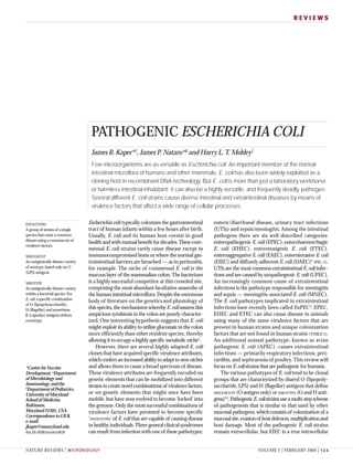
Figure 1 | Pathogenic schema of diarrhoeagenic E. coli. The six recognized categories of diarrhoeagenic E. coli each have
unique features in their interaction with eukaryotic cells. Here, the interaction of each category with a typical target cell is schematically
represented. These descriptions are largely the result of in vitro studies and might not completely reflect the phenomena that occurs in
infected humans. a | EPEC adhere to small bowel enterocytes, but destroy the normal microvillar architecture, inducing the
characteristic attaching and effacing lesion. Cytoskeletal derangements are accompanied by an inflammatory response and diarrhoea.
1. Initial adhesion, 2. Protein translocation by type III secretion, 3. Pedestal formation. b | EHEC also induce the attaching and effacing
lesion, but in the colon. The distinguishing feature of EHEC is the elaboration of Shiga toxin (Stx), systemic absorption of which leads to
potentially life-threatening complications. c | Similarly, ETEC adhere to small bowel enterocytes and induce watery diarrhoea by the
secretion of heat-labile (LT) and/or heat-stable (ST) enterotoxins. d | EAEC adheres to small and large bowel epithelia in a thick biofilm
and elaborates secretory enterotoxins and cytotoxins. e | EIEC invades the colonic epithelial cell, lyses the phagosome and moves
through the cell by nucleating actin microfilaments. The bacteria might move laterally through the epithelium by direct cell-to-cell spread
or might exit and re-enter the baso-lateral plasma membrane. f | DAEC elicits a characteristic signal transduction effect in small bowel
enterocytes that manifests as the growth of long finger-like cellular projections, which wrap around the bacteria. AAF, aggregative
adherence fimbriae; BFP, bundle-forming pilus; CFA, colonization factor antigen; DAF, decay-accelerating factor; EAST1,
enteroaggregative E. coli ST1; LT, heat-labile enterotoxin; ShET1, Shigella enterotoxin 1; ST, heat-stable enterotoxin.
3. NATURE REVIEWS | MICROBIOLOGY VOLUME 2 | FEBRUARY 2004 | 125
R E V I E W S
a (STa) and heat-stable enterotoxin b (STb), respec-
tively — all of which are produced by different strains
of ETEC (reviewed in REF. 11). The Shiga toxin (Stx) of
EHEC cleaves ribosomal RNA, thereby disrupting
protein synthesis and killing the intoxicated epithelial
or endothelial cells12
. The cytolethal distending toxin
(CDT) has DNaseI activity that ultimately blocks cell
division in the G2/M phase of the cell cycle13
.Another
toxin that blocks cell division in the same phase, called
Cif (cycle-inhibiting factor), does not possess DNaseI
activity, but might act by inhibition of Cdk1 kinase
activity14
.The cytotoxic nectrotizing factors (CNF 1 and
CNF 2) deaminate a crucial glutamine residue of RhoA,
Cdc42 and Rac, thereby locking these important sig-
nalling molecules in the ‘on’ position and leading to
marked cytoskeletal alterations, multinucleation with
cellular enlargement,and necrosis15
.The Map protein of
EPEC and EHEC has at least two independent activities
— stimulating Cdc42-dependent filopodia formation
and targeting mitochondria to disrupt membrane
potential in these organelles16
.
protein of EIEC nucleates actin filaments at one pole
of the bacterium, which allows it to move within the
cytoplasm and into adjacent epithelial cells on a‘tail’ of
polymerized actin8
. Even surface structures that are pre-
sent on commensal E. coli strains can induce signalling
cascades if the organism encounters the appropriate
receptor. The LPS of E. coli and other Gram-negative
bacteria binds to Toll-like receptor 4 (TLR4),triggering a
potent cytokine cascade that can lead to septic shock and
death9
. Flagellin, the main component of flagella, can
bind to TLR5, thereby activating interleukin (IL)-8
expression and an inflammatory response10
.
Toxins. More numerous than surface structures that
trigger signal transduction pathways are secreted toxins
and other effector proteins that affect an astonishing
variety of fundamental eukaryotic processes (TABLE 2).
Concentrations of important intracellular messengers,
such as cyclic AMP, cyclic GMP and Ca2+
, can be
increased,which leads to ion secretion by the actions of
the heat-labile enterotoxin (LT),heat-stable enterotoxin
Table 1 | E. coli virulence factors: colonization and fitness factors
Factor Pathotype Activity/effect
IcsA (VirG) EIEC Nucleation of actin filaments
Intimin EPEC, EHEC Adhesin, induces TH
1 response; 10 variants described
Dr adhesins DAEC, UPEC Adhesin, binds to decay-accelerating factor (DAF), activates
PI-3-kinase, induces MICA; >10 Dr adhesins described
P (Pap) fimbriae UPEC Adhesin; induces cytokine expression
CFAs ETEC Adhesin, >20 different factors designated CFA, CS or PCF
Type-1 fimbriae All UPEC adhesin; binds to uroplakin
F1C fimbriae UPEC Adhesin
S fimbriae UPEC, MNEC Adhesin
Bundle-forming pilus (BFP) EPEC Type IV pilus
Aggregative adherence fimbriae EAEC Adhesin; >4 subtypes
Paa EPEC, EHEC Adhesin
ToxB EHEC Adhesin
Efa-1/LifA EHEC Adhesin
Long polar fimbriae (LPF) EHEC, EPEC Adhesin
Saa EHEC Adhesin
OmpA MNEC, EHEC Adhesin
Curli Various Adhesin; binds to fibronectin
IbeA, B, C MNEC Promotes invasion
AslA MNEC Promotes invasion
Dispersin EAEC Promotes colonization; aids mucous penetration
K antigen capsules MNEC Antiphagocytic; >80 K types
Aerobactin EIEC Iron acquisition, siderophore
Yersiniabactin Various Iron acquisition, siderophore
IreA UPEC Iron acquisition, siderophore receptor
IroN UPEC Iron acquisition, siderophore receptor
Chu (Shu) EIEC, UPEC, MNEC Iron acquisition, haem transport
Flagellin All Motility; induces cytokine expression through TLR5;
>50 flagella (H) serotypes
Lipopolysaccharide All Induces cytokine expression through TLR4; >180 O types
CFA, colonization factor antigen; CS, coli surface antigen; MICA, MHC class I chain-related gene A; PCF, putative colonization factor;
PI-3-kinase, phosphatidylinositol 3-kinase; TLR, Toll-like receptor.
5. NATURE REVIEWS | MICROBIOLOGY VOLUME 2 | FEBRUARY 2004 | 127
R E V I E W S
Table 2 | E. coli virulence factors: toxins and effectors
Factor Pathotype Toxin class Target Activity/Effect
Heat-labile enterotoxin ETEC AB subunit, type II Gs
ADP ribosylates and activates adenylate
(LT) effector cyclase resulting in ion secretion
Shiga toxin EHEC AB subunit rRNA Depurinates rRNA, inhibiting protein
(Stx) synthesis; induces apoptosis
Cytolethal distending Various ABC subunit DNA DNaseI activity, blocks mitosis in G2/M
toxin (CDT) phase
Shigella enterotoxin 1 EAEC, EIEC* AB subunit – Ion secretion
(ShET1)
Urease EHEC ABC subunit Urea Cleaves urea to NH3
and CO2
EspC EPEC Autotransporter ? Serine protease; ion secretion
EspP EHEC Autotransporter ? Serine protease; cleaves coagulation
factor V
Haemoglobin-binding ExPEC, Autotransporter Haem Degrades haemoglobin to release haem/iron
protease (Tsh) APEC
Pet EAEC Autotransporter Spectrin Serine protease; ion secretion; cytotoxicity
Pic UPEC, EAEC, Autotransporter ? Protease, mucinase
EIEC*
Sat UPEC Autotransporter ? Vacuolation
SepA EIEC* Autotransporter ? Serine protease
SigA EIEC* Autotransporter ? Ion secretion
Cycle-inhibiting EPEC, EHEC Type III effector ? Blocks mitosis in G2/M phase; results in
factor (Cif) inactivation of Cdk1
EspF EPEC, EHEC Type III effector ? Opens tight junctions, induces apoptosis
EspH EPEC, EHEC Type III effector ? Modulates filopodia and pedestal formation
Map EPEC, EHEC Type III effector Mitochondria Disrupts mitochondrial membrane potential
Tir EPEC, EHEC Type III effector Nck Nucleation of cytoskeletal proteins, loss of
microvilli, GAP-like activity
IpaA EIEC Type III effector Vinculin Actin depolymerization
IpaB EIEC Type III effector Caspase 1 Apoptosis, IL-1 release; membrane
insertion
IpaC EIEC Type III effector Actin Actin polymerization, activation of Cdc42
and Rac
IpaH EIEC Type III effector Nucleus Modulates inflammation (?)
IpgD EIEC Type III effector PtdIns Inositol 4-phosphatase, membrane
(4,5)P2
blebbing
VirA EIEC Type III effector Tubulin Microtubule destabilization, membrane
ruffling
StcE EHEC Type II effector C1-esterase Cleaves C1-INH, disrupts complement
inhibitor cascade
(C1-INH)
HlyA UPEC RTX toxins Erythrocytes, Cell lysis
Leukocytes
Ehx EHEC RTX toxins Erythrocytes, Cell lysis
Leukocytes
Cytotoxic necrotizing MNEC, UPEC, RhoA, Altered cytoskeleton, necrosis
factors (CNF-1,-2) NTEC Cdc42, Rac
LifA/Efa EPEC, EHEC Lymphocytes Inhibits lymphocyte activation, adhesion
Shigella enterotoxin 2 EIEC, ETEC ? Ion secretion
(ShET2)
Heat-stable enterotoxin ETEC Heat-stable Guanylate Activates guanylate cyclase resulting in ion
a (STa) enterotoxins cyclase secretion
Heat-stable enterotoxin ETEC Heat-stable ? Increase intracellular calcium resulting in
b (STb) enterotoxins ion secretion
EAST Various Heat-stable Guanylate Activates guanylate cyclase resulting in ion
enterotoxins cyclase secretion
*These factors have been characterized in Shigella species, but their presence in EIEC has not yet been established. EAST,
enteroaggregative E.coli ST; GAP, GTPase-activating protein; IL, interleukin; PtdIns(4,5)P2
, phosphatidylinositol-4,5-bisphosphate.
6. 128 | FEBRUARY 2004 | VOLUME 2 www.nature.com/reviews/micro
R E V I E W S
Additional EPEC virulence factors that are encoded
outside the LEE have also been described.One very large
protein of ~385 kDa called lymphostatin (LifA) inhibits
lymphocyte activation33
. This protein is also present in
strains of EHEC, where it is known as Efa1, and an
adhesive property has been attributed to it34
. Typical
EPEC strains possess a plasmid of 70–100 kb called the
EAF (EPEC adherence factor) plasmid35
. This plasmid
encodes a type IV pilus called the bundle-forming
pilus (BFP)36
, which mediates interbacterial adherence
and possibly adherence to epithelial cells (FIG. 2). It also
contains the per locus (plasmid-encoded regulator),the
products of which regulate the bfp operon and most of
the genes in the LEE by the LEE-encoded regulator
(Ler). So-called atypical EPEC contain the LEE but do
not contain the EAF plasmid. In industrialized coun-
tries, atypical EPEC are more frequently isolated from
diarrhoeal cases than are typical EPEC that contain the
EAF plasmid,although typical EPEC dominate in devel-
oping countries37
.Atypical EPEC have also caused large
outbreaks of diarrhoeal disease involving both children
and adults in industrialized countries.
The model of EPEC pathogenesis is considerably
more complex than simple binding to epithelial cells by
a single adhesin and secretion of an enterotoxin that
induces diarrhoea.The emerging model,several aspects
of which are reviewed elsewhere2,38–40
, indicates that
EPEC initially adhere to epithelial cells by an adhesin,
the identity of which is not yet clearly established;
potential candidates include BFP, the EspA filament,
flagella, LifA/Efa1 and intimin (by host-cell receptors).
The type III secretion system is then activated and
various effector proteins — including Tir, EspF, EspG,
EspH and Map — are translocated into the host cell.
EPEC binds through the interaction of intimin with Tir
inserted in the membrane and numerous cytoskeletal
proteins accumulate underneath the attached bacteria.
Protein kinase C (PKC), phospholipase Cγ, myosin
light-chain kinase and mitogen-activated protein
(MAP) kinases are activated, which leads to several
downstream effects, including increased permeability
due to loosened tight junctions.Nuclear factor (NF)-κB
is activated, leading to production of IL-8 and an
inflammatory response that involves transmigration of
polymorphonuclear leukocytes (PMNs) to the lumenal
surface and activation of the adenosine receptor. The
galanin-1 receptor is upregulated41
, thereby increasing
the response of the epithelial cells to the neuropeptide
GALANIN, which is an important mediator of intestinal
secretion. Some, but not all, typical EPEC strains pro-
duce an enterotoxin, EspC, that increases short circuit
current in USSING CHAMBERS
157
. Diarrhoea probably
results from multiple mechanisms, including active
ion secretion, increased intestinal permeability,
intestinal inflammation and loss of absorptive surface
area resulting from microvillus effacement.
Enterohaemorrhagic E.coli (EHEC). First recognized as
a cause of human disease in 1982, EHEC causes bloody
diarrhoea (haemorrhagic colitis),non-bloody diarrhoea
and haemolytic uremic syndrome (HUS).The principal
as a receptor for the intimin outer-membrane protein24
.
This is a fascinating example of a pathogen that provides
its own receptor for binding to eukaryotic cells,
although additional eukaryotic proteins have also
been reported to act as receptors for intimin. A recent
study showed that EPEC can disrupt cell polarity,
causing basolateral membrane proteins, in particular
β1
-integrins, to migrate to the apical cell surface where
they can bind to intimin25
. In addition to β1
-integrin,
Tir has also been shown to bind to NUCLEOLIN
26
.In addi-
tion to its role as a receptor for intimin,Tir has important
signalling functions in epithelial cells. The portion of
Tir that is exposed to the cytosol nucleates cytoskele-
tal proteins, initially binding directly to the adaptor
protein Nck, which recruits the amino terminus of
Wiskott–Aldrich syndrome protein (N-WASP) and the
actin-related protein 2/3 (Arp2/3) complex; recruit-
ment of Arp2/3 results in actin filament nucleation
and initiation of the characteristic pedestal complex27
(FIG. 1). Interestingly, the Tir protein of EHEC O157:H7
is not functionally identical to the Tir protein of EPEC
O127:H6 because pedestals are formed independently
of Nck, which indicates that additional bacterial factors
are translocated to trigger actin signalling28
. Other
cytoskeletal proteins, such as vinculin, cortactin, talin
and α-actinin, are also recruited to the pedestal com-
plex29
. Formation of the pedestal is a dynamic process
whereby the force of actin polymerization can propel
the pedestal across the surface of ptK2 epithelial cells30
(see movement of EPEC on ptK2 cells in the Online
links). Tir also has a GAP (GTPase-activating protein)
motif that has been implicated in the ability of Tir to
downregulate filopodia formation16
.Another secreted
effector protein is EspF, which causes apoptosis31
and
induces redistribution of the tight-junction-associated
protein occludin, which leads to loss of trans-epithelial
electrical resistance32
.As noted above, the Map protein
affects mitochondrial function and filopodia forma-
tion, and additional effectors — for example, EspG and
EspH — have recently been described.
NUCLEOLIN
A nucleolar protein that
functions as a shuttle protein
between the nucleus and the
cytoplasm and is also found on
the cell surface.
GAP
GTPase-activating protein.
A family of eukaryotic proteins
that modulate the activity of
Rac,Rho and Cdc42.
GALANIN
A neuropeptide that is widely
distributed in the central
nervous system and the
gastrointestinal tract.Binding to
the galanin-1 receptor can alter
intestinal ion flux.
USSING CHAMBER
A device that is used to measure
ion flow across an epithelium.
Bacterial enterotoxins that
induce ion fluxes are frequently
studied in Ussing chambers.
Figure 3 | Attaching and effacing histopathology caused
by EPEC and EHEC. The attaching and effacing
histopathology results in pedestal-like structures, which rise
up from the epithelial cell on which the bacteria perch.
Image courtesy of J. Girón.
7. NATURE REVIEWS | MICROBIOLOGY VOLUME 2 | FEBRUARY 2004 | 129
R E V I E W S
the term EHEC is used to denote only the subset of Stx-
positive strains that also contain the LEE. However,
there are LEE-negative STEC strains that are associated
with disease — for example, O103:H21 strains —
thereby demonstrating that there are additional viru-
lence factors yet to be characterized. Several other
potential adherence factors have been described for
O157:H7 and/or non-O157:H7 strains, although the
significance of these factors in human disease is not as
well established as intimin. One potential adhesin is a
large 362-kDa protein (ToxB) encoded on the 93-kb
plasmid that is present in O157:H7 and other EHEC
strains46
. This protein shares sequence similarity with
the large Clostridium toxin family,and to the EPEC LifA
protein33
and the Efa-1 protein that has been implicated
as an adhesin in non-O157:H7 EHEC strains34
. This
plasmid (pO157)47
, also encodes an RTX (repeats in
toxin) toxin that is similar to the UPEC haemolysin, a
serine protease (EspP), a catalase and the StcE protein.
StcE cleaves the C1 esterase inhibitor (C1-INH) of the
complement pathway and could potentially contribute
to the tissue damage, intestinal oedema and throm-
botic abnormalities that are seen in EHEC infections48
.
The genome sequence of O157:H7 revealed numerous
chromosomal islands (see below) that encode addi-
tional potential virulence factors.Included among these
potential factors are novel fimbriae,iron uptake and uti-
lization systems49
, and a urease that is similar to those
produced by Klebsiella and other urinary tract
pathogens50
.
Enterotoxigenic E. coli (ETEC). ETEC causes watery
diarrhoea, which can range from mild, self-limiting
disease to severe purging disease. The organism is an
important cause of childhood diarrhoea in the devel-
oping world and is the main cause of diarrhoea in
travellers to developing countries2
.
ETEC colonizes the surface of the small bowel
mucosa and elaborates enterotoxins, which give rise to
intestinal secretion. Colonization is mediated by one or
more proteinaceous fimbrial or fibrillar colonization
factors (CFs), which are designated by CFA (coloniza-
tion factor antigen), CS (coli surface antigen) or PCF
(putative colonization factor) followed by a number.
More than 20 antigenically diverse CFs have been
characterized, yet epidemiological studies indicate that
approximately 75% of human ETEC express either
CFA/I, CFA/II or CFA/IV51
.Antibodies to CFAs might
ameliorate ETEC colonization and disease. ETEC are
also an important cause of diarrhoeal disease in animals
and these animal strains express fimbrial intestinal
colonization factors, such as K88 and K99, which are
not found in human ETEC strains.
ETEC enterotoxins belong to one of two groups: the
heat-labile enterotoxins (LTs) and the heat-stable
enterotoxins (STs).ETEC strains might express only an
LT, only an ST, or both LTs and STs. LTs are a class of
enterotoxins that are closely related in structure and
function to cholera enterotoxin (CT), which is
expressed by Vibrio cholerae 52
. The LT that is found
predominantly in human isolates (LT-I; a related
reservoir of EHEC is the bovine intestinal tract and
initial outbreaks were associated with consumption of
undercooked hamburgers. Subsequently, a wide variety
of food items have been associated with disease,
including sausages, unpasteurized milk, lettuce, can-
taloupe melon,apple juice and radish sprouts — the lat-
ter were responsible for an outbreak of 8,000 cases in
Japan. Facilitated by the extremely low infectious dose
required for infection (estimated to be <100 cells),
EHEC has also caused numerous outbreaks associated
with recreational and municipal drinking water, per-
son-to-person transmission and petting zoo and farm
visitations.A recent report indicates potential airborne
transmission after exposure to a contaminated
building42
. EHEC strains of the O157:H7 serotype are
the most important EHEC pathogens in North
America, the United Kingdom and Japan, but several
other serotypes, particularly those of the O26 and
O111 serogroups, can also cause disease and are more
prominent than O157:H7 in many countries.
The key virulence factor for EHEC is Stx, which is
also known as verocytotoxin (VT). Stx consists of five
identical B subunits that are responsible for binding the
holotoxin to the glycolipid globotriaosylceramide
(Gb3) on the target cell surface, and a single A subunit
that cleaves ribosomal RNA, causing protein synthesis
to cease12
. The Stx family contains two subgroups —
Stx1 and Stx2 — that share approximately 55% amino
acid homology.Stx is produced in the colon and travels
by the bloodstream to the kidney, where it damages
renal endothelial cells and occludes the microvascula-
ture through a combination of direct toxicity and
induction of local cytokine and chemokine production,
resulting in renal inflammation (reviewed in REF. 43).
This damage can lead to HUS,which is characterized by
haemolytic anaemia, thrombocytopoenia and poten-
tially fatal acute renal failure.Stx also induces apoptosis
in intestinal epithelial cells — a process that is regu-
lated by the Bcl-2 family44
. Stx was first purified from
Shigella dysenteriae, and HUS can also result from
infection with this species, although not with other
Shigella species or EIEC, which do not produce Stx.Stx
also mediates local damage in the colon,which results in
bloody diarrhoea, haemorrhagic colitis, necrosis and
intestinal perforation.
In addition to Stx, most EHEC strains also contain
the LEE pathogenicity island that encodes a type III
secretion system and effector proteins that are homolo-
gous to those that are produced by EPEC.Animal mod-
els have shown the importance of the intimin adhesin in
intestinal colonization, and HUS patients develop a
strong antibody response to intimin and other LEE-
encoded proteins. EHEC O157:H7 is believed to have
evolved from LEE-containing O55 EPEC strains that
acquired bacteriophage encoding Stx45
.Although more
than 200 serotypes of E. coli can produce Stx, most of
these serotypes do not contain the LEE pathogenicity
island and are not associated with human disease. This
has led to the use of Shiga toxin-producing E. coli
(STEC) or verotoxin-producing E. coli (VTEC) as gen-
eral terms for any E. coli strain that produces Stx, and
8. 130 | FEBRUARY 2004 | VOLUME 2 www.nature.com/reviews/micro
R E V I E W S
Enteroaggregative E. coli (EAEC). EAEC are increas-
ingly recognized as a cause of often persistent diar-
rhoea in children and adults in both developing and
developed countries, and have been identified as the
cause of several outbreaks worldwide.At present,EAEC
are defined as E. coli that do not secrete LT or ST and
that adhere to HEp-2 cells in a pattern known as auto-
aggregative, in which bacteria adhere to each other in
a ‘stacked-brick’ configuration2
. It is likely that this
definition encompasses both pathogenic and non-
pathogenic clones, and it remains controversial as to
whether all the EAEC have any common factors that
contribute to their shared adherence phenotype.
Nevertheless, at least a subset of EAEC are proven
human pathogens.
The basic strategy of EAEC infection seems to
comprise colonization of the intestinal mucosa, prob-
ably predominantly that of the colon, followed by
secretion of enterotoxins and cytotoxins57
. Studies on
human intestinal explants indicate that EAEC induces
mild,but significant,mucosal damage58
— these effects
are most severe in colonic sections. Mild inflammatory
changes are observed in animal models59
and evidence
indicates that at least some EAEC strains might be
capable of limited invasion of the mucosal surface60,61
.
The most dramatic histopathological finding in infected
animal models is the presence of a thick layer of auto-
aggregating bacteria adhering loosely to the mucosal
surface. EAEC prototype strains adhere to HEp-2 cells
and intestinal mucosa by virtue of fimbrial structures
known as aggregative adherence fimbriae (AAFs)62–64
,
which are related to the Dr family of adhesins.At least
four allelic variants of AAFs exist,but importantly,each
is present in only a minority of strains. It should be
noted, however, that not all EAEC strains adhere by
virtue of AAFs.A recently described protein called dis-
persin65
forms a loosely associated layer on the surface
of EAEC strains and seems to counter the strong aggre-
gating effects of the AAF adhesin, perhaps facilitating
spread across the mucosal surface or penetration of the
mucous layer. An additional surface structure that is
potentially involved in causing inflammation is a novel
EAEC flagellin protein that induces IL-8 release66
.
Release of this cytokine can stimulate neutrophil
transmigration across the epithelium, which can itself
lead to tissue disruption and fluid secretion.
SeveraltoxinshavebeendescribedforEAEC.Twosuch
toxins are encoded by the same chromosomal locus on
opposite strands. The larger gene encodes an auto-
transporter protease with mucinase activity called Pic;
the opposite strand encodes the oligomeric enterotoxin
that is known as Shigella enterotoxin 1 (ShET1), owing
to its presence in most strains of Shigella flexneri 2a67,68
.
The mode of action of ShET1 is not yet understood,
but it might contribute to the secretory diarrhoea that
accompanies EAEC and Shigella infection. A second
enterotoxin that is present in many EAEC strains is
enteroaggregative E. coli ST (EAST1), a 38-amino-acid
homologue of the ETEC STa toxin69
. It is conceivable
that EAST1 could contribute to watery diarrhoea in
EAST1-positive strains; however, the EAST1 gene
protein called LT-II is found in some animal ETEC
isolates) has ~80% amino acid identity with CT and,
like CT, consists of a single A subunit and five identical
B subunits. The B subunits mediate binding of the
holotoxin to the cell surface gangliosides GM1 and
GD1b, and the A subunit is responsible for the enzy-
matic activity of the toxin. LT has ADP-ribosyl trans-
ferase activity and transfers anADP-ribosyl moiety from
NAD to the α-subunit of the stimulatory G protein —
a regulatory protein of the basolateral membrane that
regulates adenylate cyclase. The resulting permanent
activation of adenylate cyclase leads to increased levels
of intracellular cAMP, activation of cAMP-dependent
kinases and the eventual activation of the main chloride
channel of epithelial cells — the cystic fibrosis trans-
membrane conductance regulator (CTFR). The net
result of CFTR phosphorylation is increased Cl–
secre-
tion from secretory crypt cells,which leads to diarrhoea
(reviewed in REF.11).LT can also stimulate prostaglandin
synthesis and stimulate the enteric nervous system;
both of these activities can also lead to stimulation of
secretion and inhibition of absorption11
. LT is also a
potent mucosal adjuvant independent of its toxic
activity53
and has been incorporated into numerous
vaccine candidates containing a variety of antigens,
resulting in increased antibody responses to these
antigens when they are delivered orally, nasally or even
transdermally.
STs are small, single-peptide toxins that include two
unrelated classes — STa and STb — which differ in both
structure and mechanism of action. Only toxins of the
STa class have been associated with human disease2
.The
mature STa toxin is a ~2-kDa peptide, which contains
18 or 19 amino acid residues, six of which are cysteines
that form three intramolecular disulphide bridges
(reviewed in REF. 11). The main receptor for STa is a
membrane-spanning guanylate cyclase; binding of STa
to guanylate cyclase stimulates guanylate cyclase activity,
leading to increased intracellular cGMP,which,in turn,
activates cGMP-dependent and/or cAMP-dependent
kinases and,ultimately,increases secretion.Interestingly,
intestinal guanylate cyclase is the receptor for an
endogenous ligand called guanylin54
,which has a similar
structure to that of STa.So the ST family seems to repre-
sent a case of molecular mimicry.The STb toxin is asso-
ciated with animal disease and is a 48-amino-acid pep-
tide containing two disulphide bonds (reviewed in REF.
55).STb can elevate cytosolic Ca2+
concentrations,stim-
ulate the release of prostaglandin E2
and stimulate the
release of serotonin, all of which are mechanisms that
could lead to increased ion secretion.
ETEC is largely a pathogen of developing countries,
and it is well known that these countries typically have
a low rate of colon cancer. Pitari et al.56
have reported
that STa suppresses colon cancer cell proliferation
through a guanylyl cyclase C-mediated signalling cas-
cade. So the high prevalence of ETEC in developing
countries might have a protective effect against this
important disease,and indicates that infectious diseases
might exist in a complex evolutionary balance with
their human populations.
9. NATURE REVIEWS | MICROBIOLOGY VOLUME 2 | FEBRUARY 2004 | 131
R E V I E W S
scheme are present on a large virulence plasmid that is
found in EIEC and all Shigella species. The sequence of
the 213-kb virulence plasmid of S. flexneri (pWR100)
indicates that this plasmid is a mosaic that includes
genetic elements that were initially carried by four
plasmids77
. One-third of the plasmid is composed of
insertion sequence (IS) elements,which are undoubtedly
important in the evolution of the virulence plasmid.
This plasmid encodes a type III secretion system (see
below) and a 120-kDa outer-membrane protein called
IcsA,whichnucleatesactinbythebindingof N-WASP8,78
.
The growth of actin micofilaments at only one bacterial
pole induces movement of the organism through the
epithelial cell cytoplasm.This movement culminates in
the formation of cellular protrusions that are engulfed
by neighbouring cells, after which the process is
repeated.Although EIEC are invasive,dissemination of
the organism past the submucosa is rare.
Much of EIEC/Shigella pathogenesis seems to be
the result of the multiple effects of its plasmid-borne
type III secretion system. This type III secretion system
secretes multiple proteins, such as IpaA, IpaB, IpaC
and IpgD, which mediate epithelial signalling events,
cytoskeletal rearrangements, cellular uptake, lysis of
the endocytic vacuole and other actions (reviewed in
REFS 79,80). The type III secretion system apparatus,
which is encoded by mxi and spa genes, enables the
insertion of a pore containing IpaB and IpaC proteins
into host cell membranes.In addition to pore formation,
IpaB has several functions, such as binding to the sig-
nalling protein CD44, thereby triggering cytoskeletal
rearrangements and cell entry, and binding to the
macrophage caspase 1, resulting in apoptosis and
release of IL-1 from macrophages. IpaC induces actin
polymerization, which leads to the formation of cell
extensions by activating the GTPases Cdc42 and Rac.
The actin polymerization activity resides in the carboxy
terminus of IpaC, whereas the amino terminus of this
protein is involved in lamellipodial extensions.
Conversely, IpaA binds to vinculin and induces actin
depolymerization, thereby helping to organize the
extensions that are induced by IpaC into a structure
that enables bacterial entry. The translocated effector
protein IpgD is a potent inositol 4-phosphatase that
helps to reorganize host-cell morphology by uncou-
pling the cellular plasma membrane from the actin
cytoskeleton, which leads to membrane blebbing81
.
Although the extensively characterized type III secretion
system is essential for the invasiveness characteristic
of EIEC and Shigella species, additional virulence fac-
tors have been described, including the plasmid-
encoded serine protease SepA, the chromosomally
encoded aerobactin iron-acquisition system and other
secreted proteases that are encoded by genes present
on pathogenicity islands (see below).
Diffusely adherent E.coli (DAEC).DAEC are defined by
the presence of a characteristic, diffuse pattern of
adherence to HEp-2 cell monolayers. DAEC have been
implicated as a cause of diarrhoea in several studies,
particularly in children >12 months of age2,82
.
(astA) can also be found in many commensal E. coli
isolates, and therefore the role of EAST1 in diarrhoea
remains an open question70
.Many EAEC strains secrete
an autotransporter toxin called Pet, which is encoded
on the large virulence plasmid in close proximity to
the gene encoding the AAF. Pet has enterotoxic activ-
ity and can also potentially lead to cytoskeletal
changes and epithelial-cell rounding by cleavage of
the cytoskeletal protein spectrin71
.
Although no single virulence factor has been
irrefutably associated with EAEC virulence, epidemio-
logical studies implicate a‘package’ of plasmid-borne
and chromosomal virulence factors, similar to the
virulence factors of other enteric pathogens. Several
EAEC virulence factors are regulated by a single tran-
scriptional activator called AggR, which is a member of
the AraC family of transcriptional activators64
(J.P.N.,
unpublished data). One consistent observation from
studies involving EAEC epidemiology is the associa-
tion of the AggR regulon with diarrhoeal disease.
Jiang et al. have recently shown that the presence of
genes associated with the AggR regulon is predictive
of significantly increased concentrations of faecal IL-8
and IL-1 in patients with diarrhoea caused by EAEC72
.
We suggest that the term ‘typical EAEC’ should be
reserved for strains carryingAggR and at least a subset of
AggR-regulated genes (for which the traditional EAEC
probe is an adequate marker),and that the term‘atypical
EAEC’be used for strains lacking theAggR regulon.
Enteroinvasive E.coli (EIEC). EIEC are biochemically,
genetically and pathogenically closely related to Shigella
spp. Numerous studies have shown that Shigella and
E. coli are taxonomically indistinguishable at the species
level73,74
, but, owing to the clinical significance of
Shigella, a nomenclature distinction is still maintained.
The four Shigella species that are responsible for human
disease, S. dysenteriae, S. flexneri, Shigella sonnei and
Shigella boydii, cause varying degrees of dysentery,
which is characterized by fever,abdominal cramps and
diarrhoea containing blood and mucous. EIEC might
cause an invasive inflammatory colitis,and occasionally
dysentery, but in most cases EIEC elicits watery diar-
rhoea that is indistinguishable from that due to infection
by other E. coli pathogens2
. EIEC are distinguished
from Shigella by a few minor biochemical tests, but
these pathotypes share essential virulence factors.
EIEC infection is thought to represent an inflamma-
tory colitis, although many patients seem to manifest
secretory, small bowel syndrome. The early phase of
EIEC/Shigella pathogenesis comprises epithelial cell
penetration, followed by lysis of the endocytic vacuole,
intracellular multiplication, directional movement
through the cytoplasm and extension into adjacent
epithelial cells (reviewed in REF. 75). Movement within
the cytoplasm is mediated by nucleation of cellular actin
into a‘tail’that extends from one pole of the bacterium.
In addition to invasion into and dissemination within
epithelial cells, Shigella (and presumably EIEC) also
induces apoptosis in infected macrophages76
. Genes
that are required to effect this complex pathogenetic
10. 132 | FEBRUARY 2004 | VOLUME 2 www.nature.com/reviews/micro
R E V I E W S
Uropathogenic E. coli (UPEC). The urinary tract is
among the most common sites of bacterial infection
and E. coli is by far the most common infecting agent
at this site. The subset of E. coli that causes uncom-
plicated cystitis and acute pyelonephritis is distinct
from the commensal E. coli strains that comprise
most of the E. coli populating the lower colon of
humans. E. coli from a small number of O serogroups
(six O groups cause 75% of UTIs) have phenotypes
that are epidemiologically associated with cystitis and
acute pyelonephritis in the normal urinary tract,
which include expression of P fimbriae, haemolysin,
aerobactin, serum resistance and encapsulation. Clonal
groups and epidemic strains that are associated with
UTIs have been identified88,89
.
Although many UTI isolates seem to be clonal,
there is no single phenotypic profile that causes UTIs.
Specific adhesins, including P (Pap), type 1 and other
fimbriae (such as F1C, S, M and Dr), seem to aid in
colonization90,91
. Several toxins are produced, includ-
ing haemolysin, cytotoxic necrotizing factor and an
autotransported protease known as Sat. These viru-
lence factors are found in differing percentages
among various subgroups of UPEC92
. Uropathogenic
strains possess large and small pathogenicity islands
containing blocks of genes that are not found in the
chromosome of faecal strains. Availability of the
genome sequence of E. coli CFT073 (REF. 93) and
efforts by other investigators to identify virulence
Approximately 75% of DAEC strains produce a fimbrial
adhesin called F1845 or a related adhesin (REF. 83; J.P.N.,
unpublished observations); F1845 belongs to the Dr
family of adhesins, which use DAF, a cell-surface gly-
cosylphosphatidylinositol-anchored protein, which
normally protects cells from damage by the comple-
ment system, as the receptor84–86
. DAEC strains induce
a cytopathic effect that is characterized by the devel-
opment of long cellular extensions, which wrap
around the adherent bacteria (FIG. 1). This characteris-
tic effect requires binding and clustering of the DAF
receptor by Dr fimbriae85
.All members of the Dr family
(including UPEC as well as the DAEC strain C1845)
elicit this effect83
. Binding of Dr adhesins is accompa-
nied by the activation of signal transduction cascades,
including activation of PI-3 kinase86
. Peiffer et al. have
reported that infection of an intestinal cell line by
strains of DAEC impairs the activities and reduces
the abundance of brush-border-associated sucrase-
isomaltase and dipeptidylpeptidase IV87
. This effect is
independent of the DAF-associated pathway described
above, and therefore provides a feasible mechanism for
DAEC-induced enteric disease and also indicates the
presence of virulence factors in DAEC other than Dr
adhesins. Tieng et al.7
have proposed that DAEC might
induce expression of MICA by intestinal epithelial
cells, indicating that DAEC infection could be pro-
inflammatory; this effect could potentially be important
in the induction of inflammatory bowel diseases.
SIGNATURE-TAGGED
MUTAGENESIS
(STM).A technique to screen
large numbers of distinct
mutants for those that fail to
survive an animal infection.
Each mutant is tagged with a
unique DNA sequence (called a
signature tag),which allows a
specific mutant to be tracked
within a large pool of bacteria.
E. coli crosses tubular epithelial
cell barrier to initiate bacteraemia
Haemolysin
damages
epithelium
Type 1 fimbriated E. coli
selected at high CFU
and low O2
Contamination of periurethral
area with uropathogenic E. coli
that has colonized the bowel
E. coli ascends
to kidney
Cytokines
induced
Invasion; intracellular
multiplication observed
for selected strains
P. fimbriae bind to renal
tubular epithelial cells
Urethra
Blood supply
Bladder
Kidneys
Ureters
3
6
10
8
12
1
7
9
2
4
5
11
Adherence to
uroepithelial
cells by type 1
and P fimbriae
Apoptosis and
exfoliation of
bladder epithelial
cells
Influx of
PMNs
Sat vacuolates
epithelial cell
and damages
glomeruli
Figure 4 | Pathogenesis of urinary tract infection caused by uropathogenic E. coli. The figure shows the different stages of
a urinary tract infection. Panels 2, 4, 5 and 11 are courtesy of N. Gunther, A. Jansen, X. Li and D. Auyer (University of Maryland),
respectively. CFU, colony-forming units; PMNs, polymorphonuclear leukocytes.
11. NATURE REVIEWS | MICROBIOLOGY VOLUME 2 | FEBRUARY 2004 | 133
R E V I E W S
increased production of IL-6 and IL-8 (REF. 102).
Secretion of Sat, a vacuolating cytotoxin, damages
glomeruli and is cytopathic for the surrounding
epithelium103
. In some cases, the barrier that is pro-
vided by the one-cell-thick proximal tubules can be
breached and bacteria can penetrate the endothelial
cell to enter the bloodstream, leading to bacteraemia.
Meningitis/sepsis-associated E. coli (MNEC). This
E. coli pathotype is the most common cause of Gram-
negative neonatal meningitis, with a case fatality rate of
15–40% and severe neurological defects in many of the
survivors104,105
. The incidence of infants with early-
onset sepsis owing to E. coli infection seems to be
increasing,while infection by Gram-positive organisms
decreases106
.As with E. coli pathotypes that have a well-
defined genetic basis for virulence, strains that cause
meningitis are represented by only a limited number of
O serogroups,and 80% of the strains are of the K1 cap-
sule type. One interesting difference between MNEC
and E. coli that cause intestinal or urinary tract infec-
tions is that although the latter strains can be readily
transmitted by urine or faeces, infection of the central
nervous system offers no obvious advantage for the
selection and transmission of virulent MNEC strains.
E. coli that cause meningitis are spread haematoge-
nously. Levels of bacteraemia correlate with the
development of meningitis107
; for example, bacter-
aemias of >103
colony forming units per ml of blood
are significantly more likely to lead to the develop-
ment of meningitis than in individuals with lower
colony forming units per ml in their blood. These bac-
teria translocate from the blood to the central nervous
system without apparent damage to the blood–brain
barrier, which indicates a transcytosis process.
Electron micrographs imply entry by a zippering
mechanism in a process that does not affect
transendothelial electrical resistance108
. This indicates
that the host-cell membrane is not significantly dis-
rupted during entry of the bacterium. Two models for
studying MNEC have been developed: a monolayer of
brain microvascular endothelial cells109
and an intact
animal model using 5-day-old rats110
.
As for other E. coli pathotypes,the genomes of these
extraintestinal K1 strains have additional genes that are
not found in the commensal E. coli K-12 strains. In
genomic comparisons, the genome of E. coli RS218, a
meningitis-associated strain, was found to have at least
500 kb of additional genes inserted in at least 12 loci
compared with E. coli K-12 (REFS 111,112). In addition,
strain RS218 harbours a 100-kb plasmid, on which at
least one virulence factor has been localized113
.
Some insights into the mechanism of pathogenesis
of these strains have been obtained. K1 strains use
S fimbriae to bind to the lumenal surfaces of brain
microvascular endothelium in neonatal rats114
.Invasion
requires the outer-membrane protein OmpA to bind to
the GlcNAcβ1-4GlcNAc epitope of the brain micro-
vascular endothelial cell receptor glycoprotein115
.Other
membrane proteins — for example, IbeA, IbeB, IbeC
and AslA — are also required for invasion (reviewed in
genes by SIGNATURE-TAGGED MUTAGENESIS
94
and other
methods have allowed the development of a model of
pathogenesis for UPEC (FIG. 4).
It is likely that infection begins with the colonization
of the bowel with a uropathogenic strain in addition to
the commensal flora. This strain, by virtue of factors
that are encoded in pathogenicity islands, is capable of
infecting an immunocompetent host, as it colonizes
the periurethral area and ascends the urethra to the
bladder (FIG. 4). Between 4 and 24 hours after infection,
the new environment in the bladder selects for the
expression of type 1 fimbriae95
, which have an impor-
tant role early in the development of a UTI96
. Type 1
fimbriated E. coli attach to mannose moieties of the
uroplakin receptors that coat transitional epithelial
cells97
. Attachment triggers apoptosis and exfoliation;
for at least one strain, invasion of the bladder epithe-
lium is accompanied with formation of pod-like
bulges on the bladder surface that contain bacteria
encased in a polysaccharide-rich matrix surrounded by
a shell of uroplakin98
.It is argued that invaded epithelial
cells containing a tightly packed bacterial ‘biofilm’
could act as a reservoir for recurrent infection97,98
, and
indeed, in some cases of recurrent infection, the same
serotype is encountered. However, a number of studies
have identified different serotypes as being responsible
for the recurring infection, an observation that is not
consistent with this hypothesis. Iron acquisition and
the ability to grow in urine are also crucial for survival.
In strains that cause cystitis, type 1 fimbriae are
continually expressed and the infection is confined to
the bladder96
. In pyelonephritis strains, the invertible
element that controls type 1 fimbriae expression
turns to the ‘off’ position and type 1 fimbriae are less
well expressed95
. It could be argued that this releases
the E. coli strain from bladder epithelial cell receptors
and allows the organism to ascend through the
ureters to the kidneys, where the organism can attach
by P fimbriae to digalactoside receptors that are
expressed on the kidney epithelium99,100
. At this stage,
haemolysin could damage the renal epithelium101
and, together with other bacterial products including
LPS, an acute inflammatory response recruits PMNs
to the site. Haemolysin has also been shown to induce
Ca2+
oscillations in renal epithelial cells, resulting in
Box 1 | Questions for future research
• What are the best methods for the diagnosis of intestinal E.coli pathogens so they can
be routinely diagnosed in clinical laboratories and their true significance determined?
• What are the factors that allow commensal E.coli strains to colonize the intestine and
survive so successfully in this niche?
• What is the role of E.coli in Crohn’s Disease and possibly other intestinal diseases that
were previously considered to be non-infectious in origin?
• What is the best way to treat and/or prevent enterohaemorrhagic E.coli infection to
prevent the most serious outcome — haemolytic uremic syndrome.
• What are the pathogenetic mechanisms and roles of EAEC and DAEC in enteric
disease?
• What other pathotypes of E.coli are yet to be discovered or yet to evolve?
12. 134 | FEBRUARY 2004 | VOLUME 2 www.nature.com/reviews/micro
R E V I E W S
paxillin is induced119
. In addition, a substantial list of
in vivo-induced genes, including those that encode
iron-acquisition systems, was compiled using in vivo
expression technology (IVET) in conjunction with a
murine model of septicaemic infection120
.
Other potential E. coli pathotypes. Several other
potential E. coli pathotypes have been described, but
none of these are as well established as the pathotypes
described above (BOX 1). Among the most intriguing
REF. 116). Invasion correlates with microaerobic growth
and iron supplementation117
. CNF1 is required for
invasion113
, as is the K1 capsule, which elicits serum
resistance and has antiphagocytic properties. In an
experimental model, strains that express K1 capsule
proteins and those that do not were able to cross the
blood–brain barrier, but only the K1-expressing
strains survived118
. As a consequence of invasion,
actin cytoskeletal rearrangement occurs and tyrosine
phosphorylation of focal adhesion kinase (FAK) and
IVET
In vivo expression technology
is a promoter trap technique
that uses cloned promoters
fused to a reporter gene. A
library of such constructs is
introduced into an animal
model to detect promoters that
are activated in vivo.
Deletions, point mutations, rearrangements
mxi-spa
pWR100
LT enterotoxin PAI 2PAI 1
kps PAI
ST enterotoxin
Commensal E. coli
Diarrhoea
HUS
UTI
Meningitis
Tn Plasmid
Dysentery
PAI
LEE PAI
Shi PAI
LEE PAI Shiga toxin
Phage
Figure 5 | Contribution of mobile genetic elements to the evolution of pathogenic E. coli. E. coli virulence factors can
be encoded by several mobile genetic elements, including transposons (Tn) (for example, heat stable enterotoxin (ST) of
ETEC), plasmids (for example, heat-labile enterotoxin (LT) of ETEC and invasion factors of EIEC), bacteriophage (for example,
Shiga toxin of EHEC) and pathogenicity islands (PAIs) — for example, the locus of enterocyte effacement (LEE) of
EPEC/EHEC and PAIs I and II of UPEC. Commensal E. coli can also undergo deletions resulting in ‘black holes’, point
mutations or other DNA rearrangements that can contribute to virulence. These additions, deletions and other genetic
changes can give rise to pathogenic E. coli forms capable of causing diarrhoea (EPEC, EHEC, EAEC DAEC), dysentery
(EIEC), haemolytic uremic syndrome (EHEC), urinary tract infections (UPEC) and meningitis (MNEC). HUS, haemolytic uremic
syndrome; UTI, urinary tract infection.
13. NATURE REVIEWS | MICROBIOLOGY VOLUME 2 | FEBRUARY 2004 | 135
R E V I E W S
Genetics
Mobile genetic elements. A striking feature of patho-
genic E. coli is the association of genes that encode
virulence factors with mobile genetic elements (FIG. 5).
This was first shown more than 30 years ago with ETEC
strains, in which enterotoxic activity was transferred
together with a self-transmissible plasmid. In many
cases, these‘Ent’plasmids were also shown to encode
antibiotic resistance. There are now numerous exam-
ples of plasmids that encode crucial virulence factors of
pathogenic E. coli, including plasmids in EAEC that
encode fimbriae and toxins, plasmids in EIEC/Shigella
that encode a type III secretion system and invasion
factors, the EPEC EAF plasmid, which encodes BFP,
and the pO157 plasmid of EHEC, which encodes
accessory toxins.Although many of these plasmids are
self-transmissible, some lack conjugation genes and
can only be transferred with a conjugative plasmid.
For ETEC, the genes that encode both LT and ST are
found on plasmids, but some estA genes encoding STa
are on transposons that can be inserted into either
plasmids or the chromosome. One IS element has
been described that contains the astA gene encoding
the EAST1 toxin, completely embedded in a large
putative transposase gene, the coding sequence of
which is on the same strand but in the –1 reading
frame relative to astA128
.
The main virulence factor of EHEC, Stx, is
encoded on a lambda-like bacteriophage; acquisition
of this phage was a key step in the evolution of EHEC
from EPEC45
. The EHEC EDL933 genome sequence
contains 18 regions with homology to known bacte-
riophages, but most seem to be incomplete phage
genomes49
. Although only the Stx phage seems to be
capable of lytic growth and production of infectious
particles, these cryptic phage sequences enable the
continued evolution of these strains by homologous
recombination of phages into different chromosomal
sites. The ability to produce Stx can be readily trans-
mitted by transduction of the genes encoding Stx
phage to K-12 or commensal E. coli, but this step is
probably insufficient to confer virulence because non-
O157:H7 E. coli strains containing stx genes without
other EHEC virulence factor genes can be readily iso-
lated from commercial meat products.This observation
reinforces the concept that a single gene is insufficient to
convert commensal E. coli to pathogenic E. coli, and
that instead a combination of genes encoding toxins,
colonization factors and other functions are required
to make E. coli pathogenic.
PAIs are large genomic regions (10–200 kb) that are
present in the genomes of pathogenic strains but
absent from the genomes of non-pathogenic members
of the same or related species (reviewed in REF. 129).
PAIs are typically associated with tRNA genes, have a
different G+C content compared with the host DNA
and often carry cryptic or functional genes that encode
mobility factors, such as integrases, transposases and
IS elements. PAIs were first described in pathogenic
E. coli and have subsequently been described in several
Gram-negative and Gram-positive bacteria. The first
of these potential pathogens are strains of E. coli that
are associated with Crohn’s Disease, which are known
as adherent-invasive E. coli (AIEC)121
. No unique
genetic sequences have yet been described for AIEC
strains, but such strains can invade and replicate
within macrophages without inducing host-cell
death and can induce the release of high amounts of
tumour-necrosis factor (TNF)-α, a characteristic
which could lead to the intestinal inflammation that
is characteristic of Crohn’s Disease. An inflammatory
process, together with necrosis of the intestinal
epithelium, are characteristics of necrotizing entero-
colitis (NEC), an important cause of mortality and
long-term morbidity in pre-term infants. The ability
of some E. coli strains to transcytose through epithe-
lial cell monolayers has been hypothesized to con-
tribute to NEC122
. Necrotoxic E. coli (NTEC) produce
either CNF1 or CNF2 and have been associated with
disease in both humans and animals123
. Strains that
are known as cell-detaching E. coli (CDEC) have been
isolated from children with diarrhoea and the charac-
teristic ability of these strains to detach cultured
epithelial cells from glass or plastic has been associ-
ated with the production of haemolysin124
. The rela-
tionships among the NEC-associated strains, NTEC
and CDEC, have not yet been clearly established. The
genes encoding CDT are infrequently present in
E. coli strains and no significant association with
disease has yet been found for this toxin. CDT is
usually found in strains that possess other virulence
factors, such as CNF, Stx and the LEE. However,
recent information indicates that CDT can be
encoded by four distinct genetic variants in E. coli
and so earlier epidemiological studies using only one
or two cdt genes as probes should be re-evaluated125
.
In at least one strain, the cdt genes are contained on a
bacteriophage126
, which could account for the pres-
ence of this toxin in a number of different E. coli
pathotypes.
A poorly characterized subset of E. coli infections
outside the gastrointestinal or urinary tract is a group
implicated in intra-abdominal infections (IAIs),
including abscesses, wounds, appendicitis and peri-
tonitis. The initial microflora at the site of an IAI is
polymicrobial, but E. coli and the strictly anaerobic
Bacteroides fragilis are often isolated from these
abscesses. A recent study indicates that a novel haem-
binding protein, known as the‘haemoglobin-binding
protease’ (Hbp), is significantly associated with E. coli
strains isolated from IAIs compared with those E. coli
strains isolated from blood, urine or faeces127
.
Purified Hbp was shown to be capable of delivering
haem to B. fragilis, indicating a synergy in abscess for-
mation whereby E. coli provides iron from haem to
B. fragilis to overcome iron restrictions imposed by
the host. Interestingly, Hbp is identical to Tsh, which
is an autotransporter haemagglutinin that is associ-
ated with APEC, thereby indicating that this protein
can contribute to at least two different infectious
diseases — IAIs in humans and respiratory tract
infections in poultry127
.
14. 136 | FEBRUARY 2004 | VOLUME 2 www.nature.com/reviews/micro
R E V I E W S
that are highly conserved among EPEC and EHEC
strains, as well as rabbit and other animal strains of
EPEC that produce A/E lesions. In some E. coli strains,
the LEE PAI is immediately adjacent to genes that
encode other potential virulence factors, such as the
efa1/lifA gene, to form a larger PAI of 59.5 kb131
. The
LEE of one rabbit strain is contained on a ~85-kb PAI
that contains an intact integrase gene and is flanked by
direct repeats. This PAI is capable of spontaneous dele-
tion and site-specific integration into the pheU tRNA
locus of K-12 (REF.131).The prototypic LEE of E2348/69
contains no direct repeats or mobility genes and seems
to be incapable of spontaneous deletion or transfer,
which indicates that this PAI has evolved to the point
that it has lost the genetic elements that were responsible
for the initial integration into the chromosome.
PAIs have also been described for EAEC,
EIEC/Shigella,MNEC and some ETEC strains (reviewed
in REFS 132–134). Some PAIs are unique to individual
pathotypes, whereas other PAIs are found in multiple
pathotypes. The she (Shi-I) PAI is present in EAEC,
where it encodes the ShET1 enterotoxin and the auto-
transporter toxin Pic. The high pathogenicity island
(HPI) was originally described in Yersinia, but is also
present in most strains of EAEC,DAEC and UPEC,and
in some strains of EIEC,ETEC,EPEC and EHEC,as well
as some Klebsiella and Citrobacter strains135
. The HPI
contains genes that are involved in regulation, biosyn-
thesis and uptake of the siderophore yersiniabactin.
The inverse of PAIs are‘black holes’, which refers to
the deletion of blocks of genes in commensal or K-12
E.coli that lead to increased virulence. In EIEC/Shigella,
lack of the cadA gene, which encodes lysine decarboxy-
lase (LDC) in K-12, enables activity of an enterotoxin
which is normally inhibited by the product of the LDC
reaction — cadaverine136
. In many EIEC strains, the
cadC gene that encodes a regulator of cadA is preferen-
tially mutated, which results in the same phenotype137
.
EIEC/Shigella also have a large number of pseudo-
genes (see below),which might also comprise functional
‘black holes’.Although the genes encoding E. coli viru-
lence factors are usually either present or absent, single-
nucleotide polymorphisms (SNPs) that contribute to
virulence have been found in the genes that encode the
FimH and Dr adhesins138
.
Genomic sequences. Prior to the determination of the
complete genomic sequence for a pathogenic strain of
E. coli it was anticipated that these pathogens differed
from K-12 primarily by the presence of a limited num-
ber of PAIs, plasmids and phage that encoded specific
virulence factors.However,when the first pathotype was
sequenced — namely two different strains of EHEC
O157:H7 — the extent of lateral gene transfer was
found to be far greater than had been anticipated.EHEC
strain EDL933 contains nearly 1,400 novel genes scat-
tered throughout 177 discrete regions of DNA greater
than 50 bp in size called O-islands; these regions total
1.34 Mb of DNA that is not present in K-12 (REF. 49).
Almost as surprising was the fact that although the two
strains shared a 4.1-Mb ‘backbone’ of common
PAIs were described in UPEC strain 536, which con-
tains at least four such islands130
. The PAI II536
island is
100 kb in size, is inserted at the leuX tRNA gene at
minute 97 on the E. coli chromosome and encodes
haemolysin and P fimbriae. This island is flanked by
18-bp direct repeats, which facilitate deletion of the
entire island at a relatively high frequency.
The first PAI to be described in diarrhoeagenic E. coli
was the LEE PAI in EPEC and EHEC21
. As described
above, the LEE encodes a type III secretion system and
other factors that are responsible for the A/E
histopathology. In EPEC strain E2348/69 and EHEC
strain O157:H7, the LEE is inserted at the selC tRNA
gene, which is also the site of insertion of the PAI I536
island of UPEC. The insertion of two different PAIs at
the same chromosomal site in EPEC/EHEC and
UPEC indicates the presence of ‘hot spots’ in the E.
coli chromosome into which different PAIs can insert
and give rise to different E. coli pathotypes. The 35-kb
LEE from E2348/69 contains 41 open reading frames
GadX
EAF
plasmid
Quorum
sensing
QseBC
H-NS
LEE1 LEE2 LEE3 LEE5 LEE4
flhDC
X?
QseA
IHF
FIS
BipA
Per
Ler
bfp
EspC?
perABC
Flagella
Activates
gadAB
in acid pH
Acid
resistance
Represses
per in acid pH
Figure 6 | Expression of virulence factors in pathogenic E. coli utilizes regulators that are
present only in pathogenic strains as well as regulators present in all E. coli strains,
commensals and pathogens. The attaching and effacing histopathology induced by EPEC
and EHEC is encoded by the locus of enterocyte effacement (LEE) pathogenicity island, which
contains five major polycistronic operons designated LEE1–5. Expression of the LEE genes is
regulated by EPEC-specific regulators (depicted in green) and generic E. coli regulators (depicted
in yellow). The first open reading frame of the LEE1 operon encodes the LEE-encoded regulator,
Ler, which positively regulates expression of other LEE operons by counteracting the repressive
effects of H-NS140,148
. Ler also regulates expression of the EspC enterotoxin that is produced by
many EPEC strains and potentially other virulence factors. Expression of Ler is itself regulated by
several factors, including IHF149
, FIS150
and BipA151
, and quorum sensing through the QseA
regulator152
. Quorum sensing also regulates other factors that are potentially involved in virulence,
such as flagella, through the QseBC two-component regulator153
. In EPEC, but not EHEC,
expression of Ler is positively regulated by the products of the per (plasmid-encoded regulator)154
locus, which consists of three open reading frames, perA, perB and perC; PerA (BfpT) also
regulates the bfp genes encoding a type IV pilus155
. In acidic conditions, the per genes are
repressed by GadX, which activates the gadAB genes involved in acid resistance156
. This dual
action of GadX could prevent premature expression of virulence factors in the stomach while
enhancing survival of the organism until it reaches more alkaline conditions in the small intestine
where expression of virulence factors is induced. Bip, Ig heavy chain binding protein; FIS, factor
for inversion stimulation; IHF, integration host factor.
15. NATURE REVIEWS | MICROBIOLOGY VOLUME 2 | FEBRUARY 2004 | 137
R E V I E W S
genes encoding the type III secretion system that are
also found on the LEE140
.Another example is the PapB
regulator of the pap operon encoded on PAIs in
UPEC141
.In some instances,a plasmid-encoded regula-
tor can activate transcription of chromosomal genes —
for example, regulators such as the regulatory cascade
formed by the EPEC plasmid-encoded regulator (Per)
that regulates the LEE-encoded regulator, Ler (FIG. 6).
Many pathogen-specific regulators belong to the AraC
family of transcriptional activators,such as Per (EPEC),
AggR (EAEC),VirF (EIEC) and Rns (ETEC).
Expression of E. coli virulence factors is not solely
regulated by pathogen-specific regulators.A common
theme among the various E. coli pathotypes is the
exploitation of regulators present in commensal E. coli
for the regulation of virulence factor genes that are pre-
sent only in pathogenic E. coli. For example, the stx1
gene encoding Shiga toxin is transcribed from the PR
′
promoter that also controls expression of late lambda
phage lysis genes,thereby linking toxin expression with
a lytic function,which allows release of the toxin142
.This
linkage leads to induction of transcription of both toxin
genes and lysis genes by certain antibiotics, causing
increased toxin production,increased release of toxin by
lysis and increased death in a mouse model143
.Another
example is the EPEC Ler, which in addition to being
regulated by Per is also regulated by integrative host
factor (IHF),factor for inversion stimulation (FIS) and
Ig heavy chain binding protein (BipA) — global regula-
tors of housekeeping genes in K-12 (FIG. 6). Another
regulatory system present in K-12 that regulates expres-
sion of both housekeeping and virulence factor genes is
the AI-2/luxS quorum sensing (QS) system. QS is a
method of intercellular communication that allows
unicellular organisms such as E. coli to behave as multi-
cellular organisms.A small autoinducer (AI) molecule
is produced by many organisms, including E. coli; AIs
can activate the expression of a subset of genes when the
microbial population, and therefore the AI concentra-
tion,reaches a crucial level.QS regulates the expression
of the EPEC and EHEC LEE operons by Ler as well as
flagella expression144
.As the infectious dose of EHEC
(10–100 organisms) is too low to make use of QS, a
model has been proposed in which EHEC detect the AI
signals that are produced by the large concentration of
commensal E. coli and other bacteria present in the
large intestine144
. In response to this signal, expression
of key virulence factors, including the LEE and Stx, is
induced, thereby initiating the disease process. This
regulatory mechanism can also be activated by mam-
malian hormones, such as adrenaline and noradrena-
line, in an example of regulatory‘cross-talk’ between
eukaryotic and prokaryotic organisms145
.
Regulation of virulence factor expression by physi-
cal DNA rearrangements is uncommon in pathogenic
E. coli but phase variation is seen with type 1 fimbriae.
Transcription of the fim operon that encodes type 1
fimbriae is primarily under the control of an invertible
element that contains the promoter responsible for
transcription of the main structural subunit.Individual
bacterial cells either express the fimbriae over their
sequences, EDL933 lacked 0.53 Mb of DNA that was
present in K-12 in 234 ‘K-islands’ (>50 bp). The
absence of a substantial amount of K-12 DNA in other
E. coli pathotypes was shown in a recent DNA array
study in which up to 10% of E. coli K-12 open reading
frames were not detected in several pathogenic and
non-pathogenic E. coli strains139
.
The striking mosaic structure of EHEC was further
shown by the determination of the UPEC genome
sequence, which at 5.2 Mb is similar in size to that of
EHEC93
. UPEC strain CFT073 contains 2,004 genes in
247 islands that are not present in K-12. In contrast to
the striking conservation of the core LEE PAI in EPEC
and EHEC, substantial differences were seen between
the large PAIs of CFT073 and two other well-studied
UPEC strains — J96 and 536. The analyses indicated
that extraintestinal pathogenic E. coli strains arose inde-
pendently from multiple clonal lineages. Interestingly,
when the predicted proteins from all three strains,K-12,
EHEC and UPEC, were compared, only 39.2% of the
combined (nonredundant) set of proteins are common
to all three strains93
.
As noted above, several studies using DNA
hybridization, multilocus enzyme electrophoresis and
sequencing of a small number of genes indicates that
Shigella species clearly fall taxonomically within the
E. coli species74
. The genome sequence of S. flexneri 2a
further supports this grouping and exhibits the back-
bone and island mosaic structure of the genomes of
the E. coli pathogens73
. The 4.599-Mb genome size is
closer to that of K-12 (4.639 Mb) than to EHEC and
UPEC, and the 70.6% of K-12 genes that are found in
S. flexneri is of a similar magnitude to the 74.3% of
K-12 genes that are found in UPEC CFT073. However,
UPEC contains an additional 1,827 proteins that are
not found in K-12, whereas S. flexneri contains only
205 proteins that are not found in K-12, thereby indi-
cating that S. flexneri is more similar to K-12 than is
UPEC CFT073. The S. flexneri genome is notable for
its large number of IS elements — which constitute
6.7% (309.4 kb) of the chromosome — and for the
large number (372) of pseudogenes present — which
constitute 8.1% of the genome. These pseudogenes
arose by several mechanisms, including single-
nucleotide insertions or deletions, point mutations
and IS-element insertions. Interestingly, phenotypic
tests that have traditionally been used to distinguish
E. coli from S. flexneri, such as lack of motility, utiliza-
tion of various carbon sources and the requirement for
NAD, are largely the result of pseudogenes. Whether
these pseudogenes are advantageous, disadvantageous,
or neutral cannot be stated at this time.
Regulation. Consistent with the fact that E. coli viru-
lence factors are typically encoded on ‘foreign’ DNA
that is not contained in commensal E. coli strains, the
expression of many virulence factor genes is frequently
regulated by transcriptional regulators that are also
encoded on pathogenicity islands or plasmids. One
such pathogen-specific regulator is the LEE-encoded
Ler protein,which positively regulates the EPEC/EHEC
16. 138 | FEBRUARY 2004 | VOLUME 2 www.nature.com/reviews/micro
R E V I E W S
added (PAIs, plasmids, transposons or phage) or sub-
tracted (black holes or pseudogenes) to modify the
base model to adapt to specific environments and to
enable these modified strains to cause disease in an
immunocompetent human or animal host. This
genomic plasticity complicates efforts to categorize
the various clusters of pathogenic E. coli strains into
sharply delineated pathotypes. The evolutionary
process, clearly ongoing, has resulted in a highly versa-
tile species that is capable of colonizing, multiplying
in and damaging diverse environments. The host cell
activities that are affected by these pathogenic strains
of E. coli encompass a broad spectrum of functions,
including signal transduction, protein synthesis, mito-
chondrial function, cytoskeletal function, cell division,
ion secretion, transcription and apoptosis. The ability
of various E. coli virulence factors to affect such a wide
range of cellular functions has led to the use of the
various toxins, effectors and cell surface structures as
tools to better understand these fundamental eukary-
otic processes. Our increased understanding of the
mechanisms by which E. coli can cause disease has
dramatically changed our perspective of this species
that was once dismissed as a harmless commensal of
the intestinal tract.
entire surface or do not express any fimbriae. This
phase variation of type 1 fimbriae is controlled at the
transcriptional level by the invertible element, which is
regulated by the FimB and FimE recombinases146
. The
inversion seems to be regulated during the course of
infection, and the orientation of the element correlates
with whether UPEC strains remain localized to the
bladder.In cystitis infections most of the strains have the
invertible element in the‘on’position and express type
1 fimbriae, whereas when they leave the bladder and
ascend to the kidneys to cause pyelonephritis, most of
the strains have the element in the‘off’position and do
not express type 1 fimbriae95
. The regulation of type 1
fimbriae in UPEC is further complicated by cross-talk
between two different adhesion operons, whereby
PapB, a key regulator of the pap operon, inhibits type 1
phase variation141
.
Conclusions
The evolution of pathogenic E. coli that has resulted in
formation of distinct pathotypes capable of coloniz-
ing the gastrointestinal tract, urinary tract or
meninges illustrates how key genetic elements can
adapt a strain to distinct host environments. Using
E. coli K-12 as a ‘base-model’, several features can be
1. Sweeney, N. J. et al. The Escherichia coli K-12 gntP gene
allows E. coli F-18 to occupy a distinct nutritional niche in
the streptomycin-treated mouse large intestine. Infect.
Immun. 64, 3497–3503 (1996).
2. Nataro, J. P. & Kaper, J. B. Diarrheagenic Escherichia coli.
Clin. Microbiol. Rev. 11, 142–201 (1998).
A comprehensive review of the pathogenesis,
epidemiology, diagnosis and clinical aspects of
diarrhoeagenic E. coli.
3. Russo, T. A. & Johnson, J. R. Proposal for a new inclusive
designation for extraintestinal pathogenic isolates of
Escherichia coli: ExPEC. J. Infect. Dis. 181, 1753–1754 (2000).
4. Whittam, T. S. in Escherichia coli and Salmonella (eds
Neidhardt, F. C. et al.) 2708–2720 (ASM Press, Washington
DC, USA, 1996).
5. Cassels, F. J. & Wolf, M. K. Colonization factors of
diarrheagenic E. coli and their intestinal receptors. J. Ind.
Microbiol. 15, 214–226 (1995).
6. Keller, R. et al. Afa, a diffuse adherence fibrillar adhesin
associated with enteropathogenic Escherichia coli. Infect.
Immun. 70, 2681–2689 (2002).
7. Tieng, V. et al. Binding of Escherichia coli adhesin AfaE to
CD55 triggers cell-surface expression of the MHC class
I-related molecule MICA. Proc. Natl Acad. Sci. USA 99,
2977–2982 (2002).
8. Goldberg, M. B. & Theriot, J. A. Shigella flexneri surface
protein IcsA is sufficient to direct actin-based motility. Proc.
Natl Acad. Sci. USA 92, 6572–6576 (1995).
9. Tapping, R. I., Akashi, S., Miyake, K., Godowski, P. J. &
Tobias, P. S. Toll-like receptor 4, but not toll-like receptor 2, is
a signaling receptor for Escherichia and Salmonella
lipopolysaccharides. J. Immunol. 165, 5780–5787 (2000).
10. Hayashi, F. et al. The innate immune response to bacterial
flagellin is mediated by Toll-like receptor 5. Nature 410,
1099–1103 (2001).
11. Sears, C. L. & Kaper, J. B. Enteric bacterial toxins:
mechanisms of action and linkage to intestinal secretion.
Microbiol. Rev. 60, 167–215 (1996).
12. Melton-Celsa, A. R. & O’Brien, A. D. in Escherichia coli
O157:H7 and Other Shiga Toxin-Producing E. coli Strains
(eds Kaper, J. B. & O’Brien, A. D.) 121–128 (ASM Press,
Washington DC, USA, 1998).
13. De Rycke, J. & Oswald, E. Cytolethal distending toxin (CDT):
a bacterial weapon to control host cell proliferation? FEMS
Microbiol Lett. 203, 141–148 (2001).
14. Marches, O. et al. Enteropathogenic and
enterohaemorrhagic Eschericha coli deliver a novel effector
called Cif, which blocks cell cycle G2
/M transition. Mol.
Microbiol 50, 1553–1567 (2003).
15. Lerm, M. et al. Deamidation of Cdc42 and Rac by
Escherichia coli cytotoxic necrotizing factor 1: activation of
c-Jun N-terminal kinase in HeLa cells. Infect. Immun. 67,
496–503 (1999).
16. Kenny, B. et al. Co-ordinate regulation of distinct host cell
signalling pathways by multifunctional enteropathogenic
Escherichia coli effector molecules. Mol. Microbiol. 44,
1095–1107 (2002).
17. Tauschek, M., Gorrell, R. J., Strugnell, R. A. & Robins-
Browne, R. M. Identification of a protein secretory pathway
for the secretion of heat-labile enterotoxin by an
enterotoxigenic strain of Escherichia coli. Proc. Natl Acad.
Sci. USA 99, 7066–7071 (2002).
18. Henderson, I. R., Navarro-Garcia, F. & Nataro, J. P. The
great escape: structure and function of the autotransporter
proteins. Trends Microbiol. 6, 370–378 (1998).
19. Hueck, C. J. Type III protein secretion systems in bacterial
pathogens of animals and plants. Microbiol. Mol. Biol. Rev.
62, 379–433 (1998).
20. Balakrishnan, L., Hughes, C. & Koronakis, V. Substrate-
triggered recruitment of the TolC channel-tunnel during type
I export of hemolysin by Escherichia coli. J. Mol. Biol. 313,
501–510 (2001).
21. McDaniel, T. K., Jarvis, K. G., Donnenberg, M. S. & Kaper, J. B.
A genetic locus of enterocyte effacement conserved among
diverse enterobacterial pathogens. Proc. Natl Acad. Sci.
USA 92, 1664–1668 (1995).
The first description of a pathogenicity island in
enteric E. coli pathotypes.
22. Jerse, A. E., Yu, J., Tall, B. D. & Kaper, J. B. A genetic locus
of enteropathogenic Escherichia coli necessary for the
production of attaching and effacing lesions on tissue culture
cells. Proc. Natl Acad. Sci. USA 87, 7839–7843 (1990).
23. Higgins, L. M. et al. Role of bacterial intimin in colonic
hyperplasia and inflammation. Science 285, 588–591 (1999).
24. Kenny, B. et al. Enteropathogenic E. coli (EPEC) transfers its
receptor for intimate adherence into mammalian cells. Cell
91, 511–520 (1997).
The first report of a bacterium translocating its recep-
tor into mammalian cells by a type III secretion system.
25. Muza-Moons, M. M., Koutsouris, A. & Hecht, G. Disruption
of cell polarity by enteropathogenic Escherichia coli enables
basolateral membrane proteins to migrate apically and to
potentiate physiological consequences. Infect. Immun. 71,
7069–7078 (2003).
26. Sinclair, J. F. & O’Brien, A. D. Cell surface-localized nucleolin
is a eukaryotic receptor for the adhesin intimin-γ of
enterohemorrhagic Escherichia coli O157:H7. J. Biol. Chem.
277, 2876–2885 (2002).
27. Kalman, D. et al. Enteropathogenic E. coli acts through
WASP and Arp2/3 complex to form actin pedestals. Nature
Cell Biol. 1, 389–391 (1999).
28. Campellone, K. G. & Leong, J. M. Tails of two Tirs: actin
pedestal formation by enteropathogenic E. coli and
enterohemorrhagic E. coli O157:H7. Curr. Opin. Microbiol.
6, 82–90 (2003).
29. Vallance, B. A. & Finlay, B. B. Exploitation of host cells by
enteropathogenic Escherichia coli. Proc. Natl Acad. Sci.
USA 97, 8799–8806 (2000).
30. Sanger, J. M., Chang, R., Ashton, F., Kaper, J. B. & Sanger,
J. W. Novel form of actin-based motility transports bacteria
on the surface of infected cells. Cell Motil. Cytoskeleton 34,
279–287 (1996).
31. Crane, J. K., McNamara, B. P. & Donnenberg, M. S. Role of
EspF in host cell death induced by enteropathogenic
Escherichia coli. Cell Microbiol. 3, 197–211 (2001).
32. McNamara, B. P. et al. Translocated EspF protein from
enteropathogenic Escherichia coli disrupts host intestinal
barrier function. J. Clin. Invest. 107, 621–629 (2001).
33. Klapproth, J. M. et al. A large toxin from pathogenic
Escherichia coli strains that inhibits lymphocyte activation.
Infect. Immun. 68, 2148–2155 (2000).
34. Nicholls, L., Grant, T. H. & Robins-Browne, R. M. Identification
of a novel genetic locus that is required for in vitro adhesion of
a clinical isolate of enterohaemorrhagic Escherichia coli to
epithelial cells. Mol. Microbiol. 35, 275–288 (2000).
35. Tobe, T. et al. Complete DNA sequence and structural
analysis of the enteropathogenic Escherichia coli adherence
factor plasmid. Infect. Immun. 67, 5455–5462 (1999).
36. Girón, J. A., Ho, A. S. Y. & Schoolnik, G. K. An inducible
bundle-forming pilus of enteropathogenic Escherichia coli.
Science 254, 710–713 (1991).
37. Trabulsi, L. R., Keller, R. & Tardelli Gomes, T. A. Typical and
atypical enteropathogenic Escherichia coli. Emerg. Infect.
Dis. 8, 508–513 (2002).
38. Hecht, G. Microbes and microbial toxins: paradigms for
microbial–mucosal interactions. VII. Enteropathogenic
Escherichia coli: physiological alterations from an
extracellular position. Am. J. Physiol Gastrointest. Liver
Physiol. 281, G1–G7 (2001).
39. Frankel, G. et al. Enteropathogenic and enterohaemorrhagic
Escherichia coli: more subversive elements. Mol. Microbiol.
30, 911–921 (1998).
40. Kenny, B. Mechanism of action of EPEC type III effector
molecules. Int. J. Med. Microbiol. 291, 469–477 (2002).
41. Hecht, G. et al. Pathogenic Escherichia coli increase Cl–
secretion from intestinal epithelia by upregulating galanin-1
receptor expression. J. Clin. Invest. 104, 253–262 (1999).