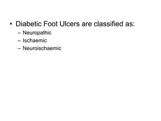
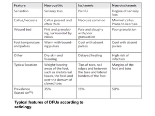
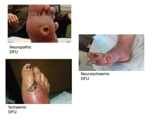
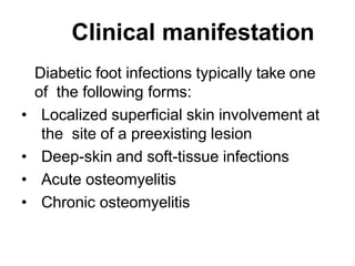
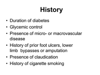
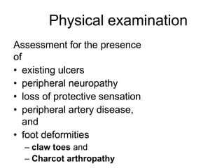
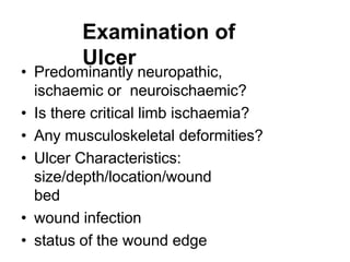
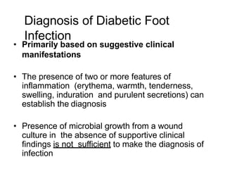
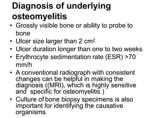
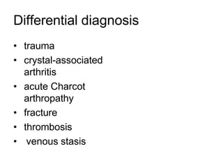
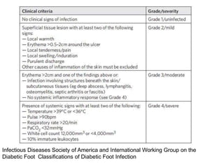
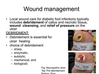
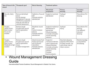
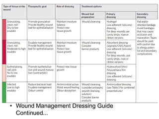
The Diabetic Foot: What You Need to KnowOmar Haqqani
Authored by Dr. Jeffrey Stone, DPM. Presented at the First Annual Omar P. Haqqani MD Vascular Symposium, November 10, 2106, Midland Country Club, Midland, MI.
The Diabetic Foot: What You Need to KnowOmar Haqqani
Authored by Dr. Jeffrey Stone, DPM. Presented at the First Annual Omar P. Haqqani MD Vascular Symposium, November 10, 2106, Midland Country Club, Midland, MI.
The presentation is for the use of Physiotherapy students. It covers a brief introduction, classification, clinical features and general principles of management.
Diabetic foot refers to a range of complications that can occur in individuals with diabetes, particularly those who have poor blood sugar control over an extended period of time. It is a serious condition that can lead to various foot problems, such as ulcers, infections, and even amputations if not properly managed.
The underlying cause of diabetic foot is neuropathy, which is nerve damage that occurs due to high blood sugar levels. Neuropathy can lead to loss of sensation in the feet, making it difficult for individuals to detect injuries or areas of pressure. Additionally, diabetes can impair blood circulation, which reduces the body's ability to heal wounds effectively.
A patient with severe limb infection in whom the amputation was the first option.
Dr Majd Alhaddadin, Consultant General and Laparoscopic Surgeon, performed a transmetatrsal amputation with extensive tissue debridement and falp creation, followed by vacuum therapy and 2 stages wound closure. Fortunately xth limb was saved and the patient returned to his normal job.
New Drug Discovery and Development .....NEHA GUPTA
The "New Drug Discovery and Development" process involves the identification, design, testing, and manufacturing of novel pharmaceutical compounds with the aim of introducing new and improved treatments for various medical conditions. This comprehensive endeavor encompasses various stages, including target identification, preclinical studies, clinical trials, regulatory approval, and post-market surveillance. It involves multidisciplinary collaboration among scientists, researchers, clinicians, regulatory experts, and pharmaceutical companies to bring innovative therapies to market and address unmet medical needs.
Lung Cancer: Artificial Intelligence, Synergetics, Complex System Analysis, S...Oleg Kshivets
RESULTS: Overall life span (LS) was 2252.1±1742.5 days and cumulative 5-year survival (5YS) reached 73.2%, 10 years – 64.8%, 20 years – 42.5%. 513 LCP lived more than 5 years (LS=3124.6±1525.6 days), 148 LCP – more than 10 years (LS=5054.4±1504.1 days).199 LCP died because of LC (LS=562.7±374.5 days). 5YS of LCP after bi/lobectomies was significantly superior in comparison with LCP after pneumonectomies (78.1% vs.63.7%, P=0.00001 by log-rank test). AT significantly improved 5YS (66.3% vs. 34.8%) (P=0.00000 by log-rank test) only for LCP with N1-2. Cox modeling displayed that 5YS of LCP significantly depended on: phase transition (PT) early-invasive LC in terms of synergetics, PT N0—N12, cell ratio factors (ratio between cancer cells- CC and blood cells subpopulations), G1-3, histology, glucose, AT, blood cell circuit, prothrombin index, heparin tolerance, recalcification time (P=0.000-0.038). Neural networks, genetic algorithm selection and bootstrap simulation revealed relationships between 5YS and PT early-invasive LC (rank=1), PT N0—N12 (rank=2), thrombocytes/CC (3), erythrocytes/CC (4), eosinophils/CC (5), healthy cells/CC (6), lymphocytes/CC (7), segmented neutrophils/CC (8), stick neutrophils/CC (9), monocytes/CC (10); leucocytes/CC (11). Correct prediction of 5YS was 100% by neural networks computing (area under ROC curve=1.0; error=0.0).
CONCLUSIONS: 5YS of LCP after radical procedures significantly depended on: 1) PT early-invasive cancer; 2) PT N0--N12; 3) cell ratio factors; 4) blood cell circuit; 5) biochemical factors; 6) hemostasis system; 7) AT; 8) LC characteristics; 9) LC cell dynamics; 10) surgery type: lobectomy/pneumonectomy; 11) anthropometric data. Optimal diagnosis and treatment strategies for LC are: 1) screening and early detection of LC; 2) availability of experienced thoracic surgeons because of complexity of radical procedures; 3) aggressive en block surgery and adequate lymph node dissection for completeness; 4) precise prediction; 5) adjuvant chemoimmunoradiotherapy for LCP with unfavorable prognosis.
Title: Sense of Taste
Presenter: Dr. Faiza, Assistant Professor of Physiology
Qualifications:
MBBS (Best Graduate, AIMC Lahore)
FCPS Physiology
ICMT, CHPE, DHPE (STMU)
MPH (GC University, Faisalabad)
MBA (Virtual University of Pakistan)
Learning Objectives:
Describe the structure and function of taste buds.
Describe the relationship between the taste threshold and taste index of common substances.
Explain the chemical basis and signal transduction of taste perception for each type of primary taste sensation.
Recognize different abnormalities of taste perception and their causes.
Key Topics:
Significance of Taste Sensation:
Differentiation between pleasant and harmful food
Influence on behavior
Selection of food based on metabolic needs
Receptors of Taste:
Taste buds on the tongue
Influence of sense of smell, texture of food, and pain stimulation (e.g., by pepper)
Primary and Secondary Taste Sensations:
Primary taste sensations: Sweet, Sour, Salty, Bitter, Umami
Chemical basis and signal transduction mechanisms for each taste
Taste Threshold and Index:
Taste threshold values for Sweet (sucrose), Salty (NaCl), Sour (HCl), and Bitter (Quinine)
Taste index relationship: Inversely proportional to taste threshold
Taste Blindness:
Inability to taste certain substances, particularly thiourea compounds
Example: Phenylthiocarbamide
Structure and Function of Taste Buds:
Composition: Epithelial cells, Sustentacular/Supporting cells, Taste cells, Basal cells
Features: Taste pores, Taste hairs/microvilli, and Taste nerve fibers
Location of Taste Buds:
Found in papillae of the tongue (Fungiform, Circumvallate, Foliate)
Also present on the palate, tonsillar pillars, epiglottis, and proximal esophagus
Mechanism of Taste Stimulation:
Interaction of taste substances with receptors on microvilli
Signal transduction pathways for Umami, Sweet, Bitter, Sour, and Salty tastes
Taste Sensitivity and Adaptation:
Decrease in sensitivity with age
Rapid adaptation of taste sensation
Role of Saliva in Taste:
Dissolution of tastants to reach receptors
Washing away the stimulus
Taste Preferences and Aversions:
Mechanisms behind taste preference and aversion
Influence of receptors and neural pathways
Impact of Sensory Nerve Damage:
Degeneration of taste buds if the sensory nerve fiber is cut
Abnormalities of Taste Detection:
Conditions: Ageusia, Hypogeusia, Dysgeusia (parageusia)
Causes: Nerve damage, neurological disorders, infections, poor oral hygiene, adverse drug effects, deficiencies, aging, tobacco use, altered neurotransmitter levels
Neurotransmitters and Taste Threshold:
Effects of serotonin (5-HT) and norepinephrine (NE) on taste sensitivity
Supertasters:
25% of the population with heightened sensitivity to taste, especially bitterness
Increased number of fungiform papillae
Report Back from SGO 2024: What’s the Latest in Cervical Cancer?bkling
Are you curious about what’s new in cervical cancer research or unsure what the findings mean? Join Dr. Emily Ko, a gynecologic oncologist at Penn Medicine, to learn about the latest updates from the Society of Gynecologic Oncology (SGO) 2024 Annual Meeting on Women’s Cancer. Dr. Ko will discuss what the research presented at the conference means for you and answer your questions about the new developments.
micro teaching on communication m.sc nursing.pdfAnurag Sharma
Microteaching is a unique model of practice teaching. It is a viable instrument for the. desired change in the teaching behavior or the behavior potential which, in specified types of real. classroom situations, tends to facilitate the achievement of specified types of objectives.
263778731218 Abortion Clinic /Pills In Harare ,sisternakatoto
263778731218 Abortion Clinic /Pills In Harare ,ABORTION WOMEN’S CLINIC +27730423979 IN women clinic we believe that every woman should be able to make choices in her pregnancy. Our job is to provide compassionate care, safety,affordable and confidential services. That’s why we have won the trust from all generations of women all over the world. we use non surgical method(Abortion pills) to terminate…Dr.LISA +27730423979women Clinic is committed to providing the highest quality of obstetrical and gynecological care to women of all ages. Our dedicated staff aim to treat each patient and her health concerns with compassion and respect.Our dedicated group ABORTION WOMEN’S CLINIC +27730423979 IN women clinic we believe that every woman should be able to make choices in her pregnancy. Our job is to provide compassionate care, safety,affordable and confidential services. That’s why we have won the trust from all generations of women all over the world. we use non surgical method(Abortion pills) to terminate…Dr.LISA +27730423979women Clinic is committed to providing the highest quality of obstetrical and gynecological care to women of all ages. Our dedicated staff aim to treat each patient and her health concerns with compassion and respect.Our dedicated group of receptionists, nurses, and physicians have worked together as a teamof receptionists, nurses, and physicians have worked together as a team wwww.lisywomensclinic.co.za/
Flu Vaccine Alert in Bangalore Karnatakaaddon Scans
As flu season approaches, health officials in Bangalore, Karnataka, are urging residents to get their flu vaccinations. The seasonal flu, while common, can lead to severe health complications, particularly for vulnerable populations such as young children, the elderly, and those with underlying health conditions.
Dr. Vidisha Kumari, a leading epidemiologist in Bangalore, emphasizes the importance of getting vaccinated. "The flu vaccine is our best defense against the influenza virus. It not only protects individuals but also helps prevent the spread of the virus in our communities," he says.
This year, the flu season is expected to coincide with a potential increase in other respiratory illnesses. The Karnataka Health Department has launched an awareness campaign highlighting the significance of flu vaccinations. They have set up multiple vaccination centers across Bangalore, making it convenient for residents to receive their shots.
To encourage widespread vaccination, the government is also collaborating with local schools, workplaces, and community centers to facilitate vaccination drives. Special attention is being given to ensuring that the vaccine is accessible to all, including marginalized communities who may have limited access to healthcare.
Residents are reminded that the flu vaccine is safe and effective. Common side effects are mild and may include soreness at the injection site, mild fever, or muscle aches. These side effects are generally short-lived and far less severe than the flu itself.
Healthcare providers are also stressing the importance of continuing COVID-19 precautions. Wearing masks, practicing good hand hygiene, and maintaining social distancing are still crucial, especially in crowded places.
Protect yourself and your loved ones by getting vaccinated. Together, we can help keep Bangalore healthy and safe this flu season. For more information on vaccination centers and schedules, residents can visit the Karnataka Health Department’s official website or follow their social media pages.
Stay informed, stay safe, and get your flu shot today!
Knee anatomy and clinical tests 2024.pdfvimalpl1234
This includes all relevant anatomy and clinical tests compiled from standard textbooks, Campbell,netter etc..It is comprehensive and best suited for orthopaedicians and orthopaedic residents.
Basavarajeeyam is an important text for ayurvedic physician belonging to andhra pradehs. It is a popular compendium in various parts of our country as well as in andhra pradesh. The content of the text was presented in sanskrit and telugu language (Bilingual). One of the most famous book in ayurvedic pharmaceutics and therapeutics. This book contains 25 chapters called as prakaranas. Many rasaoushadis were explained, pioneer of dhatu druti, nadi pareeksha, mutra pareeksha etc. Belongs to the period of 15-16 century. New diseases like upadamsha, phiranga rogas are explained.
Explore natural remedies for syphilis treatment in Singapore. Discover alternative therapies, herbal remedies, and lifestyle changes that may complement conventional treatments. Learn about holistic approaches to managing syphilis symptoms and supporting overall health.
2. Introduction: Diabetic Foot
Ulcers and Infections
• Most common problem in persons
with diabetes.
• Lifetime risk of a foot ulcer in
Diabetes patients: 25 %
• Account for approximately 25 percent of
all hospital stays for patients with
diabetes
3. Risk factors
• Local trauma and/or pressure
• Prior ulcers or amputations
• Infection
• Effects of chronic ischemia, due to
peripheral artery disease
• Patients with diabetes also have an
increased risk for nonhealing related to
mechanical and cytogenic factors
8. Microbiology
• Most diabetic foot infections are
polymicrobial
• Superficial diabetic foot infections
:likely due to aerobic gram-positive cocci.
• Ulcers that are deep, chronically
infected and/or previously treated with
antibiotics are more likely to be
polymicrobial.
9. • Wounds with extensive local
inflammation, necrosis, malodorous
drainage, or gangrene with signs of
systemic toxicity should be presumed to
have anaerobic organisms in addition to
the above pathogens.
10. Ulcer classification
University of Texas system
Grade 0: Pre- or postulcerative
Grade 1:Full-thickness ulcer not involving tendon, capsule, or
bone
Grade 2: Tendon or capsular involvement without bone
palpable
Grade 3: Probes to bone
19. Clinical manifestation
Diabetic foot infections typically take one
of the following forms:
• Localized superficial skin involvement at
the site of a preexisting lesion
• Deep-skin and soft-tissue infections
• Acute osteomyelitis
• Chronic osteomyelitis
20. History
• Duration of diabetes
• Glycemic control
• Presence of micro- or macrovascular
disease
• History of prior foot ulcers, lower
limb bypasses or amputation
• Presence of claudication
• History of cigarette smoking
21. Physical examination
Assessment for the presence
of
• existing ulcers
• peripheral neuropathy
• loss of protective sensation
• peripheral artery disease,
and
• foot deformities
– claw toes and
– Charcot arthropathy
22. Examination of
Ulcer
• Predominantly neuropathic,
ischaemic or neuroischaemic?
• Is there critical limb ischaemia?
• Any musculoskeletal deformities?
• Ulcer Characteristics:
size/depth/location/wound
bed
• wound infection
• status of the wound edge
24. • Procedure:
– Quiet Surrounding
– Eyes Closed for the test
– Supine position
– Testing in inner aspect of arm/hand
– Apply the monofilament perpendicular to the
skin surface with sufficient force to bend it
– Ask: whether they felt it?/Where they felt it?
– Duration: 2 secs
– 3 applications in each site with at least 1
mock
25. • Inference:
– Protective sensation is present at each site if
the patient correctly answers two out of
three applications
– Protective sensation is absent with two
out of three incorrect answers
26. Physical signs of peripheral artery
disease
• diminished foot pulses,
• decrease in skin
temperature,
• thin skin,
• lack of skin hair, and
• bluish skin color
27. Diagnosis of Diabetic Foot
Infection
• Primarily based on suggestive clinical
manifestations
• The presence of two or more features of
inflammation (erythema, warmth, tenderness,
swelling, induration and purulent secretions) can
establish the diagnosis
• Presence of microbial growth from a wound
culture in the absence of supportive clinical
findings is not sufficient to make the diagnosis of
infection
28. Diagnosis of underlying
osteomyelitis
• Grossly visible bone or ability to probe to
bone
• Ulcer size larger than 2 cm2
• Ulcer duration longer than one to two weeks
• Erythrocyte sedimentation rate (ESR) >70
mm/h
• A conventional radiograph with consistent
changes can be helpful in making the
diagnosis ((MRI), which is highly sensitive
and specific for osteomyelitis )
• Culture of bone biopsy specimens is also
important for identifying the causative
organisms
30. Infectious Diseases Society of America and International Working Group on the
Diabetic Foot Classifications of Diabetic Foot Infection
31. Management
Management of diabetic foot
infections requires:
• Attentive wound management
• Good nutrition
• Appropriate antimicrobial therapy
• Glycemic control, and
• fluid and electrolyte balance.
32. Wound management
• Local wound care for diabetic foot infections typically
includes debridement of callus and necrotic tissue,
wound cleansing, and relief of pressure on the
ulcer
DEBRIDMENT:
• Debridement is essential for
ulcer healing
• choice of debridement
– sharp,
– enzymatic,
– autolytic,
– mechanical, and
– biological)
Fig: Neuropathic ulcer
Top: Pre debridement
33. DRESSINGS
• After debridement, ulcers should be kept
clean and moist but free of excess fluids
• Dressings should be selected based upon
ulcer characteristics, such as the extent of
exudate, desiccation, or necrotic tissue
Adjunctive local therapies :
• negative pressure wound therapy (NPWT)
• use of custom-fit semipermeable
polymeric membrane dressings
• cultured human dermal grafts
• application of growth factors
34. • Wound Management Dressing
Guide
International Best Practice Guidelines: Wound Management in Diabetic Foot Ulcers.
36. Surgery
Required for cure of infections complicated by
• abscess,
• extensive bone or joint involvement,
• crepitus, necrosis, gangrene or necrotizing fasciitis
• And for source control in patients with severe
sepsis
In addition to surgical debridement, revascularization
(via angioplasty or bypass grafting) and/or
amputation may be necessary.
37. Antimicrobial therapy
EMPERIC THERAPY:
Mild infection: Outpatient oral
antimicrobial therapy.
Should include activity against skin
flora including streptococci and S.
aureus
Agents with activity against methicillin-
resistant S. aureus (MRSA) should be used
in patients with purulent infections and those
at risk for MRSA infection
38. • Moderate infection: Deep ulcers with
extension to fascia. Should include activity
against streptococci, S. aureus (and MRSA
if risk factors are present), aerobic gram-
negative bacilli and anaerobes
– can be administered orally
• Empiric coverage for P. aeruginosa may not
always be necessary unless the patient has
particular risk for involvement with this
organism, such as a macerated wound or
one with significant water exposure
39. Severe infection: Limb-threatening diabetic
foot infections and those that are
associated with systemic toxicity should
be treated with broad-spectrum
parenteral antibiotic therapyIn most cases, surgical debridement is
also necessary.
40.
41. Duration of therapy
• Mild infection should receive oral antibiotic
therapy in conjunction with attentive
wound care until there is evidence that
the infection has resolved (usually about
one to two weeks)
• Patients with infection also requiring
surgical debridement or amputation
should receive intravenous antibiotic
therapy perioperatively
42. • In case of osteomyelitis:
• No data support the superiority of
specific antimicrobial agents for
osteomyelitis
• Appropriate regimens for empiric therapy
are similar to that for moderate to severe
diabetic foot infections
• Therapy should be tailored to culture
and sensitivity results, ideally from
bone biopsy.
• Patients who were initiated on
parenteral therapy, a switch to an oral
regimen is reasonable following
43. Extensive surgical debridement or resection is
preferable in the following clinical
circumstances
• Persistent sepsis without an alternate source
• Inability to receive or tolerate
appropriate antibiotic therapy
• Progressive bone deterioration
despite appropriate antibiotic
therapy
• Mechanics of the foot are compromised by
extensive bony destruction requiring
correction
• Surgery is needed to achieve soft tissue
wound or primary closure
45. Follow-
up
• Close follow-up is important to ensure
continued improvement and to evaluate
the need for modification of antimicrobial
therapy, further imaging, or additional
surgical intervention
46. Summar
y• Hyperglycemia, sensory and autonomic
neuropathy, and peripheral arterial
disease all contribute to the pathogenesis
of lower extremity infections in diabetic
patients
• Evaluation of a patient with a diabetic foot
infection involves determining the extent
and severity of infection through clinical
and radiographic assessment
47. • The presence of two or more features of
inflammation (erythema, warmth, tenderness,
swelling, induration, or purulent secretions) can
establish the diagnosis of a diabetic foot
infection. The definitive diagnosis of osteomyelitis
is made through histologic and microbiologic
evaluation of a bone biopsy sample
• Management of diabetic foot infections requires
attentive wound management, good nutrition,
antimicrobial therapy, glycemic control, and fluid
and electrolyte balance
48. Reference
s::• Lipsky BA, et al. 2012 Infectious Diseases Society
of America clinical practice guideline for the
diagnosis and treatment of diabetic foot infections.
Clin Infect Dis 2012; 54:e132.
• International Best Practice Guidelines:
Wound Management in Diabetic Foot
Ulcers. Wounds International, 2013.
• Gulf Diabetic Foot Working Group. Identification
and management of infection in diabetic foot
ulcers: International consensus. Wounds
International 2017.
• www.uptodate.com
• Internet