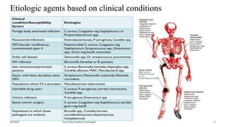
SlideShare a Scribd company logo
1 of 76
MANAGEMENT OF OSTEOMYELITIS
Presentation
Contents
Objectives
Introduction
What is
Osteomyelitis?
• Suppurative process in a bone caused by a
pyogenic organism.
myelos
Osteitis -
Centripedal Osteomyelitis
-centrifugal
Incidence & Epidemiology of OM
Incidence & Epidemiology of OM….
Classifications of Osteomyelitis
• Acute
• Subacute
• Chronic
Duration of
Symptoms
• Pyogenic
• Non pyogenic
Host response for
infection
• Hematogenous
• Contiguous spread
• Direct inoculation
Infection
mechanism/route
Early Acute
Late Acute
Acute on Chronic
Exogenous
WaldVogel's classification of OM
Etiologic agents of OM
• Staph aureus
Etiologic agents based on clinical conditions
Why is Staph aureus the commonest & the most
destructive?
SPECIFIC FEATURES
Risk Factors for OM
• Recent trauma or surgery
• Patients with Prosthetic joints
• Extremes of age
• Infection (Local or Systemic)
• Previous history
• Animal bite
• Immunocompromised
patients
• Illicit IV drug abusers
• Poor Vascular state
• Peripheral neuropathy
• Sickle cell disease
Acute Osteomyelitis
hematogenous route
Acute Hematogenous Osteomyelitis
AHOM………
Pathophysiology of AHOM
• Response increase vascular permeability, resulting
in edema, increased vascularity, and the influx of
polymorphonuclear leukocytes
• Pressure increases as pus collects and is confined
within the rigid bone.
• Exudation through Volkmann's canals and the
Haversian canal affords little relief, although the
relatively inelastic periosteum may become
elevated.
• The blood supply to the area of involvement is
decreased secondary to the pressure; necrosis of
the infected bone may result in the formation of
a sequestrum.
Pathophysiology of AHOM
The effects in children vary with age
• In <2yrs- blood vessels cross physis & infection
spreads into the epiphysis and there might be joint
involvement (Suppurative SA).
The hip joint is commonly affected and other
bones with an intraarticular metaphysis
In severe infection, epiphyseal separation occurs
and,
Can result in limb shortening or angular deformity
Pathophysiology of AHOM
• In older children-the physis acts as a barrier
The diaphysis is at greater risk with spread
of infection to the diaphysis compromising
the endosteal blood supply & resulting in
sequestration.
• In adults, there is ossification of the growth plate & the
metaphyseal-epiphyseal circulation merges
 So, there is no sluggish blood flow
 Hematogenous OM is Uncommon
 Infection may extend to the epiphysis & can result in
SA
Hematogenous OM after Physis
Closure
 Gram-negative bacteria
Progression of AOM to COM
• After the vascular supply to the involved
area has been interrupted and necrosis has
occurred, the chronic phase of
osteomyelitis is established.
• The residual dead bone acts as a foreign
body, making the eradication of bacteria
impossible until the sequestrum is
removed.
• If the infected area becomes well-
demarcated and the infection is contained,
the acute inflammatory process may
subside, leaving a subperiosteal
accumulation of pus which may be
discovered by tenderness on palpation.
• This relatively quiescent form of
subperiosteal infection is
termed Brodie's abscess.
• After some time, there is deposition of
new bone, the involucrum, under the
elevated periosteum.
Events in AHOM………
Bacterial seeding
Inflammatory reaction
Intravascular
thrombosis and
obstruction of blood
flow
Abscess formation
Intramedullary
pressure increases
Extension of abscess to
Subperiosteal space
Cortical ischemia (from ↑↑
pressure, vascular obstruction
& infective thrombus)
Bone necrosis & Sequestrum
formation
Involucrum formation with
Cloaca
CHRONIC OSTEOMYELITIS
Clinical Features of AOM
History
Signs and symptoms of bacteremia
Clinical Features of AOM
Lab. findings
Aspiration Biopsy
Imaging
Imaging….
Algorithm for radiologic evaluation and Rx when AHOM is suspected.
Differential Diagnosis
Important differential diagnoses
to consider are:
• Septic arthritis
• Acute rheumatic arthritis
• Hemarthrosis
• Ewing's sarcoma
• Osteosarcoma
Treatment of Acute OM
• The treatment of AHO demands appropriate
antimicrobial therapy in all cases and may
require surgical incision and drainage.
Treatment….. Surgery







Treatment……
 Antibiotics that have proven efficacy
against S. aureus bone and joint infections
include nafcillin, clindamycin, first-generation
cephalosporins and vancomycin
Treatment……
 Intravenous antibiotics are traditionally
prescribed for 3 weeks, followed by 3
weeks of oral antibiotics. (A minimum
of 4-6 weeks of antimicrobial therapy
Treatment….
• successful response is indicated by a fall in CRP, ESR,
temperature and improvement of local symptoms/signs.
Complications of AOM
septicemia and
metastatic abscesses
with a risk of severe
illness
secondary
involvement of the
adjacent joint (SA)
DVT (Surgery,
Age>8, CRP>6,
MRSA)
chronic
osteomyelitis
(in 5-10%)
Brodie’s abscess
Growth
disturbances and
limb-length
discrepancies
Pathologic fractures Recurrence of OM
Subacute
osteomyelitis
Subacute Osteomyelitis
• Subacute osteomyelitis is a chronic low-grade infection of bone
characterized by a lack of systemic manifestations.
• Staph aureus remains the most commonly identified organism
with Kingella kingae considered an important alternative pathogen to
consider, especially in apparent culture negative cases in younger children
Clinical presentation
• The diagnosis of subacute osteomyelitis should be considered in the
limping child with focal tenderness, especially in the patient who has
received antibiotics for an unrelated infection in the weeks prior.
Clinical presentation….
Lab workups
• Purulent material is not always obtained on biopsy, but granulation tissue is
a common finding.
Radiography
• Correct diagnosis is largely dependent on clinical suspicion and radiographic
findings.
• In patients older than 18 months of age, AHO rarely crosses the physis;
however, subacute osteomyelitis frequently does cross the physis. There is rarely
permanent damage to the growth plate in these patients.
Classification
• Gledhill
• Modified by Roberts et al
Radiologic classification of SAO
with Ddx
Robert’s classification
Type 1-central metaphyseal lesion;
• I A-punched out radiolucency
• I B-punched out radiolucency with a sclerotic rim
Type 2-eccentric metaphyseal lesion with cortical
erosion;
Type 3-diaphyseal cortical lesion; with periosteal
reaction
Type 4-diaphyseal lesion with periosteal new bone
formation but without definite bony lesion; Onion
skinning
Type 5-primary subacute epiphyseal osteomyelitis; and
Type 6-Vertebral body lesion with punched out or
sclerotic margin
Treatment
-Oral anti-staphylococcal antibiotics for 6 weeks
-For lesions that appear to be a simple abscess in the epiphysis or metaphysis should be treated
with intravenous antibiotics for 48 hours followed by a 6-week course of oral antibiotics.
Operative
-Debridement & Curettage followed by treatment with appropriate antibiotics for all lesions that
appear aggressive & if symptoms persist after 2wks of antibiotics.
-In cases in which extensive destruction is seen, postoperative cast immobilization should be
considered to prevent fractures
Brodie Abscess
• A localized form of subacute osteomyelitis that occurs most often in the long
bones of the lower extremities of young adults.
• A well-defined cavity in cancellous bone.
• A chronic abscess of bone surrounded by dense fibrous tissue and sclerotic bone.
• Before physeal closure, the metaphysis is most often affected.
• In adults the metaphyseal-epiphyseal area is involved.
• Intermittent pain of long duration is the presenting complaint, along with local
tenderness over the affected area.
• S. aureus is cultured in 50% of pts
• In 20%, the culture is negative.
Brodie Abscess
•
•
•
•
•
•
•
•
Chronic Osteomyelitis
Chronic Osteomyelitis……
COM
Predisposing factors for COM development
• A history of trauma,
• Open fractures
• Surgery
• Presence of implants
• Diabetes
• Peripheral vascular disease
• Malnutrition
• Dialysis
• Chronic steroid use
• Malignancy
• Alcoholism
• Smoking
• Systemic or local immunocompromise,
• Intravenous drug use and development of
decubitus ulcers.
Etiologic agents of Chronic Osteomyelitis
• The type of pathogen isolated is highly
dependent on patient-related factors such
as age, immune status, history of trauma
and geographical location.
• Contiguous-focus osteomyelitis is
generally polymicrobial.
• The most commonly involved pathogen is
by far Staphylococcus aureus.
• Methicillin-resistant S. aureus (MRSA)
has also been increasingly isolated from
chronic osteomyelitis lesions.
Clinical manifestations
9/2/2023 mng Osteomyelites dr.temesgen 55
The Cierny and Mader Staging system for COM
• Classification system for chronic osteomyelitis, based on physiological and anatomical
criteria, to determine the stage of infection and management options.
Physiological criteria.
 Class A hosts have a normal response to infection and surgery.
 Class B hosts are compromised and have deficient wound healing capabilities.
(BL,BS,BSL)
 Class C host when the results of treatment are potentially more damaging than the
presenting condition
Classification of COM

•
•
•
•
9/2/2023 mng Osteomyelites dr.temesgen 57
Diagnosis…

•
•
•
•

• biopsy for histological and microbiological
evaluation of the infected bone.




9/2/2023 mng Osteomyelites dr.temesgen 58
Imaging studies
• Plain radiography.
Signs of cortical destruction and periosteal
reaction strongly suggest the diagnosis of
osteomyelitis.
To assess overall bony architecture, limb
length& alignment, fracture malunion or
non union.
• Computed tomography
Provides excellent definition of cortical
bone and a fair evaluation of the
surrounding soft tissues and is especially
useful in identification of sequestra.
• Nuclear medicine studies
• Three phases Tc 99m
• Gallium Scintigraphy
• MRI
• More useful for soft tissue evaluation than
CT.
• Shows areas of bony edema quite well.
• Reveal a well-defined rim of high signal
intensity surrounding the focus of active
disease.
• Sinus tracks and cellulitis appear as areas
of increased signal intensity on T2-
weighted imaging.
9/2/2023 mng Osteomyelites dr.temesgen 59
Imaging…
9/2/2023 mng Osteomyelites dr.temesgen 61
• The management of chronic osteomyelitis depends on
• The condition of the host
• The duration and severity of symptoms (the functional impairment caused by the disease)
• The site of involvement
• The extent of bony necrosis,
• The main goal of treatment is to
 Eliminate the inflammatory process by removing all the pathogens and the devitalized tissue, and
 If healing has not occurred, promote healing by optimizing the mechanical and biological environment.
• This can be achieved with a combination of treatment with antibiotic agents, surgical
debridement, and management of the dead space.
Treatment of COM
9/2/2023 mng Osteomyelites dr.temesgen 62
• The curative approach to chronic
osteomyelitis has the following goals:
• Arrest the infection
• Reduce pain
• Retain limb and function.
Treatment of COM
1 UTMB Treatment protocol for adult COM
2 UTMB Treatment protocol for adult COM
3 UTMB Treatment protocol for adult COM
Surgery
•
•
•
•
Limb salvage surgery steps
Radical debridement
Dead space mgt
Four basic methods of immediate, biological
management of dead space using living tissue or
cancellous bone grafts.
The methods to eliminate dead space are
1.
2.
3.
4.
5.
9/2/2023 mng Osteomyelites dr.temesgen 69
Instrumentation for Bony Stability
Soft tissue coverage
9/2/2023 mng Osteomyelites dr.temesgen 72
Antibiotics
• Initial empiric antibiotic treatment should be commenced as soon as the culture samples have been
obtained.
• The antibiotic regime used later should be based on the results of the cultures and sensitivities obtained by
bone biopsies.
Treatment of COM….
Complications
Pathological fracture
Acute exacerbation
Sepsis
Growth disturbance
Deformities
Malignant transformation
Persistence or extension of infection
Amputation
• Despite surgical debridement and long-
term antibiotics, recurrence rate of
chronic osteomyelitis in adults is 30%
• Has poor prognosis in patients with
major nutritional or systemic disorders
summary
References
• Cierny III G, Mader JT, Penninck JJ. A clinical staging for adult
osteomyelitis. Cont Orthop. 1985;10:5
•Thank You

More Related Content

Similar to OSTEOMYELITIS .pptx

14. Osteomyelitis...pptx
14. Osteomyelitis...pptx14. Osteomyelitis...pptx
14. Osteomyelitis...pptxNoelMabele
 
Bone and joint infection
Bone and joint infectionBone and joint infection
Bone and joint infectionSherif El Aidy
 
Osteomyelitis march 2018
Osteomyelitis  march 2018Osteomyelitis  march 2018
Osteomyelitis march 2018Meher Rizvi
 
Infected Fractures - Principles, com.ppt
Infected Fractures - Principles, com.pptInfected Fractures - Principles, com.ppt
Infected Fractures - Principles, com.pptBedrumohammed2
 
Chronic Osteomyelitis of bone and marrow cavity.pptx
Chronic Osteomyelitis of bone and marrow cavity.pptxChronic Osteomyelitis of bone and marrow cavity.pptx
Chronic Osteomyelitis of bone and marrow cavity.pptxBinod Chaudhary
 
CHRONIC OSTEOMYELITIS (1).pptx
CHRONIC OSTEOMYELITIS (1).pptxCHRONIC OSTEOMYELITIS (1).pptx
CHRONIC OSTEOMYELITIS (1).pptxKeyaArere
 
Hiv and orthopaedics
Hiv and orthopaedicsHiv and orthopaedics
Hiv and orthopaedicsLibin Thomas
 
BONE INFECTIONS.pptx
BONE INFECTIONS.pptxBONE INFECTIONS.pptx
BONE INFECTIONS.pptxkmachy
 
Paediatric musculoskeletal infections
Paediatric musculoskeletal infectionsPaediatric musculoskeletal infections
Paediatric musculoskeletal infectionsRITESHJAISWAL57
 
14. Osteomyelitis.pptx
14. Osteomyelitis.pptx14. Osteomyelitis.pptx
14. Osteomyelitis.pptxSani191640
 

Similar to OSTEOMYELITIS .pptx (20)

14. Osteomyelitis...pptx
14. Osteomyelitis...pptx14. Osteomyelitis...pptx
14. Osteomyelitis...pptx
 
Bone and joint infection
Bone and joint infectionBone and joint infection
Bone and joint infection
 
Osteomyelitis march 2018
Osteomyelitis  march 2018Osteomyelitis  march 2018
Osteomyelitis march 2018
 
Osteomyelitis In Adults
Osteomyelitis In AdultsOsteomyelitis In Adults
Osteomyelitis In Adults
 
Chronic Osteomyelitis
Chronic OsteomyelitisChronic Osteomyelitis
Chronic Osteomyelitis
 
Osteomyelitis
OsteomyelitisOsteomyelitis
Osteomyelitis
 
osteomyelitis
osteomyelitisosteomyelitis
osteomyelitis
 
Infected Fractures - Principles, com.ppt
Infected Fractures - Principles, com.pptInfected Fractures - Principles, com.ppt
Infected Fractures - Principles, com.ppt
 
Chronic Osteomyelitis of bone and marrow cavity.pptx
Chronic Osteomyelitis of bone and marrow cavity.pptxChronic Osteomyelitis of bone and marrow cavity.pptx
Chronic Osteomyelitis of bone and marrow cavity.pptx
 
Osteomyelitis seminar
Osteomyelitis seminarOsteomyelitis seminar
Osteomyelitis seminar
 
Specific osteomyelitis
Specific osteomyelitisSpecific osteomyelitis
Specific osteomyelitis
 
CHRONIC OSTEOMYELITIS (1).pptx
CHRONIC OSTEOMYELITIS (1).pptxCHRONIC OSTEOMYELITIS (1).pptx
CHRONIC OSTEOMYELITIS (1).pptx
 
Chronic infections of jaws
Chronic infections of jaws  Chronic infections of jaws
Chronic infections of jaws
 
Hiv and orthopaedics
Hiv and orthopaedicsHiv and orthopaedics
Hiv and orthopaedics
 
BONE INFECTIONS.pptx
BONE INFECTIONS.pptxBONE INFECTIONS.pptx
BONE INFECTIONS.pptx
 
OSTEOMYELITIS
OSTEOMYELITISOSTEOMYELITIS
OSTEOMYELITIS
 
Osteomyelitis
OsteomyelitisOsteomyelitis
Osteomyelitis
 
Paediatric musculoskeletal infections
Paediatric musculoskeletal infectionsPaediatric musculoskeletal infections
Paediatric musculoskeletal infections
 
14. Osteomyelitis.pptx
14. Osteomyelitis.pptx14. Osteomyelitis.pptx
14. Osteomyelitis.pptx
 
maliganancy in osteomyeitis
maliganancy in osteomyeitismaliganancy in osteomyeitis
maliganancy in osteomyeitis
 

More from Bedrumohammed2

distal tibia malunion orthopedics sem.pptx
distal tibia malunion orthopedics sem.pptxdistal tibia malunion orthopedics sem.pptx
distal tibia malunion orthopedics sem.pptxBedrumohammed2
 
1principles of Chronic dislocations.pptx
1principles of Chronic dislocations.pptx1principles of Chronic dislocations.pptx
1principles of Chronic dislocations.pptxBedrumohammed2
 
neglected supracondylarbest slide pres.pptx
neglected supracondylarbest slide pres.pptxneglected supracondylarbest slide pres.pptx
neglected supracondylarbest slide pres.pptxBedrumohammed2
 
Germ cell tumorsby alexanderarnold2.pptx
Germ cell tumorsby alexanderarnold2.pptxGerm cell tumorsby alexanderarnold2.pptx
Germ cell tumorsby alexanderarnold2.pptxBedrumohammed2
 
management of benign and malignant disease of breast.pptx
management of benign and malignant disease of breast.pptxmanagement of benign and malignant disease of breast.pptx
management of benign and malignant disease of breast.pptxBedrumohammed2
 
Daniel EMBRYOLOGY OF GUT MALFORMATIONS 2023.pptx
Daniel EMBRYOLOGY OF GUT MALFORMATIONS 2023.pptxDaniel EMBRYOLOGY OF GUT MALFORMATIONS 2023.pptx
Daniel EMBRYOLOGY OF GUT MALFORMATIONS 2023.pptxBedrumohammed2
 
undescended testis and acute scrotum 2023.pptx
undescended testis and acute scrotum 2023.pptxundescended testis and acute scrotum 2023.pptx
undescended testis and acute scrotum 2023.pptxBedrumohammed2
 
Bisrat Edit Approach to Hydronephrosis in children.pptx
Bisrat Edit Approach to Hydronephrosis in children.pptxBisrat Edit Approach to Hydronephrosis in children.pptx
Bisrat Edit Approach to Hydronephrosis in children.pptxBedrumohammed2
 
Volkmann Ischemic Contracture plastic .pptx
Volkmann Ischemic Contracture plastic .pptxVolkmann Ischemic Contracture plastic .pptx
Volkmann Ischemic Contracture plastic .pptxBedrumohammed2
 
Pressure Sore mgt by bedru mohammed.pptx
Pressure Sore mgt by bedru mohammed.pptxPressure Sore mgt by bedru mohammed.pptx
Pressure Sore mgt by bedru mohammed.pptxBedrumohammed2
 
Compartment Syndrome by dr bedru mo.pptx
Compartment Syndrome by dr bedru mo.pptxCompartment Syndrome by dr bedru mo.pptx
Compartment Syndrome by dr bedru mo.pptxBedrumohammed2
 
Anorectal Malformation tghytefbjeme.pptx
Anorectal Malformation tghytefbjeme.pptxAnorectal Malformation tghytefbjeme.pptx
Anorectal Malformation tghytefbjeme.pptxBedrumohammed2
 
Management of Elbow Fracture Dislocation.pptx
Management of Elbow Fracture Dislocation.pptxManagement of Elbow Fracture Dislocation.pptx
Management of Elbow Fracture Dislocation.pptxBedrumohammed2
 
hand infectionby drbedrumohanmedgsr.pptx
hand infectionby drbedrumohanmedgsr.pptxhand infectionby drbedrumohanmedgsr.pptx
hand infectionby drbedrumohanmedgsr.pptxBedrumohammed2
 
pancreatic cas managementby bedrumoh.pptx
pancreatic cas managementby bedrumoh.pptxpancreatic cas managementby bedrumoh.pptx
pancreatic cas managementby bedrumoh.pptxBedrumohammed2
 
colorectal ca presented by bedu mohammed.pptx
colorectal ca presented by bedu mohammed.pptxcolorectal ca presented by bedu mohammed.pptx
colorectal ca presented by bedu mohammed.pptxBedrumohammed2
 
GI8. Abdominal Traumtmnagement in adulta.pptx
GI8. Abdominal Traumtmnagement in adulta.pptxGI8. Abdominal Traumtmnagement in adulta.pptx
GI8. Abdominal Traumtmnagement in adulta.pptxBedrumohammed2
 
ABDOMINAL TRAUMA part 2bedrumohs, 2023.pptx
ABDOMINAL TRAUMA part 2bedrumohs, 2023.pptxABDOMINAL TRAUMA part 2bedrumohs, 2023.pptx
ABDOMINAL TRAUMA part 2bedrumohs, 2023.pptxBedrumohammed2
 
Lisfranc fracture of lower extrimities.pptx
Lisfranc fracture of lower extrimities.pptxLisfranc fracture of lower extrimities.pptx
Lisfranc fracture of lower extrimities.pptxBedrumohammed2
 
Calcaneal fracture 101 bedru mohammed15168tyr42.pptx
Calcaneal fracture 101 bedru mohammed15168tyr42.pptxCalcaneal fracture 101 bedru mohammed15168tyr42.pptx
Calcaneal fracture 101 bedru mohammed15168tyr42.pptxBedrumohammed2
 

More from Bedrumohammed2 (20)

distal tibia malunion orthopedics sem.pptx
distal tibia malunion orthopedics sem.pptxdistal tibia malunion orthopedics sem.pptx
distal tibia malunion orthopedics sem.pptx
 
1principles of Chronic dislocations.pptx
1principles of Chronic dislocations.pptx1principles of Chronic dislocations.pptx
1principles of Chronic dislocations.pptx
 
neglected supracondylarbest slide pres.pptx
neglected supracondylarbest slide pres.pptxneglected supracondylarbest slide pres.pptx
neglected supracondylarbest slide pres.pptx
 
Germ cell tumorsby alexanderarnold2.pptx
Germ cell tumorsby alexanderarnold2.pptxGerm cell tumorsby alexanderarnold2.pptx
Germ cell tumorsby alexanderarnold2.pptx
 
management of benign and malignant disease of breast.pptx
management of benign and malignant disease of breast.pptxmanagement of benign and malignant disease of breast.pptx
management of benign and malignant disease of breast.pptx
 
Daniel EMBRYOLOGY OF GUT MALFORMATIONS 2023.pptx
Daniel EMBRYOLOGY OF GUT MALFORMATIONS 2023.pptxDaniel EMBRYOLOGY OF GUT MALFORMATIONS 2023.pptx
Daniel EMBRYOLOGY OF GUT MALFORMATIONS 2023.pptx
 
undescended testis and acute scrotum 2023.pptx
undescended testis and acute scrotum 2023.pptxundescended testis and acute scrotum 2023.pptx
undescended testis and acute scrotum 2023.pptx
 
Bisrat Edit Approach to Hydronephrosis in children.pptx
Bisrat Edit Approach to Hydronephrosis in children.pptxBisrat Edit Approach to Hydronephrosis in children.pptx
Bisrat Edit Approach to Hydronephrosis in children.pptx
 
Volkmann Ischemic Contracture plastic .pptx
Volkmann Ischemic Contracture plastic .pptxVolkmann Ischemic Contracture plastic .pptx
Volkmann Ischemic Contracture plastic .pptx
 
Pressure Sore mgt by bedru mohammed.pptx
Pressure Sore mgt by bedru mohammed.pptxPressure Sore mgt by bedru mohammed.pptx
Pressure Sore mgt by bedru mohammed.pptx
 
Compartment Syndrome by dr bedru mo.pptx
Compartment Syndrome by dr bedru mo.pptxCompartment Syndrome by dr bedru mo.pptx
Compartment Syndrome by dr bedru mo.pptx
 
Anorectal Malformation tghytefbjeme.pptx
Anorectal Malformation tghytefbjeme.pptxAnorectal Malformation tghytefbjeme.pptx
Anorectal Malformation tghytefbjeme.pptx
 
Management of Elbow Fracture Dislocation.pptx
Management of Elbow Fracture Dislocation.pptxManagement of Elbow Fracture Dislocation.pptx
Management of Elbow Fracture Dislocation.pptx
 
hand infectionby drbedrumohanmedgsr.pptx
hand infectionby drbedrumohanmedgsr.pptxhand infectionby drbedrumohanmedgsr.pptx
hand infectionby drbedrumohanmedgsr.pptx
 
pancreatic cas managementby bedrumoh.pptx
pancreatic cas managementby bedrumoh.pptxpancreatic cas managementby bedrumoh.pptx
pancreatic cas managementby bedrumoh.pptx
 
colorectal ca presented by bedu mohammed.pptx
colorectal ca presented by bedu mohammed.pptxcolorectal ca presented by bedu mohammed.pptx
colorectal ca presented by bedu mohammed.pptx
 
GI8. Abdominal Traumtmnagement in adulta.pptx
GI8. Abdominal Traumtmnagement in adulta.pptxGI8. Abdominal Traumtmnagement in adulta.pptx
GI8. Abdominal Traumtmnagement in adulta.pptx
 
ABDOMINAL TRAUMA part 2bedrumohs, 2023.pptx
ABDOMINAL TRAUMA part 2bedrumohs, 2023.pptxABDOMINAL TRAUMA part 2bedrumohs, 2023.pptx
ABDOMINAL TRAUMA part 2bedrumohs, 2023.pptx
 
Lisfranc fracture of lower extrimities.pptx
Lisfranc fracture of lower extrimities.pptxLisfranc fracture of lower extrimities.pptx
Lisfranc fracture of lower extrimities.pptx
 
Calcaneal fracture 101 bedru mohammed15168tyr42.pptx
Calcaneal fracture 101 bedru mohammed15168tyr42.pptxCalcaneal fracture 101 bedru mohammed15168tyr42.pptx
Calcaneal fracture 101 bedru mohammed15168tyr42.pptx
 

Recently uploaded

💎VVIP Kolkata Call Girls Parganas🩱7001035870🩱Independent Girl ( Ac Rooms Avai...
💎VVIP Kolkata Call Girls Parganas🩱7001035870🩱Independent Girl ( Ac Rooms Avai...💎VVIP Kolkata Call Girls Parganas🩱7001035870🩱Independent Girl ( Ac Rooms Avai...
💎VVIP Kolkata Call Girls Parganas🩱7001035870🩱Independent Girl ( Ac Rooms Avai...Taniya Sharma
 
Artifacts in Nuclear Medicine with Identifying and resolving artifacts.
Artifacts in Nuclear Medicine with Identifying and resolving artifacts.Artifacts in Nuclear Medicine with Identifying and resolving artifacts.
Artifacts in Nuclear Medicine with Identifying and resolving artifacts.MiadAlsulami
 
Call Girls Horamavu WhatsApp Number 7001035870 Meeting With Bangalore Escorts
Call Girls Horamavu WhatsApp Number 7001035870 Meeting With Bangalore EscortsCall Girls Horamavu WhatsApp Number 7001035870 Meeting With Bangalore Escorts
Call Girls Horamavu WhatsApp Number 7001035870 Meeting With Bangalore Escortsvidya singh
 
VIP Service Call Girls Sindhi Colony 📳 7877925207 For 18+ VIP Call Girl At Th...
VIP Service Call Girls Sindhi Colony 📳 7877925207 For 18+ VIP Call Girl At Th...VIP Service Call Girls Sindhi Colony 📳 7877925207 For 18+ VIP Call Girl At Th...
VIP Service Call Girls Sindhi Colony 📳 7877925207 For 18+ VIP Call Girl At Th...jageshsingh5554
 
College Call Girls Pune Mira 9907093804 Short 1500 Night 6000 Best call girls...
College Call Girls Pune Mira 9907093804 Short 1500 Night 6000 Best call girls...College Call Girls Pune Mira 9907093804 Short 1500 Night 6000 Best call girls...
College Call Girls Pune Mira 9907093804 Short 1500 Night 6000 Best call girls...Miss joya
 
High Profile Call Girls Coimbatore Saanvi☎️ 8250192130 Independent Escort Se...
High Profile Call Girls Coimbatore Saanvi☎️  8250192130 Independent Escort Se...High Profile Call Girls Coimbatore Saanvi☎️  8250192130 Independent Escort Se...
High Profile Call Girls Coimbatore Saanvi☎️ 8250192130 Independent Escort Se...narwatsonia7
 
♛VVIP Hyderabad Call Girls Chintalkunta🖕7001035870🖕Riya Kappor Top Call Girl ...
♛VVIP Hyderabad Call Girls Chintalkunta🖕7001035870🖕Riya Kappor Top Call Girl ...♛VVIP Hyderabad Call Girls Chintalkunta🖕7001035870🖕Riya Kappor Top Call Girl ...
♛VVIP Hyderabad Call Girls Chintalkunta🖕7001035870🖕Riya Kappor Top Call Girl ...astropune
 
Russian Escorts Girls Nehru Place ZINATHI 🔝9711199012 ☪ 24/7 Call Girls Delhi
Russian Escorts Girls  Nehru Place ZINATHI 🔝9711199012 ☪ 24/7 Call Girls DelhiRussian Escorts Girls  Nehru Place ZINATHI 🔝9711199012 ☪ 24/7 Call Girls Delhi
Russian Escorts Girls Nehru Place ZINATHI 🔝9711199012 ☪ 24/7 Call Girls DelhiAlinaDevecerski
 
Call Girls Service Navi Mumbai Samaira 8617697112 Independent Escort Service ...
Call Girls Service Navi Mumbai Samaira 8617697112 Independent Escort Service ...Call Girls Service Navi Mumbai Samaira 8617697112 Independent Escort Service ...
Call Girls Service Navi Mumbai Samaira 8617697112 Independent Escort Service ...Call girls in Ahmedabad High profile
 
Call Girl Number in Vashi Mumbai📲 9833363713 💞 Full Night Enjoy
Call Girl Number in Vashi Mumbai📲 9833363713 💞 Full Night EnjoyCall Girl Number in Vashi Mumbai📲 9833363713 💞 Full Night Enjoy
Call Girl Number in Vashi Mumbai📲 9833363713 💞 Full Night Enjoybabeytanya
 
Lucknow Call girls - 8800925952 - 24x7 service with hotel room
Lucknow Call girls - 8800925952 - 24x7 service with hotel roomLucknow Call girls - 8800925952 - 24x7 service with hotel room
Lucknow Call girls - 8800925952 - 24x7 service with hotel roomdiscovermytutordmt
 
Russian Call Girls in Pune Riya 9907093804 Short 1500 Night 6000 Best call gi...
Russian Call Girls in Pune Riya 9907093804 Short 1500 Night 6000 Best call gi...Russian Call Girls in Pune Riya 9907093804 Short 1500 Night 6000 Best call gi...
Russian Call Girls in Pune Riya 9907093804 Short 1500 Night 6000 Best call gi...Miss joya
 
Call Girls Ludhiana Just Call 9907093804 Top Class Call Girl Service Available
Call Girls Ludhiana Just Call 9907093804 Top Class Call Girl Service AvailableCall Girls Ludhiana Just Call 9907093804 Top Class Call Girl Service Available
Call Girls Ludhiana Just Call 9907093804 Top Class Call Girl Service AvailableDipal Arora
 
Call Girl Number in Panvel Mumbai📲 9833363713 💞 Full Night Enjoy
Call Girl Number in Panvel Mumbai📲 9833363713 💞 Full Night EnjoyCall Girl Number in Panvel Mumbai📲 9833363713 💞 Full Night Enjoy
Call Girl Number in Panvel Mumbai📲 9833363713 💞 Full Night Enjoybabeytanya
 
Russian Call Girls in Pune Tanvi 9907093804 Short 1500 Night 6000 Best call g...
Russian Call Girls in Pune Tanvi 9907093804 Short 1500 Night 6000 Best call g...Russian Call Girls in Pune Tanvi 9907093804 Short 1500 Night 6000 Best call g...
Russian Call Girls in Pune Tanvi 9907093804 Short 1500 Night 6000 Best call g...Miss joya
 
Aspirin presentation slides by Dr. Rewas Ali
Aspirin presentation slides by Dr. Rewas AliAspirin presentation slides by Dr. Rewas Ali
Aspirin presentation slides by Dr. Rewas AliRewAs ALI
 
Bangalore Call Girl Whatsapp Number 100% Complete Your Sexual Needs
Bangalore Call Girl Whatsapp Number 100% Complete Your Sexual NeedsBangalore Call Girl Whatsapp Number 100% Complete Your Sexual Needs
Bangalore Call Girl Whatsapp Number 100% Complete Your Sexual NeedsGfnyt
 
Call Girls Colaba Mumbai ❤️ 9920874524 👈 Cash on Delivery
Call Girls Colaba Mumbai ❤️ 9920874524 👈 Cash on DeliveryCall Girls Colaba Mumbai ❤️ 9920874524 👈 Cash on Delivery
Call Girls Colaba Mumbai ❤️ 9920874524 👈 Cash on Deliverynehamumbai
 
Call Girls Service Pune Vaishnavi 9907093804 Short 1500 Night 6000 Best call ...
Call Girls Service Pune Vaishnavi 9907093804 Short 1500 Night 6000 Best call ...Call Girls Service Pune Vaishnavi 9907093804 Short 1500 Night 6000 Best call ...
Call Girls Service Pune Vaishnavi 9907093804 Short 1500 Night 6000 Best call ...Miss joya
 
CALL ON ➥9907093804 🔝 Call Girls Baramati ( Pune) Girls Service
CALL ON ➥9907093804 🔝 Call Girls Baramati ( Pune)  Girls ServiceCALL ON ➥9907093804 🔝 Call Girls Baramati ( Pune)  Girls Service
CALL ON ➥9907093804 🔝 Call Girls Baramati ( Pune) Girls ServiceMiss joya
 

Recently uploaded (20)

💎VVIP Kolkata Call Girls Parganas🩱7001035870🩱Independent Girl ( Ac Rooms Avai...
💎VVIP Kolkata Call Girls Parganas🩱7001035870🩱Independent Girl ( Ac Rooms Avai...💎VVIP Kolkata Call Girls Parganas🩱7001035870🩱Independent Girl ( Ac Rooms Avai...
💎VVIP Kolkata Call Girls Parganas🩱7001035870🩱Independent Girl ( Ac Rooms Avai...
 
Artifacts in Nuclear Medicine with Identifying and resolving artifacts.
Artifacts in Nuclear Medicine with Identifying and resolving artifacts.Artifacts in Nuclear Medicine with Identifying and resolving artifacts.
Artifacts in Nuclear Medicine with Identifying and resolving artifacts.
 
Call Girls Horamavu WhatsApp Number 7001035870 Meeting With Bangalore Escorts
Call Girls Horamavu WhatsApp Number 7001035870 Meeting With Bangalore EscortsCall Girls Horamavu WhatsApp Number 7001035870 Meeting With Bangalore Escorts
Call Girls Horamavu WhatsApp Number 7001035870 Meeting With Bangalore Escorts
 
VIP Service Call Girls Sindhi Colony 📳 7877925207 For 18+ VIP Call Girl At Th...
VIP Service Call Girls Sindhi Colony 📳 7877925207 For 18+ VIP Call Girl At Th...VIP Service Call Girls Sindhi Colony 📳 7877925207 For 18+ VIP Call Girl At Th...
VIP Service Call Girls Sindhi Colony 📳 7877925207 For 18+ VIP Call Girl At Th...
 
College Call Girls Pune Mira 9907093804 Short 1500 Night 6000 Best call girls...
College Call Girls Pune Mira 9907093804 Short 1500 Night 6000 Best call girls...College Call Girls Pune Mira 9907093804 Short 1500 Night 6000 Best call girls...
College Call Girls Pune Mira 9907093804 Short 1500 Night 6000 Best call girls...
 
High Profile Call Girls Coimbatore Saanvi☎️ 8250192130 Independent Escort Se...
High Profile Call Girls Coimbatore Saanvi☎️  8250192130 Independent Escort Se...High Profile Call Girls Coimbatore Saanvi☎️  8250192130 Independent Escort Se...
High Profile Call Girls Coimbatore Saanvi☎️ 8250192130 Independent Escort Se...
 
♛VVIP Hyderabad Call Girls Chintalkunta🖕7001035870🖕Riya Kappor Top Call Girl ...
♛VVIP Hyderabad Call Girls Chintalkunta🖕7001035870🖕Riya Kappor Top Call Girl ...♛VVIP Hyderabad Call Girls Chintalkunta🖕7001035870🖕Riya Kappor Top Call Girl ...
♛VVIP Hyderabad Call Girls Chintalkunta🖕7001035870🖕Riya Kappor Top Call Girl ...
 
Russian Escorts Girls Nehru Place ZINATHI 🔝9711199012 ☪ 24/7 Call Girls Delhi
Russian Escorts Girls  Nehru Place ZINATHI 🔝9711199012 ☪ 24/7 Call Girls DelhiRussian Escorts Girls  Nehru Place ZINATHI 🔝9711199012 ☪ 24/7 Call Girls Delhi
Russian Escorts Girls Nehru Place ZINATHI 🔝9711199012 ☪ 24/7 Call Girls Delhi
 
Call Girls Service Navi Mumbai Samaira 8617697112 Independent Escort Service ...
Call Girls Service Navi Mumbai Samaira 8617697112 Independent Escort Service ...Call Girls Service Navi Mumbai Samaira 8617697112 Independent Escort Service ...
Call Girls Service Navi Mumbai Samaira 8617697112 Independent Escort Service ...
 
Call Girl Number in Vashi Mumbai📲 9833363713 💞 Full Night Enjoy
Call Girl Number in Vashi Mumbai📲 9833363713 💞 Full Night EnjoyCall Girl Number in Vashi Mumbai📲 9833363713 💞 Full Night Enjoy
Call Girl Number in Vashi Mumbai📲 9833363713 💞 Full Night Enjoy
 
Lucknow Call girls - 8800925952 - 24x7 service with hotel room
Lucknow Call girls - 8800925952 - 24x7 service with hotel roomLucknow Call girls - 8800925952 - 24x7 service with hotel room
Lucknow Call girls - 8800925952 - 24x7 service with hotel room
 
Russian Call Girls in Pune Riya 9907093804 Short 1500 Night 6000 Best call gi...
Russian Call Girls in Pune Riya 9907093804 Short 1500 Night 6000 Best call gi...Russian Call Girls in Pune Riya 9907093804 Short 1500 Night 6000 Best call gi...
Russian Call Girls in Pune Riya 9907093804 Short 1500 Night 6000 Best call gi...
 
Call Girls Ludhiana Just Call 9907093804 Top Class Call Girl Service Available
Call Girls Ludhiana Just Call 9907093804 Top Class Call Girl Service AvailableCall Girls Ludhiana Just Call 9907093804 Top Class Call Girl Service Available
Call Girls Ludhiana Just Call 9907093804 Top Class Call Girl Service Available
 
Call Girl Number in Panvel Mumbai📲 9833363713 💞 Full Night Enjoy
Call Girl Number in Panvel Mumbai📲 9833363713 💞 Full Night EnjoyCall Girl Number in Panvel Mumbai📲 9833363713 💞 Full Night Enjoy
Call Girl Number in Panvel Mumbai📲 9833363713 💞 Full Night Enjoy
 
Russian Call Girls in Pune Tanvi 9907093804 Short 1500 Night 6000 Best call g...
Russian Call Girls in Pune Tanvi 9907093804 Short 1500 Night 6000 Best call g...Russian Call Girls in Pune Tanvi 9907093804 Short 1500 Night 6000 Best call g...
Russian Call Girls in Pune Tanvi 9907093804 Short 1500 Night 6000 Best call g...
 
Aspirin presentation slides by Dr. Rewas Ali
Aspirin presentation slides by Dr. Rewas AliAspirin presentation slides by Dr. Rewas Ali
Aspirin presentation slides by Dr. Rewas Ali
 
Bangalore Call Girl Whatsapp Number 100% Complete Your Sexual Needs
Bangalore Call Girl Whatsapp Number 100% Complete Your Sexual NeedsBangalore Call Girl Whatsapp Number 100% Complete Your Sexual Needs
Bangalore Call Girl Whatsapp Number 100% Complete Your Sexual Needs
 
Call Girls Colaba Mumbai ❤️ 9920874524 👈 Cash on Delivery
Call Girls Colaba Mumbai ❤️ 9920874524 👈 Cash on DeliveryCall Girls Colaba Mumbai ❤️ 9920874524 👈 Cash on Delivery
Call Girls Colaba Mumbai ❤️ 9920874524 👈 Cash on Delivery
 
Call Girls Service Pune Vaishnavi 9907093804 Short 1500 Night 6000 Best call ...
Call Girls Service Pune Vaishnavi 9907093804 Short 1500 Night 6000 Best call ...Call Girls Service Pune Vaishnavi 9907093804 Short 1500 Night 6000 Best call ...
Call Girls Service Pune Vaishnavi 9907093804 Short 1500 Night 6000 Best call ...
 
CALL ON ➥9907093804 🔝 Call Girls Baramati ( Pune) Girls Service
CALL ON ➥9907093804 🔝 Call Girls Baramati ( Pune)  Girls ServiceCALL ON ➥9907093804 🔝 Call Girls Baramati ( Pune)  Girls Service
CALL ON ➥9907093804 🔝 Call Girls Baramati ( Pune) Girls Service
 

OSTEOMYELITIS .pptx

  • 5. What is Osteomyelitis? • Suppurative process in a bone caused by a pyogenic organism. myelos Osteitis - Centripedal Osteomyelitis -centrifugal
  • 8. Classifications of Osteomyelitis • Acute • Subacute • Chronic Duration of Symptoms • Pyogenic • Non pyogenic Host response for infection • Hematogenous • Contiguous spread • Direct inoculation Infection mechanism/route Early Acute Late Acute Acute on Chronic Exogenous
  • 10. Etiologic agents of OM • Staph aureus
  • 11. Etiologic agents based on clinical conditions
  • 12. Why is Staph aureus the commonest & the most destructive? SPECIFIC FEATURES
  • 13. Risk Factors for OM • Recent trauma or surgery • Patients with Prosthetic joints • Extremes of age • Infection (Local or Systemic) • Previous history • Animal bite • Immunocompromised patients • Illicit IV drug abusers • Poor Vascular state • Peripheral neuropathy • Sickle cell disease
  • 17. Pathophysiology of AHOM • Response increase vascular permeability, resulting in edema, increased vascularity, and the influx of polymorphonuclear leukocytes • Pressure increases as pus collects and is confined within the rigid bone. • Exudation through Volkmann's canals and the Haversian canal affords little relief, although the relatively inelastic periosteum may become elevated. • The blood supply to the area of involvement is decreased secondary to the pressure; necrosis of the infected bone may result in the formation of a sequestrum.
  • 18. Pathophysiology of AHOM The effects in children vary with age • In <2yrs- blood vessels cross physis & infection spreads into the epiphysis and there might be joint involvement (Suppurative SA). The hip joint is commonly affected and other bones with an intraarticular metaphysis In severe infection, epiphyseal separation occurs and, Can result in limb shortening or angular deformity
  • 19. Pathophysiology of AHOM • In older children-the physis acts as a barrier The diaphysis is at greater risk with spread of infection to the diaphysis compromising the endosteal blood supply & resulting in sequestration. • In adults, there is ossification of the growth plate & the metaphyseal-epiphyseal circulation merges  So, there is no sluggish blood flow  Hematogenous OM is Uncommon  Infection may extend to the epiphysis & can result in SA
  • 20. Hematogenous OM after Physis Closure  Gram-negative bacteria
  • 21. Progression of AOM to COM • After the vascular supply to the involved area has been interrupted and necrosis has occurred, the chronic phase of osteomyelitis is established. • The residual dead bone acts as a foreign body, making the eradication of bacteria impossible until the sequestrum is removed. • If the infected area becomes well- demarcated and the infection is contained, the acute inflammatory process may subside, leaving a subperiosteal accumulation of pus which may be discovered by tenderness on palpation. • This relatively quiescent form of subperiosteal infection is termed Brodie's abscess. • After some time, there is deposition of new bone, the involucrum, under the elevated periosteum.
  • 22. Events in AHOM……… Bacterial seeding Inflammatory reaction Intravascular thrombosis and obstruction of blood flow Abscess formation Intramedullary pressure increases Extension of abscess to Subperiosteal space Cortical ischemia (from ↑↑ pressure, vascular obstruction & infective thrombus) Bone necrosis & Sequestrum formation Involucrum formation with Cloaca CHRONIC OSTEOMYELITIS
  • 23. Clinical Features of AOM History Signs and symptoms of bacteremia
  • 29. Algorithm for radiologic evaluation and Rx when AHOM is suspected.
  • 30. Differential Diagnosis Important differential diagnoses to consider are: • Septic arthritis • Acute rheumatic arthritis • Hemarthrosis • Ewing's sarcoma • Osteosarcoma
  • 31. Treatment of Acute OM • The treatment of AHO demands appropriate antimicrobial therapy in all cases and may require surgical incision and drainage.
  • 33. Treatment……  Antibiotics that have proven efficacy against S. aureus bone and joint infections include nafcillin, clindamycin, first-generation cephalosporins and vancomycin
  • 34. Treatment……  Intravenous antibiotics are traditionally prescribed for 3 weeks, followed by 3 weeks of oral antibiotics. (A minimum of 4-6 weeks of antimicrobial therapy
  • 35. Treatment…. • successful response is indicated by a fall in CRP, ESR, temperature and improvement of local symptoms/signs.
  • 36. Complications of AOM septicemia and metastatic abscesses with a risk of severe illness secondary involvement of the adjacent joint (SA) DVT (Surgery, Age>8, CRP>6, MRSA) chronic osteomyelitis (in 5-10%) Brodie’s abscess Growth disturbances and limb-length discrepancies Pathologic fractures Recurrence of OM
  • 38. Subacute Osteomyelitis • Subacute osteomyelitis is a chronic low-grade infection of bone characterized by a lack of systemic manifestations. • Staph aureus remains the most commonly identified organism with Kingella kingae considered an important alternative pathogen to consider, especially in apparent culture negative cases in younger children
  • 40. • The diagnosis of subacute osteomyelitis should be considered in the limping child with focal tenderness, especially in the patient who has received antibiotics for an unrelated infection in the weeks prior. Clinical presentation….
  • 41. Lab workups • Purulent material is not always obtained on biopsy, but granulation tissue is a common finding.
  • 42. Radiography • Correct diagnosis is largely dependent on clinical suspicion and radiographic findings. • In patients older than 18 months of age, AHO rarely crosses the physis; however, subacute osteomyelitis frequently does cross the physis. There is rarely permanent damage to the growth plate in these patients. Classification • Gledhill • Modified by Roberts et al
  • 44. Robert’s classification Type 1-central metaphyseal lesion; • I A-punched out radiolucency • I B-punched out radiolucency with a sclerotic rim Type 2-eccentric metaphyseal lesion with cortical erosion; Type 3-diaphyseal cortical lesion; with periosteal reaction Type 4-diaphyseal lesion with periosteal new bone formation but without definite bony lesion; Onion skinning Type 5-primary subacute epiphyseal osteomyelitis; and Type 6-Vertebral body lesion with punched out or sclerotic margin
  • 45. Treatment -Oral anti-staphylococcal antibiotics for 6 weeks -For lesions that appear to be a simple abscess in the epiphysis or metaphysis should be treated with intravenous antibiotics for 48 hours followed by a 6-week course of oral antibiotics. Operative -Debridement & Curettage followed by treatment with appropriate antibiotics for all lesions that appear aggressive & if symptoms persist after 2wks of antibiotics. -In cases in which extensive destruction is seen, postoperative cast immobilization should be considered to prevent fractures
  • 46. Brodie Abscess • A localized form of subacute osteomyelitis that occurs most often in the long bones of the lower extremities of young adults. • A well-defined cavity in cancellous bone. • A chronic abscess of bone surrounded by dense fibrous tissue and sclerotic bone. • Before physeal closure, the metaphysis is most often affected. • In adults the metaphyseal-epiphyseal area is involved. • Intermittent pain of long duration is the presenting complaint, along with local tenderness over the affected area. • S. aureus is cultured in 50% of pts • In 20%, the culture is negative.
  • 51. COM
  • 52. Predisposing factors for COM development • A history of trauma, • Open fractures • Surgery • Presence of implants • Diabetes • Peripheral vascular disease • Malnutrition • Dialysis • Chronic steroid use • Malignancy • Alcoholism • Smoking • Systemic or local immunocompromise, • Intravenous drug use and development of decubitus ulcers.
  • 53. Etiologic agents of Chronic Osteomyelitis • The type of pathogen isolated is highly dependent on patient-related factors such as age, immune status, history of trauma and geographical location. • Contiguous-focus osteomyelitis is generally polymicrobial. • The most commonly involved pathogen is by far Staphylococcus aureus. • Methicillin-resistant S. aureus (MRSA) has also been increasingly isolated from chronic osteomyelitis lesions.
  • 55. 9/2/2023 mng Osteomyelites dr.temesgen 55 The Cierny and Mader Staging system for COM • Classification system for chronic osteomyelitis, based on physiological and anatomical criteria, to determine the stage of infection and management options. Physiological criteria.  Class A hosts have a normal response to infection and surgery.  Class B hosts are compromised and have deficient wound healing capabilities. (BL,BS,BSL)  Class C host when the results of treatment are potentially more damaging than the presenting condition Classification of COM
  • 58. Diagnosis…  • • • •  • biopsy for histological and microbiological evaluation of the infected bone.     9/2/2023 mng Osteomyelites dr.temesgen 58
  • 59. Imaging studies • Plain radiography. Signs of cortical destruction and periosteal reaction strongly suggest the diagnosis of osteomyelitis. To assess overall bony architecture, limb length& alignment, fracture malunion or non union. • Computed tomography Provides excellent definition of cortical bone and a fair evaluation of the surrounding soft tissues and is especially useful in identification of sequestra. • Nuclear medicine studies • Three phases Tc 99m • Gallium Scintigraphy • MRI • More useful for soft tissue evaluation than CT. • Shows areas of bony edema quite well. • Reveal a well-defined rim of high signal intensity surrounding the focus of active disease. • Sinus tracks and cellulitis appear as areas of increased signal intensity on T2- weighted imaging. 9/2/2023 mng Osteomyelites dr.temesgen 59
  • 61. 9/2/2023 mng Osteomyelites dr.temesgen 61 • The management of chronic osteomyelitis depends on • The condition of the host • The duration and severity of symptoms (the functional impairment caused by the disease) • The site of involvement • The extent of bony necrosis, • The main goal of treatment is to  Eliminate the inflammatory process by removing all the pathogens and the devitalized tissue, and  If healing has not occurred, promote healing by optimizing the mechanical and biological environment. • This can be achieved with a combination of treatment with antibiotic agents, surgical debridement, and management of the dead space. Treatment of COM
  • 62. 9/2/2023 mng Osteomyelites dr.temesgen 62 • The curative approach to chronic osteomyelitis has the following goals: • Arrest the infection • Reduce pain • Retain limb and function. Treatment of COM
  • 63. 1 UTMB Treatment protocol for adult COM
  • 64. 2 UTMB Treatment protocol for adult COM
  • 65. 3 UTMB Treatment protocol for adult COM
  • 69. Dead space mgt Four basic methods of immediate, biological management of dead space using living tissue or cancellous bone grafts. The methods to eliminate dead space are 1. 2. 3. 4. 5. 9/2/2023 mng Osteomyelites dr.temesgen 69
  • 72. 9/2/2023 mng Osteomyelites dr.temesgen 72 Antibiotics • Initial empiric antibiotic treatment should be commenced as soon as the culture samples have been obtained. • The antibiotic regime used later should be based on the results of the cultures and sensitivities obtained by bone biopsies. Treatment of COM….
  • 73. Complications Pathological fracture Acute exacerbation Sepsis Growth disturbance Deformities Malignant transformation Persistence or extension of infection Amputation • Despite surgical debridement and long- term antibiotics, recurrence rate of chronic osteomyelitis in adults is 30% • Has poor prognosis in patients with major nutritional or systemic disorders
  • 75. References • Cierny III G, Mader JT, Penninck JJ. A clinical staging for adult osteomyelitis. Cont Orthop. 1985;10:5

Editor's Notes

  1. The term osteomyelitis was coined by Nelaton. It is an inflammation of the bone caused by infective organisms. The infection may be limited to a single portion of the bone or may involve numerous regions. (May remain localized or it may spread through the bone to involve the bone marrow, cortex, periosteum and soft tissue surrounding the bone.) In the literature, the term osteomyelitis is not universally used in the same context. Some authors differentiate bone infection according to the way that the infection affects the bone: Osteitis refers to the infection of the bone mostly caused by bacteria and that can lead to the complete destruction of the affected bone and of the surrounding tissues (centripedal infection), while osteomyelitis refers to the primary infection of the bone marrow with subsequent involvement of the cortical bone and the periosteum (centrifugal infection)- [RockWoods]
  2. Incidence Article used, title={The changing epidemiology of osteomyelitis in children.}, author={M. A. C. Craigen and John P Watters and Janine Hackett}, journal={The Journal of bone and joint surgery. British volume}, year={1992}, volume={74 4}, pages={ 541-5 } } Incidence of infection increases with the grade of compounding of a fracture GA I&2-2%, GA 3-10-50%
  3. Acute<14 days, Subacute <21 days (2wk-6wk), Chronic >21days(>6wks)-the time limits are arbitrary There are other classification systems for subacute and Chronic Osteomyelitis based on different criterias.
  4. Overall, around 80% of AOM is caused by S. aureus. There has been a tremendous increase in infections caused by community-acquired, methicillin-resistant S aureus (have genes encoding for Panton-Valentine leucocidin PVL cytotoxin)-and are more associated with complex infections, multifocal infections, prolonged fever, abscess, DVT & Sepsis with septic emboli. GB Streptococcus-S. agalactiae –common organism in neonates Kingella Kingae-is becoming more common in younger age groups.
  5. S. aureus is the most prevalent and most destructive pathogen in skeletal infections. From asymptomatic skin colonization to life-threatening disease, this Gram-positive bacterium has evolved to infect nearly every human tissue type. S. aureus is specifically pathogenic in skeletal infections because of its unique ability to invade, colonize and thrive within bone. This organism elaborates a range of extracellular and cell-associated factors contributing to its virulence. First are factors promoting attachment to extracellular matrix proteins, called bacterial, microbial surface components recogadhesins. The ability of Staph aureus to adhere is thought to be crucial for the early colonisation of host tissues, implanted biomaterials, or both. Staph aureus expresses several adhesins (MSCRAMMnising adhesive matrix molecules) on its surface, each specifically interacting with one host protein component, such as fibrinogen, fibronectin, collagen, vitronectin, laminin, thrombospondin, bone sialoprotein, elastin, or von Willebrand factor. The second set of factors promote evasion from host defences (protein A, some toxins, capsular polysaccharides). The third set promote invasion or tissue penetration by specifically attacking host cells (exotoxins) or degrading components of extracellular matrix (various hydrolases). Finally, the ability of Staph aureus to invade mammalian cells may explain its capacity to colonise tissues and to persist after bacteraemia.13 Staph aureus can promote its endocytic uptake by epithelial or endothelial cells.
  6. Hemoglobinopathy-sickle cell disease Chronic renal disease Juvenile RA Varicella infection
  7. History of trauma is reported in 30% of patients.
  8. This age-dependent preference for bone relates to the vasculature and blood flow to the site. In children, the metaphysis is very active metabolic tissue with a large blood flow and with vasculature predisposed to infection. Phagocytes lining the capillaries in this region are deficient in number and function. The nutrient arteries near the epiphyseal cartilage are non-anastomosing, thereby allowing any blockage to produce tissue necrosis and the sinusoids (venous side of the capillary) have slow, turbulent flow predisposing to thrombosis. As aging occurs metaphysis metabolism slows down, blood flow decreases, and phagocytic activity increases. Locus minoris resistantae-place of less resistance-more vulnerable than others. In about 25% of patients with osteomyelitis, the predisposing factor is trauma to the bone at or near the site of infection  
  9. A protein-rich liquid containing inflammatory cells may collect in an adjacent joint but such effusions are sterile.
  10. The effects of osteomyelitis in children vary with age based on differences in blood supply and the anatomical structure of the bone. the physes of the proximal humerus, radial neck, & distal fibula are intraarticular, and infection in these areas can lead to septic arthritis as well In older children since the metaphyseal cortex is thicker, the diaphysis is at greater risk
  11. In adults and neonates, Infection can extend directly from the metaphysis into the epiphysis and involve the joint.
  12. By changes associated with aging the vertebrae become more vascular with maturation (senile hyperemia) and bacteremias seed vertebral bodies preferentially at the more vascular anterior vertebral end plates. In addition, lumbar paracentral veins communicate freely with pelvic veins by valveless anastomoses. Theoretically, retrograde flow from pelvic tissues (urethra, prostate, bladder) to lumbar vertebrae explains the spread of pelvic infections preferentially to lumbar vertebrae. It spreads readily through the anastomotic venous system to adjacent ligaments and vertebral bodies. It is common for more than one vertebral body to be involved. Pus may accumulate between the vertebral periosteum and dura mater, forming an extradural abscess. Compression of the spinal cord may result, yielding a paraplegia. If a subdural abscess ruptures into the subarachnoid space, meningitis results.
  13. If the subperiosteal abscess is left untreated, extensive sequester formation occurs and there will be reactive new bone formation leading to Chronic Osteomyelitis.
  14. Inflammation: This stage represents initial inflammation with vascular congestion and increased intraosseous pressure; obstruction to blood flow occurs with intravascular thrombosis Suppuration: Pus within the bones forces its way through the Haversian system and forms a Subperiosteal abscess in 2-3 days. Sequestrum: Increased pressure, vascular obstruction, and infective thrombus compromise the periosteal & endosteal blood supply, causing bone necrosis & sequestrum formation in approximately 7 days. Involucrum: This is new bone formation from the stripped surface of the periosteum. Resolution or Progression to complications: With antibiotics and surgical treatment early in the course of disease
  15. Hematogenous osteomyelitis is often preceded by the signs and symptoms of bacteremia. This phase of the illness may last for several days Pain is the first and most important symptom Previous trauma to the affected site is present in 30% to 50% of pediatric patients. The majority of infections involve only a single bone; involvement at two or more sites is very uncommon except in neonatal infections.
  16. The majority of infections involve only a single bone; involvement at two or more sites is very uncommon except in neonatal infections. Increased effusion in the adjacent joint is from sympathetic synovitis with sterile clear fluid
  17. The white blood cell count often is normal, but the erythrocyte sedimentation rate and C-reactive protein level usually are elevated. The C-reactive protein is a measurement of the acute phase response and is especially useful in monitoring the course of treatment of acute osteomyelitis. An elevated peripheral blood white blood cell (WBC) count with increased polymorphonuclear cells is indicative of infection but is only elevated in up to 50% of patients; therefore, its absence does not rule out infection and correlates poorly with treatment responses. The ESR is elevated in approximately 92% of pediatric patients with osteomyelitis. It rises within 2 days from the onset of infection continues to rise for 3 to 5 days after appropriate antibiotic treatment is instituted and returns to normal after approximately 3 weeks. In contrast, the CRP is elevated in approximately 98% of pediatric patients with osteomyelitis, begins rising within 6 hours, reaches a peak within 36 to 50 hours, and returns to normal approximately 1 week after successful therapy. CRP: > 100 mg/L values seem to be indicative of associated arthritis and of the need for longer IV antibiotic therapy Surgical treatment prolongs the peak and normalization times of both the ESR and CRP
  18. Using large bore needle for aspirating subperiosteal & intraosseous spaces.
  19. Abnormal shadows can be seen on X-ray if compared with the normal side. Skeletal changes take longer to be seen on radiograph in flat bones (>4wks) 30-50% reduction of bone density must occur before radiographic changes appear Ultrasound been used to detect intra-articular, soft-tissue, and subperiosteal fluid collections prior to their appearance on plain radiographs. On US-Deep soft-tissue swelling is an early sign of OM; periosteal elevation > 2 mm on US indicates a periosteal abscess.
  20. A negative Tech99m bone scan effectively rules out the diagnosis. Ga & In labeled leukocyte- more specific MRI image-Acute osteomyelitis secondary to methicillin-resistant Staphylococcus aureus infection. A: Coronal T2-weighted image of the left humerus shows bone marrow edema (solid arrow) as well as a periosteal fluid collection (dashed arrow) consistent with periosteal abscess; B: Axial T1-weighted post-contrast image shows both enhancement of the bone marrow (solid arrow) as well as extensive periosteal and soft tissue abscess formation (dashed arrows) which is characteristic of methicillin-resistant Staphylococcus aureus infection. MRI 2 (Early acute osteomyelitis. A: Coronal T1-weighted image of the right knee of a 4-year-old male shows ill-defined areas of low T1 signal in the bone marrow in the lateral femoral metaphysis (arrow) and lateral epiphysis (arrowhead); B: Coronal T1-weighted post-contrast image in the same patient shows associated enhancement in these areas (white arrow and arrowhead) as well as some periosteal reaction, indicated by periosteal enhancement (black arrow)
  21. For a successful outcome, prompt diagnosis and aggressive management of osteomyelitis are of paramount importance
  22. Rheumatic fever - there is severe pain and limitation of joint motion in this disease but there is no bone tenderness. Monoarthritic rheumatoid arthritis - the major swelling and tenderness is limited to the joint, without local tenderness on palpation over the adjacent metaphysis. Septic arthritis - joints are exquisitely tender and painful, whereas the swollen joint associated with osteomyelitis may be gently manipulated through a limited range of motion. Bacterial cellulitis - there is warmth, erythema, pain and edema of the soft tissue but it is clearly demarcated whereas in osteomyelitis it is not clearly demarcated
  23. Proper immobilization of the involved extremity is essential part of the treatment. A posterior plaster splint or a bivalved case affords rest and permits frequent easy inspection and examination of the extremity. Immobilization can reduce dissemination of the infection by lymphatic or venous routes. Moreover, bone and joint deformity, pathological fractures and dislocation are more likely to occur in the limb that is not protected. Winters and Cahen6 noted 60 per cent complications in a group of patients treated without immobilization, this was higher than complication in their patients as a whole.
  24. Incision and drainage should be performed whenever an abscess (intra-osseous, subperiosteal and/or soft-tissue) exists Antibiotics do not sterilize avascular tissues/ abscesses; Such areas require surgical removal. The medullary canal should be decompressed in patients with continuing bone pain, sepsis and failure to respond to antibiotic therapy. Multiple drill holes, Small bone window-if no obvious abscess upon opening Surgery should not damage further already ischemic bone & soft tissue Antibiotics should be continued after surgery-prevents abscess reformation
  25. The duration and route of administration of antibiotic treatment have previously been empirical, with the length of intravenous therapy ranging from 4 to 8 weeks. Success of treatment correlates most closely with an adequate serum level of the antibiotic, rather than the route of administration. There are no reports of neonates with osteomyelitis being treated by intravenous-oral regimens.
  26. Early treatment protocols suggested transition to oral antibiotics once clinical improvement was observed, with treatment continuing until normalization of the ESR Surgical management may be required if improvement is not seen within 36 hours of treatment, as this indicates the presence of pus in the metaphysis and immediate drainage is necessary. Inability to reliably take oral medications, poor oral absorption, poor response to intravenous therapy, inadequate monitoring of antibiotic levels, and inadequate improvement of the local environment by surgery have been implicated in treatment failures.
  27. Septicemia and Meningitis Excessive surgical debridement can also cause pathologic fracture and growth arrest with subsequent limbleng Serious permanent sequelae occur in 6% to 50% of affected children due to the multiple sites of involvement (in 20% to 50% of cases) and the high rate of concomitant septic arthritis. Poor prognostic signs in OM OM in children S. Aureus infection with culture positive result OM around the trunk Concomitant SA, Pyomyositis Treatment delays (worse results in Rx after 5 days of symptom onset.
  28. Relatively common. 35% of patients with primary bone infections. Entity described 1st by Brodie in 1836 Subacute was added by Bill Roth in 1881
  29. When the cortex is destroyed (Gledhill type II) or when there is extensive cortical reaction (Gledhill type IV), neoplasms such as eosinophilic granuloma, Ewing’s sarcoma, and osteogenic sarcoma should be considered in the differential diagnosis. In as many as 50% of cases, subacute osteomyelitis is confused with tumor. A biopsy is usually needed for a definitive diagnosis. Differentiating these lesions from a primary bone tumor sometimes can be difficult The importance of ruling out the differential diagnosis of subacute osteomyelitis far outweighs the diagnosis of the same.
  30. Robert modified and expanded Gledhill’s classification on the basis of morphology, location and similarity to neoplasms.
  31. Since Brodie described localized metaphyseal tibial abscesses without any associated systemic illness in 1832, these lesions have been known as Brodie’s abscesses. Brodie’s abscess may be thought of as a form of subacute pyogenic osteomyelitis. Most Brodie’s abscesses occur in the metaphysis (Gledhill type I lesions) and usually respond well to surgical debridement and postoperative antibiotics.
  32. Roberts I B
  33. Chronic osteomyelitis is difficult to eradicate. The infected foci within the bone are surrounded by sclerotic, relatively avascular bone covered by a thickened periosteum and scarred muscle and subcutaneous tissue. This avascular envelope of scar tissue leaves systemic antibiotics essentially ineffective.
  34. In the post-traumatic milieu, opportunistic bacteria can be thought of as ‘‘taking advantage’’ of bone that has been devitalized by injury (quite the opposite from acute osteomyelitis, wherein microbes seed previously healthy bone).
  35. These include a large inoculum size (>105 organisms per gram of tissue), an environment of ischemic bone and surrounding soft tissue, or the presence of a foreign body. Unfortunately for the individual with a contaminated open fracture, virtually all of these conditions are present. prosthetic joint infections represent a relatively new entity of chronic osteomyelitis with a reported incidence of 1% to 2.5% in primary arthroplasty and as high as 20% in revision arthroplasty.
  36. Location spine and ribs in dialysis patients medial or lateral clavicle in IV drug abusers foot and decubitus ulcers in diabetics
  37. Other causative pathogens include Staphylococcus epidermidis, Pseudomonas aeruginosa, Serratia marcescens and Escherichia coli. Mycobacterial and fungal infections are generally uncommon and are often associated with immunodeficiency.
  38. but one or more foci in the bone may contain purulent material, infected granulation tissue, or a sequestrum. The presentation of chronic osteomyelitis may be as a recurrent or intermittent disease, with considerable variation between the symptoms and their duration. In chronic osteomyelitis secondary infections are common, and sinus track cultures usually do not correlate with cultures obtained at bone biopsy The clinical features of chronic osteomyelitis are usually not specific and therefore difficult to recognise. It can also be difficult to differentiate signs of osteomyelitis from soft tissue infection, especially in diabetic patients. A variety of symptoms have been reported, ranging from no skin lesions to open wounds over fractured bones. Chronic pain, an area of erythema around the affected bone, swelling and bone tenderness, impaired wound healing often associated with tissue necrosis, increased drainage or persistent sinus tracts, chills, low grade fever and general malaise are some of the most commonly reported clinical symptoms. In neglected cases, patients typically report a cyclical pain that increases in severity, is associated with fever and subsides when pus breaks out through the fistula.
  39. The immunocompetence portion of the Cierny-Mader classification stratifies patients according to their ability to mount an immune response.
  40. Anatomical criteria. Type I, a medullary lesion, is characterized by endosteal disease. Type II, superficial osteomyelitis is limited to the surface of the bone, and infection is secondary to a coverage defect. Type III is a localized infection involving a stable, well-demarcated lesion characterized by full-thickness cortical sequestration and cavitation (in this type, complete debridement of the area will not lead to instability). Type IV is a diffuse osteomyelitic lesion that creates mechanical instability, either at presentation or after appropriate treatment. Patients with post-traumatic osteomyelitis almost always have type III or type IV disease.
  41. dead space management does not play a large role in any infection deemed to be either a medullary (type I) or superficial (type II) osteomyelitis. However, if an infection is labeled a localized (type III) osteomyelitis, there is usually the need for simple measures to stabilize the bone and dead space management. Any infection classified as diffuse (type IV) osteomyelitis would require extensive skeletal stabilization and extensive dead space management.
  42. Because acute osteomyelitis is a disease characterized by a vigorous influx of phagocytes and other inflammatory cells into hematogenously seeded bone, it follows that a laboratory test that indicates systemic inflammation should be a sensitive marker for infection. However, this should not necessarily be the case in chronic osteomyelitis, which is a disease characterized by devitalized tissues and a muted inflammatory response. It makes sense, therefore, that WBC counts and acute phase reactants, such as ESR and CRP, are often normal in cases of chronic osteomyelitis. The ‘gold standard’ for the diagnosis of chronic osteomyelitis is the presence of positive bone cultures and histopathologic examination of the bone
  43. Plain radiographs play an important role in the workup of chronic osteomyelitis, because they provide the clinician with a sense of the overall bony architecture, limb length and alignment and the presence of orthopaedic implants, as well as any fractures, malunions, or nonunions. MRI is highly beneficial in surgical planning by providing information regarding the extent of soft tissue edema as well as the location of hidden sinus tracts and abscesses.
  44. Radiographic findings of chronic osteomyelitis can be subtle and include osteopenia, thinning of the cortices,and loss of the trabecular architecture in cancellous bone. Once the bone becomes necrotic and separated from normal bone by an involucrum sheath, it becomes easier to identify on plain film.
  45. The Cierny-Mader COM classification system incorporates the four prognostic factors, delineates treatment for progressive stages of the disease, and provides guidelines for the use of adjunctive therapies. Surgical debridement constitutes the cornerstone of management with antibiotic therapy playing only an adjunctive role. Assumed ‘remission’ should only be claimed after at least 12 months of follow-up, while ‘cure’ of the disease cannot be safely declared.
  46. The first question to be asked, when dealing with a patient with chronic osteomyelitis is precise, whether the limb can be salvaged or not. If limb salvage is deemed possible, the second question to ask is whether the patient can tolerate the procedure (or, as is so often the case, multiple procedures) required for complete debridementAssumed ‘remission’ should only be claimed after at least 12 months of follow-up, while ‘cure’ of the disease cannot be safely declared.
  47. The goal of surgery is eradication of the infection by achieving a viable and vascular environment.
  48. Careful preoperative planning is critical to achieve a high rate of success and to minimize wound complications Direct, atraumatic, and executed with the reconstruction in mind. Whenever possible, the incisions are laced between myocutaneous territories. Soft tissue retraction is minimized by careful wound planning.
  49. A complete wound closure is secured whenever possible. Cancellous bone grafts are placed beneath local or transferred tissues when structural augmentation is necessary or a significant dead space will otherwise persist in the bone. Bypass grafts are performed when an in situ reconstruction will prove inadequate or is not feasible
  50. What is most important for the antibiotic agent used is the bone penetration it can achieve, as well as if it exceeds the minimum inhibitory concentrations for the isolated pathogen. the role for systemic antibiotics in the management of chronic osteomyelitis is mostly adjunctive, helping to keep the surrounding, viable tissues infection-free after debridement.
  51. Malignant transformation incidence 1% in chronic osteomyelitis most commonly squamous cell carcinoma (Marjolin's ulcer)      risk factors chronic draining sinus treatment wide surgical resection