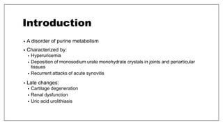
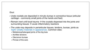
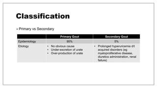
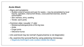
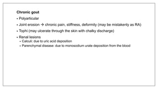
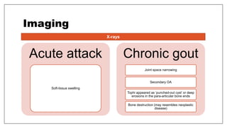
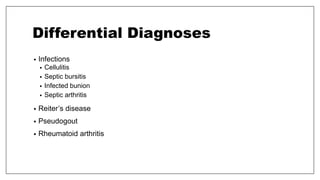
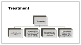
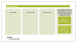
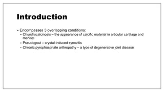
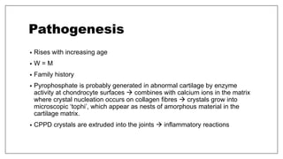
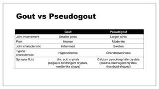
ORTHOPAEDICS NOTES
CRYSTAL DEPOSITION DISORDERS
Crystal deposition disorders around joints, bursae or tendons.
Outlines
• Gout
• Pseudogout (Calcium Pyrophosphate Dihydrate Arthropathy, CPPD)
• Calcium hydroxyapatite (HA) deposition disorder
GOUT
Introduction
• A disorder of purine metabolism
• Characterized by:
• Hyperuricemia
• Deposition of monosodium urate monohydrate crystals in joints and periarticular
tissues
• Recurrent attacks of acute synovitis
• Late changes:
• Cartilage degeneration
• Renal dysfunction
• Uric acid urolithiasis
Pathology
Hyperuricemia
• Serum urate concentration
which is significantly higher
than that of the population to
which they belong.
Gout
• Urate crystals are deposited in minute clumps in connective tissue (articular
cartilage – commonly small joints of the hands and feet)
• Remain inert until local trauma  the crystals dispersed into the joints and
surrounding tissues  acute inflammatory reactions
• The urate may deposits in periarticular tissues, tendons, bursae, joints as
‘tophi’ (chalky materials in appearance). Common sites:
• Metatarsophalangeal joints of the big toes
• Achilles tendons
• Olecranon bursae
• Pinnae of the ears
Classification
• Primary vs Secondary
Primary Gout Secondary Gout
Epidemiology 95% 5%
Etiology • No obvious cause
• Under-excretion of urate
• Over-production of urate
• Prolonged hyperuricemia d/t
acquired disorders (eg
myeloproliferative disease,
diuretics administration, renal
failure)
Clinical Features
Risk factors of hyperuricemia
Acute Attack
• Signs and symptoms:
• Sudden onset of severe joint pain for weeks – may be precipitated by local
trauma, operation, intercurrent illness, unaccustomed exercise, alcohol
consumption
• Skin redness, shiny, swelling
• Tender, warm joints
• Common sites: (usually >1 site)
• Metatarsophalangeal joints of the big toes
• Ankle
• Finger joints
• Olecranon bursa
• Uric acid level may be normal! (hyperuricemia is not diagnostic)
• So, examine the synovial fluid by using polarizing microscopy:
• Negatively birefringent urate crystals (needle-like shape)
Chronic gout
• Polyarticular
• Joint erosion  chronic pain, stiffness, deformity (may be mistakenly as RA)
• Tophi (may ulcerate through the skin with chalky discharge)
• Renal lesions
• Calculi: due to uric acid deposition
• Parenchymal disease: due to monosodium urate deposition from the blood
Imaging
Acute attack
Soft-tissue swelling
Chronic gout
Joint space narrowing
Secondary OA
Tophi appeared as ‘punched-out cyst’ or deep
erosions in the para-articular bone ends
Bone destruction (may resembles neoplastic
disease)
X-rays
Differential Diagnoses
• Infections
• Cellulitis
• Septic bursitis
• Infected bunion
• Septic arthritis
• Reiter’s disease
• Pseudogout
• Rheumatoid arthritis
Treatment
Acute attack
Joint resting, ice-
pack
NSAIDs or oral
corticosteroid in pt
who unable to
tolerate NSAIDs
Cholcicine – SE:
Nausea, vomiting,
diarrhoea
Corticosteroid
intra-articular
injection – for
tense joint effusion
Losing weight Cutting alcohols Eliminate diuretics
Urate-lowering drug therapy –
recurrent attacks, tophi, renal
complication, do not start
before acute attack symptoms
subside (may prolong or
precipitate acute attck)
Uricosuric drugs
(probenecid,
sulfinpyrazone) – if no
renal complication
Xanthine oxidase
inhibitor (allopurinol) –
preferred in pt with
renal complication +
chronic tophaceous
gout allopurinol
Interval therapy
Surgery
• In ulcerating tophi
PSEUDOGOUT
(Calcium Pyrophosphate Dihydrate Arthropathy, CPPD)
Introduction
• Encompasses 3 overlapping conditions:
• Chondrocalcinosis – the appearance of calcific material in articular cartilage and
menisci
• Pseudogout – crystal-induced synovitis
• Chronic pyrophosphate arthropathy – a type of degenerative joint disease
Pathogenesis
• Rises with increasing age
• W = M
• Family history
• Pyrophosphate is probably generated in abnormal cartilage by enzyme
activity at chondrocyte surfaces  combines with calcium ions in the matrix
where crystal nucleation occurs on collagen fibres  crystals grow into
microscopic ‘tophi’, which appear as nests of amorphous material in the
cartilage matrix.
• CPPD crystals are extruded into the joints  inflammatory reactions
Clinical Features
Asymptomatic chondrocalcinosis
• Elderly
• Calcification of the menisci, usually asymptomatic
• If seen <50y, suggest the possibility of underlying metabolic disease or familial disorder.
Acute synovitis (pseudogout)
• Middle-aged women
• Acute pain and swelling in large joints – usually the knee
• Sometimes attack is precipitated by minor illness or operation
• Tense, inflamed joint but not as acute as gout
• Lasts for few weeks, subsides spontaneously
• X-rays: Chondrocalcinosis
• Synovial fluid aspiration: positive biferingent crystals
Chronic pyrophosphate arthropathy
• Elderly women
• Polyarticular OA affecting large joints (hip, knees)
• Pain, stiffness, swelling, joint crepitus, loss of movement
• Often diagnosed as ‘generalized OA’
Imaging
X-rays
• Intra-articular & peri-articular calcification
• Knees, wrists, shoulders, hips, pubic symphysis, intervertebral discs
• Less common sites: joint synovium, capsules, ligaments, tendon, bursae
• Bilateral, symmetrical
• In articular cartilage: appeared as thin line parallel to the joint
• In fibrocartilagenous menisci and discs: appeared cloudy, irregular opacities
• Degenerative arthritis in distinctive sites
• Same as osteoarthritis BUT in distinctive uncommon sites – in non-weightbearing
joints
• Eg: isolated patellofemoral compartment, talonavicular joint
• Loose bodies might be seen as well
Treatment
Pseudogout
• Treatment is the same as acute gout – rest, high dose anti-inflammatory
therapy
• Joint aspiration & intra-articular steroid – in elderly to reduce the side effects
of NSAIDs
Chronic chondrocalcinosis
• Irreversible but few symptoms
• Treat as advanced OA
Gout vs Pseudogout
Gout Pseudogout
Joint involvement Smaller joints Larger joints
Pain Intense Moderate
Joint characteristic Inflammed Swollen
Typical
characteristic
Hyperuricemia Chondrocalcinosis
Synovial fluid Uric acid crystals
(negative birefringent crystals,
needle-like shape)
Calcium pyrophosphate crystals
(positive brefringent crystals,
rhomboid-shaped)

Orthopaedics notes - Crystal Deposition Disorder

  • 1.
    ORTHOPAEDICS NOTES CRYSTAL DEPOSITIONDISORDERS Crystal deposition disorders around joints, bursae or tendons.
  • 2.
    Outlines • Gout • Pseudogout(Calcium Pyrophosphate Dihydrate Arthropathy, CPPD) • Calcium hydroxyapatite (HA) deposition disorder
  • 3.
  • 4.
    Introduction • A disorderof purine metabolism • Characterized by: • Hyperuricemia • Deposition of monosodium urate monohydrate crystals in joints and periarticular tissues • Recurrent attacks of acute synovitis • Late changes: • Cartilage degeneration • Renal dysfunction • Uric acid urolithiasis
  • 5.
    Pathology Hyperuricemia • Serum urateconcentration which is significantly higher than that of the population to which they belong.
  • 6.
    Gout • Urate crystalsare deposited in minute clumps in connective tissue (articular cartilage – commonly small joints of the hands and feet) • Remain inert until local trauma  the crystals dispersed into the joints and surrounding tissues  acute inflammatory reactions • The urate may deposits in periarticular tissues, tendons, bursae, joints as ‘tophi’ (chalky materials in appearance). Common sites: • Metatarsophalangeal joints of the big toes • Achilles tendons • Olecranon bursae • Pinnae of the ears
  • 7.
    Classification • Primary vsSecondary Primary Gout Secondary Gout Epidemiology 95% 5% Etiology • No obvious cause • Under-excretion of urate • Over-production of urate • Prolonged hyperuricemia d/t acquired disorders (eg myeloproliferative disease, diuretics administration, renal failure)
  • 8.
  • 9.
    Acute Attack • Signsand symptoms: • Sudden onset of severe joint pain for weeks – may be precipitated by local trauma, operation, intercurrent illness, unaccustomed exercise, alcohol consumption • Skin redness, shiny, swelling • Tender, warm joints • Common sites: (usually >1 site) • Metatarsophalangeal joints of the big toes • Ankle • Finger joints • Olecranon bursa • Uric acid level may be normal! (hyperuricemia is not diagnostic) • So, examine the synovial fluid by using polarizing microscopy: • Negatively birefringent urate crystals (needle-like shape)
  • 10.
    Chronic gout • Polyarticular •Joint erosion  chronic pain, stiffness, deformity (may be mistakenly as RA) • Tophi (may ulcerate through the skin with chalky discharge) • Renal lesions • Calculi: due to uric acid deposition • Parenchymal disease: due to monosodium urate deposition from the blood
  • 11.
    Imaging Acute attack Soft-tissue swelling Chronicgout Joint space narrowing Secondary OA Tophi appeared as ‘punched-out cyst’ or deep erosions in the para-articular bone ends Bone destruction (may resembles neoplastic disease) X-rays
  • 12.
    Differential Diagnoses • Infections •Cellulitis • Septic bursitis • Infected bunion • Septic arthritis • Reiter’s disease • Pseudogout • Rheumatoid arthritis
  • 13.
    Treatment Acute attack Joint resting,ice- pack NSAIDs or oral corticosteroid in pt who unable to tolerate NSAIDs Cholcicine – SE: Nausea, vomiting, diarrhoea Corticosteroid intra-articular injection – for tense joint effusion
  • 14.
    Losing weight Cuttingalcohols Eliminate diuretics Urate-lowering drug therapy – recurrent attacks, tophi, renal complication, do not start before acute attack symptoms subside (may prolong or precipitate acute attck) Uricosuric drugs (probenecid, sulfinpyrazone) – if no renal complication Xanthine oxidase inhibitor (allopurinol) – preferred in pt with renal complication + chronic tophaceous gout allopurinol Interval therapy Surgery • In ulcerating tophi
  • 15.
  • 16.
    Introduction • Encompasses 3overlapping conditions: • Chondrocalcinosis – the appearance of calcific material in articular cartilage and menisci • Pseudogout – crystal-induced synovitis • Chronic pyrophosphate arthropathy – a type of degenerative joint disease
  • 17.
    Pathogenesis • Rises withincreasing age • W = M • Family history • Pyrophosphate is probably generated in abnormal cartilage by enzyme activity at chondrocyte surfaces  combines with calcium ions in the matrix where crystal nucleation occurs on collagen fibres  crystals grow into microscopic ‘tophi’, which appear as nests of amorphous material in the cartilage matrix. • CPPD crystals are extruded into the joints  inflammatory reactions
  • 18.
    Clinical Features Asymptomatic chondrocalcinosis •Elderly • Calcification of the menisci, usually asymptomatic • If seen <50y, suggest the possibility of underlying metabolic disease or familial disorder. Acute synovitis (pseudogout) • Middle-aged women • Acute pain and swelling in large joints – usually the knee • Sometimes attack is precipitated by minor illness or operation • Tense, inflamed joint but not as acute as gout • Lasts for few weeks, subsides spontaneously • X-rays: Chondrocalcinosis • Synovial fluid aspiration: positive biferingent crystals
  • 19.
    Chronic pyrophosphate arthropathy •Elderly women • Polyarticular OA affecting large joints (hip, knees) • Pain, stiffness, swelling, joint crepitus, loss of movement • Often diagnosed as ‘generalized OA’
  • 20.
    Imaging X-rays • Intra-articular &peri-articular calcification • Knees, wrists, shoulders, hips, pubic symphysis, intervertebral discs • Less common sites: joint synovium, capsules, ligaments, tendon, bursae • Bilateral, symmetrical • In articular cartilage: appeared as thin line parallel to the joint • In fibrocartilagenous menisci and discs: appeared cloudy, irregular opacities • Degenerative arthritis in distinctive sites • Same as osteoarthritis BUT in distinctive uncommon sites – in non-weightbearing joints • Eg: isolated patellofemoral compartment, talonavicular joint • Loose bodies might be seen as well
  • 21.
    Treatment Pseudogout • Treatment isthe same as acute gout – rest, high dose anti-inflammatory therapy • Joint aspiration & intra-articular steroid – in elderly to reduce the side effects of NSAIDs Chronic chondrocalcinosis • Irreversible but few symptoms • Treat as advanced OA
  • 22.
    Gout vs Pseudogout GoutPseudogout Joint involvement Smaller joints Larger joints Pain Intense Moderate Joint characteristic Inflammed Swollen Typical characteristic Hyperuricemia Chondrocalcinosis Synovial fluid Uric acid crystals (negative birefringent crystals, needle-like shape) Calcium pyrophosphate crystals (positive brefringent crystals, rhomboid-shaped)