Diabetic foot ulcers are a major complication of diabetes, affecting around 15% of people with the disease. They are caused by neuropathy, peripheral vascular disease, and foot deformities resulting from diabetes. Treatment involves wound debridement, managing any infection, revascularization if needed, and strict offloading of pressure on the affected foot to aid healing. Left untreated, diabetic foot ulcers can lead to amputation in around 50-70% of cases.

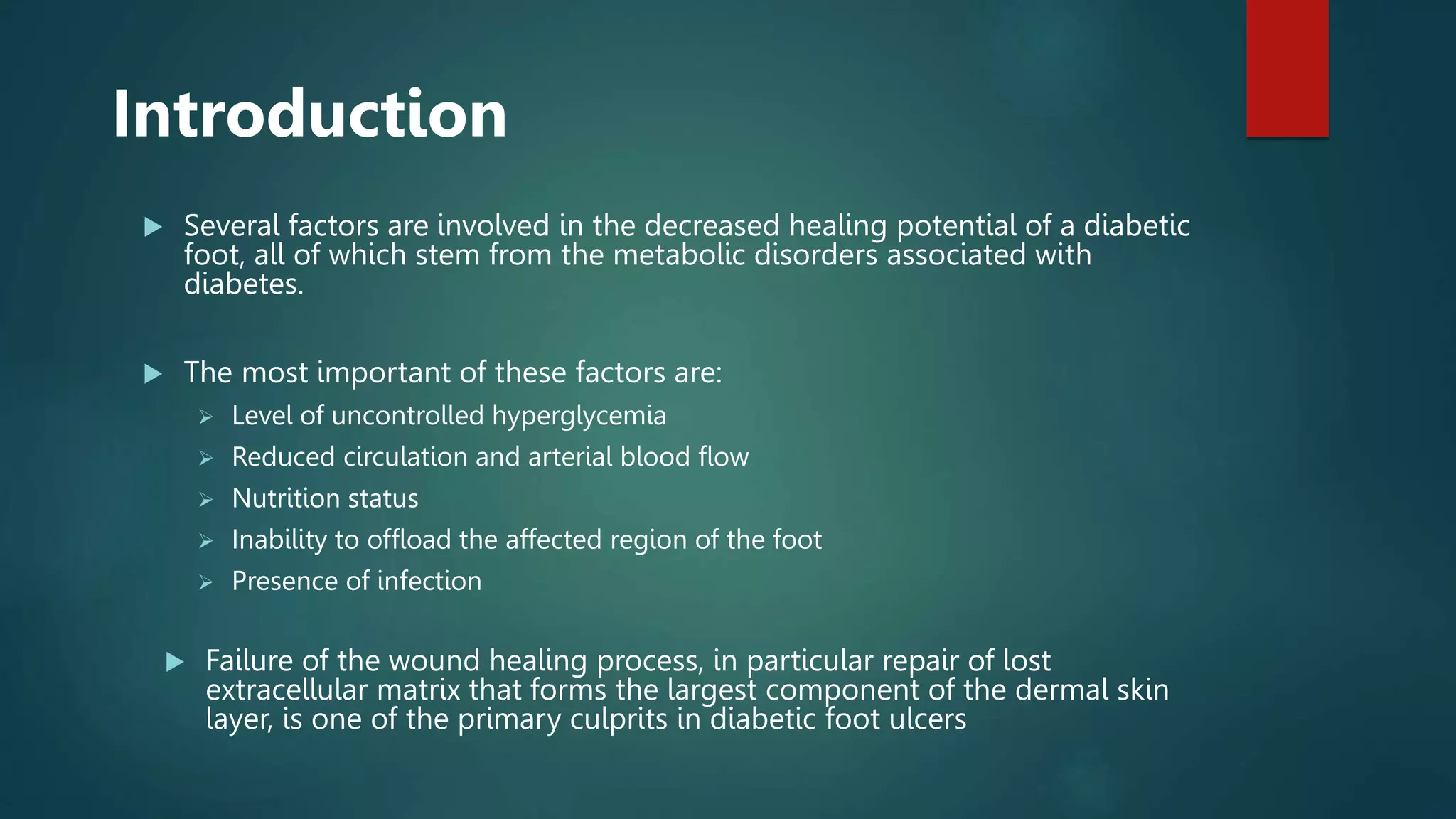
![Introduction
Diabetes mellitus (DM) is one of the main problems in health systems and a global public
health threat that has increased dramatically over the past 2 decades.
Foot ulcers are the most common medical complications of patients with diabetes,
estimated prevalence of 12-15% among all individuals with diabetes.[1]
It is estimated that approximately 20% of hospital admissions among patients with DM are
the result of DFU.
Ulcerations can have potential devastating complications as they cause lower extremity
amputations in patients with diabetes for about 50%-70%.
Furthermore, DFU is responsible for substantial emotional and physical distress as well as
productivity and financial losses that lower the quality of life.
(1) American Diabetes Association: Diabetes statistics. Available at: www.diabetes.org/diabetes-
statistics.jsp](https://image.slidesharecdn.com/diabeticfootulcer-221102034116-4142973a/75/Diabetic-Foot-Ulcer-pptx-2-2048.jpg)


![Microbiology
Most diabetic foot ulcers present as polymicrobial
infections.
The most common pathogens seen are aerobic
Gram-positive cocci, in particular Staphylococcus
aureus, and Gram-negative rods such as
Pseudomonas aeruginosa.[3]
Infection with anaerobic organisms such as
Clostridium perfringes may lead to foot ischemia or
gangrene.
Deep wound cultures and blood cultures are useful
to help direct antibiotic therapy and monitor the
presence of early sepsis.](https://image.slidesharecdn.com/diabeticfootulcer-221102034116-4142973a/75/Diabetic-Foot-Ulcer-pptx-11-2048.jpg)