Download to read offline

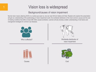


Vision impairment affects approximately 285 million people globally, with major causes including uncorrected refractive errors, cataracts, and age-related macular degeneration. The financial burden of vision impairments is significant, costing the U.S. around $139 billion in 2013 alone. The International Agency for the Prevention of Blindness has proposed a Global Action Plan to eliminate avoidable blindness and visual impairment.


