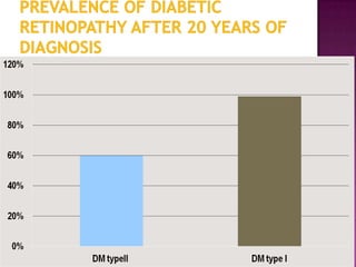
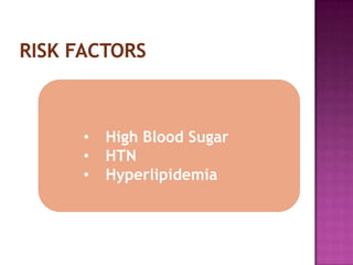
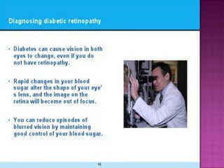
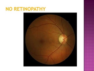
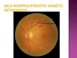
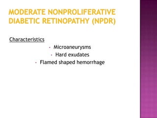
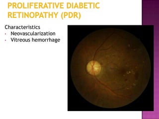
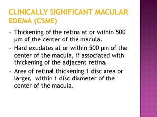
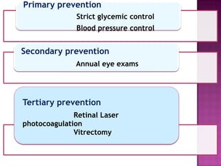
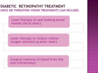
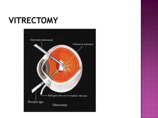
Syeda Baseerat
•   Recognize the importance of diabetic
    retinopathy as a public health problem
•   Discuss diabetic retinopathy as a leading cause
    of blindness in developed countries
•   Identify the risk factors for diabetic retinopathy
•   Describe and distinguish between the stages of
    diabetic retinopathy
Diabetes Mellitus is a group of diseases characterized by high blood
glucose levels. Diabetes results from defects in the body's ability to
produce and/or use insulin.
• Type 1 diabetes is usually diagnosed in children and young
   adults, and was previously known as juvenile diabetes. In type 1
   diabetes, the body does not produce insulin. 5% of people with
   diabetes have this form of the disease.
• In Type 2 diabetes, either the body does not produce enough
   insulin or the cells ignore the insulin. This is the most common
   form of diabetes.
 Blindness
 Kidney  failure
 Myocardial infarction
 Stroke
 Hypertension
 dyslipidemia
 Anyone  with diabetes is at risk of diabetic
  retinopathy.
 Approximately 45% of diabetic have diabetic
  retinopathy.
 The severity of hyperglycemia is the key
  alterable risk factor associated with the
  development of diabetic retinopathy
Definition:
Progressive dysfunction of the retinal blood vessels
          caused by chronic hyperglycemia.
•   DR can be a complication of diabetes type
    1 or diabetes type 2.
•   Initially, DR is asymptomatic, if not
    treated though it can cause low vision and
    blindness.
 Diabetic retinopathy is responsible for 1.8
  million of the 37 million cases of blindness
  throughout the world .
 Diabetic retinopathy (DR) is also the
  leading cause of blindness
•   After 20 years of diabetes, nearly 99% of
    patients with type 1 diabetes and 60% with
    type 2 have some degree on diabetic
    retinopathy
•   33% of patients with diabetes have signs of
    diabetic retinopathy
•   High Blood Sugar
•   HTN
•   Hyperlipidemia
   Having difficulty with fine details (e.g. when reading or
    watching television)
   Having difficulty with outdoor travel
   Experiencing visual fluctuations from hour to hour or day
    to day
   Seeing images as rippled (e.g. straight lines appear bent)
   Experiencing blurred, hazy or double vision
   Losing some visual field
   Having difficulty seeing at night or in low light
   Being particularly sensitive to glare and light
   Having difficulty focusing.
 1. Mild Nonproliferative Retinopathy
 2. Moderate Nonproliferative Retinopathy
  3. Severe Nonproliferative Retinopathy
       4. Proliferative Retinopathy
Characteristics
            • Microaneurysms only
 Thishappens when the tiny blood vessels of
 the retina begin to swell.
Microaneurysms
Characteristics
                  • Microaneurysms
                   • Hard exudates
           •   Flamed shaped hemorrhage
Damage to the blood vessels growth.

     The blood vessels become blocked
Microaneurysm


Hard exudates


Flamed shaped
hemorrhage
Hard exudates


microaneurysm
Any of the following:
•    More than 20 intraretinal hemorrhages in each of four
     quadrants
•    Definite venous beading in two or more quadrants
•    Prominent Intraretinal Microvascular Abnormalities
     (IRMA) in one or more quadrants
•    And no signs of proliferative retinopathy
 -Blood supply is blocked causing the eye to
 signal the need for new blood vessels.
IRMA
Venous beading
Venous beading
Characteristics
• Neovascularization
• Vitreous hemorrhage
   The retina sends signals so new blood vessels are created.
    These blood vessels are abnormal and more likely to
    hemorrhage due to the thin walls of the vessel. The walls
    of these vessels are weak and soon begin to hemorrhage
Cotton-wool
                        spot


Neovascularization

Neovascularization
                        Hard exudate
                     Blot hemorrhage
At risk for serious vision loss
 Any combination of three of the following four
 findings
• Presence of vitreous or preretinal hemorrhage.
• Presence of new vessels (neovascularization, NV)
• Location of NV on or near the optic disc.
• Moderate to severe extent of new vessels.
•   Diabetic macular edema is the leading
    cause of legal blindness in diabetics.
•   Diabetic macular edema can be present
    at any stage of the disease, but is more
    common in patients with proliferative
    diabetic retinopathy.
 The  macula is responsible for central vision.
 Diabetic macular edema may be
  asymptomatic at first. As the edema moves
  in to the fovea (the center of the macula)
  the patient will notice blurry central vision.
  The ability to read and recognize faces will
  be compromised.

                                      Macula
                                      Fovea
Normal   Macular Edema
•   Thickening of the retina at or within 500
    µm of the center of the macula.
•   Hard exudates at or within 500 µm of the
    center of the macula, if associated with
    thickening of the adjacent retina.
•   Area of retinal thickening 1 disc area or
    larger, within 1 disc diameter of the
    center of the macula.
The best measure for prevention
of loss of vision from diabetic
retinopathy is strict glycemic
control
90 percent of diabetic eye disease can
be prevented simply by proper regular
   examinations, treatment and by
       controlling blood sugar.
Primary prevention
               Strict glycemic control
               Blood pressure control


Secondary prevention
               Annual eye exams


Tertiary prevention
              Retinal Laser
photocoagulation
              Vitrectomy
 Laser photocoagulation is a therapy for
  numerous retinal diseases.
 Photocoagulation uses light to coagulate
  tissue. When energy from a strong light source is
  absorbed by tissue and is converted into thermal
  energy, coagulation necrosis occurs with
  denaturation of cellular proteins as temperature
  rises above 65 degrees C
Indications:
 Laser Photocoagulation is recommended for
 eyes with:
• Clinical significant macular edema (CSME)
• High risk Proliferative diabetic
   retinopathy
• Ocular tumors
Laser therapy to seal leaking blood
vessels (focal laser)


Laser therapy to reduce retinal
oxygen demand (scatter laser)


Surgical removal of blood from the
eye (vitrectomy)
Vitrectomy is surgery to remove some or all of
  the vitreous humor from the eye. Anterior
  vitrectomy entails removing small portions of
  the vitreous from the front structures of the eye
Pars plana vitrectomy is a general term for a
  group of operations accomplished in the deeper
  part of the eye, all of which involve removing
  some or all of the vitreous—the eye's clear
  internal jelly.
Diabetic Retinopathy is
preventable through strict
glycemic control and annual
dilated eye exams by an
ophthalmologist.
diabetic retinopathy

diabetic retinopathy

  • 1.
  • 2.
    Recognize the importance of diabetic retinopathy as a public health problem • Discuss diabetic retinopathy as a leading cause of blindness in developed countries • Identify the risk factors for diabetic retinopathy • Describe and distinguish between the stages of diabetic retinopathy
  • 3.
    Diabetes Mellitus isa group of diseases characterized by high blood glucose levels. Diabetes results from defects in the body's ability to produce and/or use insulin. • Type 1 diabetes is usually diagnosed in children and young adults, and was previously known as juvenile diabetes. In type 1 diabetes, the body does not produce insulin. 5% of people with diabetes have this form of the disease. • In Type 2 diabetes, either the body does not produce enough insulin or the cells ignore the insulin. This is the most common form of diabetes.
  • 4.
     Blindness  Kidney failure  Myocardial infarction  Stroke  Hypertension  dyslipidemia
  • 5.
     Anyone with diabetes is at risk of diabetic retinopathy.  Approximately 45% of diabetic have diabetic retinopathy.  The severity of hyperglycemia is the key alterable risk factor associated with the development of diabetic retinopathy
  • 7.
    Definition: Progressive dysfunction ofthe retinal blood vessels caused by chronic hyperglycemia.
  • 8.
    DR can be a complication of diabetes type 1 or diabetes type 2. • Initially, DR is asymptomatic, if not treated though it can cause low vision and blindness.
  • 9.
     Diabetic retinopathyis responsible for 1.8 million of the 37 million cases of blindness throughout the world .  Diabetic retinopathy (DR) is also the leading cause of blindness
  • 10.
    After 20 years of diabetes, nearly 99% of patients with type 1 diabetes and 60% with type 2 have some degree on diabetic retinopathy • 33% of patients with diabetes have signs of diabetic retinopathy
  • 13.
    High Blood Sugar • HTN • Hyperlipidemia
  • 21.
    Having difficulty with fine details (e.g. when reading or watching television)  Having difficulty with outdoor travel  Experiencing visual fluctuations from hour to hour or day to day  Seeing images as rippled (e.g. straight lines appear bent)  Experiencing blurred, hazy or double vision  Losing some visual field  Having difficulty seeing at night or in low light  Being particularly sensitive to glare and light  Having difficulty focusing.
  • 23.
     1. MildNonproliferative Retinopathy  2. Moderate Nonproliferative Retinopathy  3. Severe Nonproliferative Retinopathy  4. Proliferative Retinopathy
  • 24.
    Characteristics • Microaneurysms only
  • 25.
     Thishappens whenthe tiny blood vessels of the retina begin to swell.
  • 27.
  • 28.
    Characteristics • Microaneurysms • Hard exudates • Flamed shaped hemorrhage
  • 29.
    Damage to theblood vessels growth. The blood vessels become blocked
  • 30.
  • 31.
  • 32.
    Any of thefollowing: • More than 20 intraretinal hemorrhages in each of four quadrants • Definite venous beading in two or more quadrants • Prominent Intraretinal Microvascular Abnormalities (IRMA) in one or more quadrants • And no signs of proliferative retinopathy
  • 33.
     -Blood supplyis blocked causing the eye to signal the need for new blood vessels.
  • 34.
  • 35.
  • 36.
  • 37.
  • 38.
    The retina sends signals so new blood vessels are created. These blood vessels are abnormal and more likely to hemorrhage due to the thin walls of the vessel. The walls of these vessels are weak and soon begin to hemorrhage
  • 39.
    Cotton-wool spot Neovascularization Neovascularization Hard exudate Blot hemorrhage
  • 40.
    At risk forserious vision loss Any combination of three of the following four findings • Presence of vitreous or preretinal hemorrhage. • Presence of new vessels (neovascularization, NV) • Location of NV on or near the optic disc. • Moderate to severe extent of new vessels.
  • 41.
    Diabetic macular edema is the leading cause of legal blindness in diabetics. • Diabetic macular edema can be present at any stage of the disease, but is more common in patients with proliferative diabetic retinopathy.
  • 43.
     The macula is responsible for central vision.  Diabetic macular edema may be asymptomatic at first. As the edema moves in to the fovea (the center of the macula) the patient will notice blurry central vision. The ability to read and recognize faces will be compromised. Macula Fovea
  • 44.
    Normal Macular Edema
  • 45.
    Thickening of the retina at or within 500 µm of the center of the macula. • Hard exudates at or within 500 µm of the center of the macula, if associated with thickening of the adjacent retina. • Area of retinal thickening 1 disc area or larger, within 1 disc diameter of the center of the macula.
  • 47.
    The best measurefor prevention of loss of vision from diabetic retinopathy is strict glycemic control
  • 48.
    90 percent ofdiabetic eye disease can be prevented simply by proper regular examinations, treatment and by controlling blood sugar.
  • 49.
    Primary prevention Strict glycemic control Blood pressure control Secondary prevention Annual eye exams Tertiary prevention Retinal Laser photocoagulation Vitrectomy
  • 50.
     Laser photocoagulationis a therapy for numerous retinal diseases.  Photocoagulation uses light to coagulate tissue. When energy from a strong light source is absorbed by tissue and is converted into thermal energy, coagulation necrosis occurs with denaturation of cellular proteins as temperature rises above 65 degrees C
  • 51.
    Indications: Laser Photocoagulationis recommended for eyes with: • Clinical significant macular edema (CSME) • High risk Proliferative diabetic retinopathy • Ocular tumors
  • 53.
    Laser therapy toseal leaking blood vessels (focal laser) Laser therapy to reduce retinal oxygen demand (scatter laser) Surgical removal of blood from the eye (vitrectomy)
  • 54.
    Vitrectomy is surgeryto remove some or all of the vitreous humor from the eye. Anterior vitrectomy entails removing small portions of the vitreous from the front structures of the eye Pars plana vitrectomy is a general term for a group of operations accomplished in the deeper part of the eye, all of which involve removing some or all of the vitreous—the eye's clear internal jelly.
  • 56.
    Diabetic Retinopathy is preventablethrough strict glycemic control and annual dilated eye exams by an ophthalmologist.

Editor's Notes

  • #4 http://www.diabetes.org/diabetes-basics/?loc=GlobalNavDB
  • #9 http://www.mdconsult.com/das/book/pdf/282715756-3/978-0-323-04332-8/4-u1.0-B978-0-323-04332-8..00092-5..DOCPDF.pdf?isbn=978-0-323-04332-8&eid=4-u1.0-B978-0-323-04332-8..00092-5..DOCPDF
  • #11 Ophthalmology Myron Handoff MD and Jay S. Duke Basic and Clinical Science Course, Section 12: Retina and Vitreous AAOhttp://www.aao.org/eyecare/news/upload/Eye-Health-Fact-Sheet.pdf -
  • #49 http://www.aao.org/newsroom/release/20091030.cfm