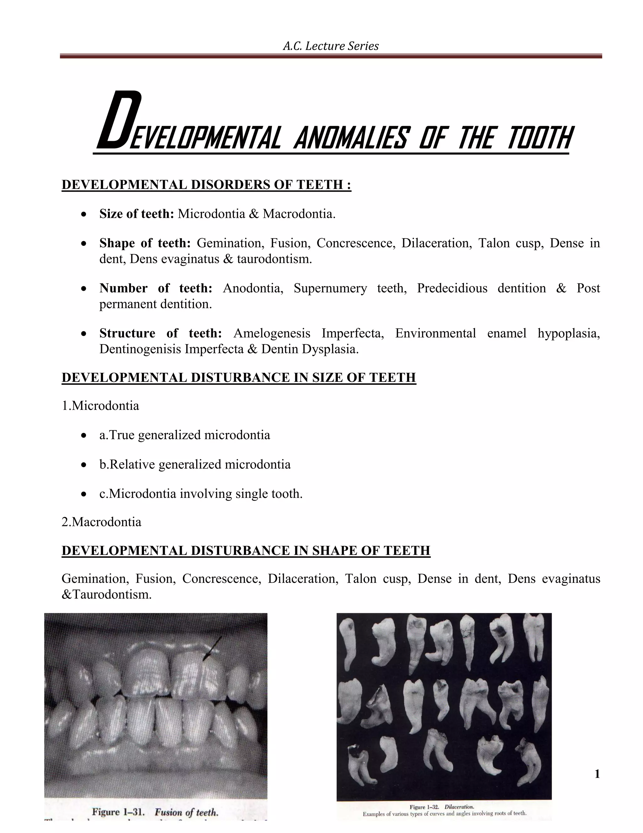
The document discusses various developmental anomalies of teeth, including disturbances in size, shape, number, and structure. It covers conditions like microdontia, macrodontia, gemination, fusion, anodontia, amelogenesis imperfecta, enamel hypoplasia, dentinogenesis imperfecta, and dentin dysplasia. It also discusses agents that can affect tooth development, such as vitamin deficiencies, tetracycline, and fluoride, and how they may incorporate into developing teeth and influence mineralization. Clinical applications regarding how various disturbances can result from disruptions at different stages of tooth development are also mentioned.
This presentation specifically deals with the maxillary and mandibular Major connectors used in a cast partial denture. it also mentions the uses, advantages and disadvantages of each,
In this lecture I explain in step-by-step fashion the basics of Endodontic Diagnosis: Pulp Vitality Tests. a photo guide is attached to the guide to aid in better understanding of the topic
Dentin dysplasia (DD) is a rare hereditary disturbance is inherited as an autosomal dominant trait.
unknown etiology that affects approximately 1 :100,000.
In 1972, Witkop classified it into type I and type II which affect both dentitions.DD Type I
Radicular dentin dysplasia
Characterized by:-
1.Both dentitions are affected.
2.Normal appearing crowns
3.No or only rudimentary root development (rootless teeth)
4.Incomplete or total obliteration of the pulp chamber.
5.Teeth may exhibit extreme mobility and exfoliate prematurely.DD type II
coronal dentin dysplasia
Characterized by:-
1.partial pulpal obliteration.
2.Thistle-tube-or flame-shaped coronal pulp chambers
3. Thread-like root canals
4. Usually the absence of periapical radiolucencies.
5. In this type of anomaly, teeth roots are of normal shape and contour.The enamel and the immediately subjacent dentin appear normal.
Deeper layers of dentin show an atypical tubular pattern with an amorphous, atubular area, and irregular organization.
Normal dentinal tubule formation appears to have been blocked so that new dentine forms around obstacles and takes on the characteristic appearances described as “lava flowing around boulders”The radiograph revealed features of dentine dysplasia type I with normal appearance of crown but no root development Autosomal Dominant Disorder:
Manifested in heterozygous states
At least one parent of index case is usually affected
Both males and females are affected.
Clinical feature can be modified by variation in penetrance and expressivity. Some individual inherit the mutant gene but are phenotpically normal. This is reffered to as “incomplete penetrance”.
In many condition the age of onset is delayed.
Inheritance Pattern:
Typical pattern is a heterozygous affected parent with a homozygous unaffected parent.
Every child has one chance in two of having the disease
Both sexes are affected equally..Autosomal Recessive Disorder
Largest category of Mendelian disorder
Usually does not affect the parent of the affected individual, but sibling may show the disease.
Complete penetrance is common.
Onset is frequently early in life.
Usually affect enzymatic proteins.
Pattern Of Inheritance:
Typical pattern is two heterozygous unaffected (carrier) parent.
The triat does not usually affect the parent, but siblings may show the disease
Siblings have one chance in four of being affected
Both sexes affected equally.
this contains the steps for the class 1 cavity preparation for amalgam in detail. also contains the difference between composite and amalgam cavity preparation.
etiology of malocclusion for general practitioners.docxDr.Mohammed Alruby
Etiology of Malocclusion
For general practitioners
Prepared by
Dr. M Alruby
Etiology in orthodontics is the study of actual causes of dento – facial abnormalities.
Malocclusion is the condition where there is a deviation from the usual or accepted relationship, dental malocclusion exists when the individual teeth within one or both jaws are abnormally related to each other, this condition may be limited to a couple of teeth or involving the majority of teeth present.
Development of normal dentition and occlusion depends on a number of interrelated factors that include the dento alveolar, skeletal and the neuromuscular factors. Thus localization of the possible etiology may be a very difficult task.
A- Extrinsic factors:
1- Evolution:
With evolution, the jaws become smaller, reduction in the number and size of teeth and diminution of jaw projection together with increased in vertical height of the face and there is retrognathic tendency in man as he ascends the evolutionary scale.
2- Heredity:
Transmission of dento facial characteristic through generation by genes. The child is a product of parents who have dissimilar genetic material. Thus the child may inherit conflicting traits from both the parents resulting in abnormalities of the dentofacial region. Another reason attributed for genetically determined malocclusion is the racial, ethnic and regional intermixer, which might have led to uncoordinated inheritance of teeth and jaws.
There are three types of transmission of malocclusion from the stand point of genetics:
1- Repetitive: the recurrence of single dentofacial deviation within the immediate family.
2- Discontinuous: a tendency for a malocclusion trait to reappear within the family over several generations.
3- Variable: the occurrence of different but related types of malocclusion within several generation of the same family.
Dental defect of genetic origin include the following:
= Crowding and spacing of teeth.
= Size and characteristic of soft tissue including muscles and frenum.
= Macrognathia and micrognathia.
= Macrodontia and microdontia.
= Oligodontia.
= Tooth shape variations.
= Median diastemas.
= upper face height, nose height, and bigonial width.
= Bimaxillary protrusion.
4- Congenital:
Those are deformities of hereditary or non-hereditary origin but exciting at birth.
The congenital abnormalities that cause malocclusion:
= Cleft lip and palate:
lack of fusion between the two palatal processes to each other. From one third to one half of all cleft palate children have familial history of this deformity.
As with the non-cleft child, palatal, pharyngeal and perioral musculature is well developed at birth to meet the demand of suckling, deglutition and mastication. While the complete unilateral or complete bilateral cleft break the continuity of the upper lip and disturbs the functional pattern and significantly reduce the restraining effect of the buccinators mechanism that pro
I am Dr. Karrar Ghaffar. I am a dentist. Dental stain is a problem for alot of people so in this presentation I explained the main categories and main causes
Primary etiologic sites:
1- Neuromuscular system:
The muscle group that serve most frequently as primary etiologic sites are:
== muscles of mastication
== muscles of facial expression
== tongue
The neuromuscular system plays its primary role in the etiology of dentofacial deformity by the effect of abnormal contraction of bony skeleton and the dentition. Both bones and teeth are affected by the many functional activities of orofacial region
2- Bone:
Since the bone pf maxilla and mandible serve as bases of dental arches, changes in dental arches growth may alter the occlusal and functional relationship.
3- Teeth;
The teeth may be primary sites in the etiology of dentofacial deformity in many ways
Gross variation in size and shape are encountered frequently and always are of concern
Decrease or increase in the regular number of teeth will give rise malocclusion
Etiologic factors:
A- Extrinsic factors:
1- Evolution:
With evolution, the jaws become smaller, reduction in number and size of teeth and diminution of jaw projections together with increased in vertical height of the face and there is a retrognathic tendency in mans as he ascends the evolutionary scale
2- Heredity:
Transmission of dentofacial characteristics through generations by genes. Most authors between 1900-- 1920 did not completely determine the role of inheritance in determination of the form, size and proportion of dentofacial skeleton, but they stress their work upon the effect of the environmental factors, and at this time they were hardly belief that the effect of local lack of function is more important.
Bennet statement: the size, form and density of bones such as maxilla and mandible varies according to the extent to which these structure are used during period of growth – (function stimulate growth)
Walk Joff statement: the form and degree of development of maxilla and mandible depends upon the magnitude of functional stimuli of muscles acting upon these structures.
Baker: his study was performed on animals by unilateral amputation of muscles of mastication, he found lack of growth on the affected side.
Brash: studied the facial form and the dental development in twins on genetic bases, he also emphasized the genetic facial pattern of some royal families in Europe where they had been inter-marriage, his studies gave the best evidence to support the role of inheritance
Axel Lundstorm:1925 showed that, the form and size of dental bases and the teeth are genetically determined, when the size of the teeth and their basal arches are not correlated, problems of crowding or spacing will be arising.
Broadbent and Hofrath 1931: developed standardized cephalometric x-ray technique which permit serial longitudinal studies of facial growth, by this studies the concept of inheritance growth pattern arises
There are three types of transmission of malocclusion from the standpoint of genetics:
a- Repetitive: the recurrence of single dentofacial deviation within the immediate famil
The wasting diseases of teeth, namely attrition, abrasion and dental erosion have taken their toll in the population around the world due to the changing lifestyles, increase in the stress levels and many others factors that were persistent earlier but have suddenly increased drastically. This presentation brings to light the new factors that have attributed to this condition as well as discusses the previous ones.
Anomalies of tooth formation and eruption, MISSING TEETH, EXTRA TEETH, ABNORMALITY OF TOOTH SIZE, Crown size, Root size, Abnormality of crown form, ABNORMALITY OF ROOT FORM, All tissues, Cementum defects, Dentine defects, enamel defects, DISTURBANCES OF ERUPTION & EXFOLIATION,
The Indian Dental Academy is the Leader in continuing dental education , training dentists in all aspects of dentistry and
offering a wide range of dental certified courses in different formats.for more details please visit
www.indiandentalacademy.com
Similar to Developmental anomalies of the tooth (20)
Read| The latest issue of The Challenger is here! We are thrilled to announce that our school paper has qualified for the NATIONAL SCHOOLS PRESS CONFERENCE (NSPC) 2024. Thank you for your unwavering support and trust. Dive into the stories that made us stand out!
Model Attribute Check Company Auto PropertyCeline George
In Odoo, the multi-company feature allows you to manage multiple companies within a single Odoo database instance. Each company can have its own configurations while still sharing common resources such as products, customers, and suppliers.
Palestine last event orientationfvgnh .pptxRaedMohamed3
An EFL lesson about the current events in Palestine. It is intended to be for intermediate students who wish to increase their listening skills through a short lesson in power point.
Synthetic Fiber Construction in lab .pptxPavel ( NSTU)
Synthetic fiber production is a fascinating and complex field that blends chemistry, engineering, and environmental science. By understanding these aspects, students can gain a comprehensive view of synthetic fiber production, its impact on society and the environment, and the potential for future innovations. Synthetic fibers play a crucial role in modern society, impacting various aspects of daily life, industry, and the environment. ynthetic fibers are integral to modern life, offering a range of benefits from cost-effectiveness and versatility to innovative applications and performance characteristics. While they pose environmental challenges, ongoing research and development aim to create more sustainable and eco-friendly alternatives. Understanding the importance of synthetic fibers helps in appreciating their role in the economy, industry, and daily life, while also emphasizing the need for sustainable practices and innovation.
Operation “Blue Star” is the only event in the history of Independent India where the state went into war with its own people. Even after about 40 years it is not clear if it was culmination of states anger over people of the region, a political game of power or start of dictatorial chapter in the democratic setup.
The people of Punjab felt alienated from main stream due to denial of their just demands during a long democratic struggle since independence. As it happen all over the word, it led to militant struggle with great loss of lives of military, police and civilian personnel. Killing of Indira Gandhi and massacre of innocent Sikhs in Delhi and other India cities was also associated with this movement.
Embracing GenAI - A Strategic ImperativePeter Windle
Artificial Intelligence (AI) technologies such as Generative AI, Image Generators and Large Language Models have had a dramatic impact on teaching, learning and assessment over the past 18 months. The most immediate threat AI posed was to Academic Integrity with Higher Education Institutes (HEIs) focusing their efforts on combating the use of GenAI in assessment. Guidelines were developed for staff and students, policies put in place too. Innovative educators have forged paths in the use of Generative AI for teaching, learning and assessments leading to pockets of transformation springing up across HEIs, often with little or no top-down guidance, support or direction.
This Gasta posits a strategic approach to integrating AI into HEIs to prepare staff, students and the curriculum for an evolving world and workplace. We will highlight the advantages of working with these technologies beyond the realm of teaching, learning and assessment by considering prompt engineering skills, industry impact, curriculum changes, and the need for staff upskilling. In contrast, not engaging strategically with Generative AI poses risks, including falling behind peers, missed opportunities and failing to ensure our graduates remain employable. The rapid evolution of AI technologies necessitates a proactive and strategic approach if we are to remain relevant.
Unit 8 - Information and Communication Technology (Paper I).pdfThiyagu K
This slides describes the basic concepts of ICT, basics of Email, Emerging Technology and Digital Initiatives in Education. This presentations aligns with the UGC Paper I syllabus.
Biological screening of herbal drugs: Introduction and Need for
Phyto-Pharmacological Screening, New Strategies for evaluating
Natural Products, In vitro evaluation techniques for Antioxidants, Antimicrobial and Anticancer drugs. In vivo evaluation techniques
for Anti-inflammatory, Antiulcer, Anticancer, Wound healing, Antidiabetic, Hepatoprotective, Cardio protective, Diuretics and
Antifertility, Toxicity studies as per OECD guidelines
Welcome to TechSoup New Member Orientation and Q&A (May 2024).pdfTechSoup
In this webinar you will learn how your organization can access TechSoup's wide variety of product discount and donation programs. From hardware to software, we'll give you a tour of the tools available to help your nonprofit with productivity, collaboration, financial management, donor tracking, security, and more.
The French Revolution, which began in 1789, was a period of radical social and political upheaval in France. It marked the decline of absolute monarchies, the rise of secular and democratic republics, and the eventual rise of Napoleon Bonaparte. This revolutionary period is crucial in understanding the transition from feudalism to modernity in Europe.
For more information, visit-www.vavaclasses.com
The French Revolution Class 9 Study Material pdf free download
Developmental anomalies of the tooth
1. A.C. Lecture Series
D EVELOPMENTAL ANOMALIES OF THE TOOTH
DEVELOPMENTAL DISORDERS OF TEETH :
Size of teeth: Microdontia & Macrodontia.
Shape of teeth: Gemination, Fusion, Concrescence, Dilaceration, Talon cusp, Dense in
dent, Dens evaginatus & taurodontism.
Number of teeth: Anodontia, Supernumery teeth, Predecidious dentition & Post
permanent dentition.
Structure of teeth: Amelogenesis Imperfecta, Environmental enamel hypoplasia,
Dentinogenisis Imperfecta & Dentin Dysplasia.
DEVELOPMENTAL DISTURBANCE IN SIZE OF TEETH
1.Microdontia
a.True generalized microdontia
b.Relative generalized microdontia
c.Microdontia involving single tooth.
2.Macrodontia
DEVELOPMENTAL DISTURBANCE IN SHAPE OF TEETH
Gemination, Fusion, Concrescence, Dilaceration, Talon cusp, Dense in dent, Dens evaginatus
&Taurodontism.
1
2. A.C. Lecture Series
DEVELOPMENTAL DISTURBANCE IN SHAPE OF TEETH
Anodontia, Supernumery teeth, Predecidious dentition & Post permanent dentition.
2
3. A.C. Lecture Series
DEVELOPMENTAL DISTURBANCE IN STRUCTURE OF TEETH
Amelogenesis Imperfecta
It is an hereditary defects of enamel. It is ectodermal disturbance, since the mesodermal
components of the teeth are normal.
Development of enamel occurs in three stages : a. Formative stages, b. Calcification
stage, c. Maturation stage.
Accordingly 3 basic types of Amelogenesis Imperfecta are recognized :
a. Hypoplastic type, b. Hypocalcification type, c. Hypomaturation type. (C/F)
3
4. A.C. Lecture Series
2. Environmental Enamel Hypoplasia :
“May be defined as an in complete or defective formation of organic enamel matrix of teeth”.
To basic types of Enamel Hypoplasia exist
a. Hereditary type,
b. Those caused by environmental factor.
In hereditary type both decidious & permanent dentitions are usually involved & generally
only the enamel is affected. In contrast when the defect is caused by environmental factor,
either dentition may be involved & some times only a single tooth. Both enamel & dentin
are affected.
Environmental factor which are responsible for causing enamel hypoplasia are.
1.Nutritional deficiency:Vit-A,C & D.
2.Exanthematous disease: measles, chicken pox, scarlet fever.
3.Congenital syphilis
4.Hypocalcemia
5.Birth injury, Rh incompatibility
6.Local infection or trauma
4
5. A.C. Lecture Series
7.Fluorides
Enamel Hypoplasia due to Fluoride
(Mottled Enamel):
It is caused by ingestion of fluoride containing drinking water during the time of tooth
formation. Severity of mottling increases with increasing amount of fluoride in water.
There is little mottling of any significance at a level below 0.9 to 1 ppm.
This type of hypoplasia is due to a disturbance of the ameloblast during the formative stage
of tooth development.
Depending upon the level of fluoride in the water supply. There is a wide range of severity
in the appearance of mottled teeth varying from
White flecking or spotting of enamel.
Mild changes manifested by white opaque areas involving more of the tooth surface area.
Moderate & sever changes showing pitting & brownish staining of the surface.
Corrode appearance of the teeth.
5
6. A.C. Lecture Series
3. Dentinogenisis Imperfecta :expired
Type I: always occurs in families with osteogenesis Imperfecta.
Type II: never occurs in families with osteogenesis imperfecta. Most frequently referred as
Hereditary opalesent dentin. It is most common dominant inherited disorder in human.
Type III: Is Brandy wine type, characterized bymultiple pulp exposure in decidious teeth.
Decidious teeth are more severely affected than the permanent teeth in type I. In type II &
III both dentition are affected.
Colour of teeth may range from brownish yellow & exhibit a characteristic translucent or
opalescent hue.
R/F most striking feature is the partial or total obliteration of the pulp chamber & root
canals by continued formation of dentin.
6
7. A.C. Lecture Series
The affected patients in type III DI had features characterised as “Shell Teeth.” enamel of
the tooth appears normal, while dentin is extremely thin, & the pulp chamber are
enormous.
4. Dentin Dysplasia: Characterized by normal enamel but atypical dentin formation with
abnormal pulp morphology. (hereditary)
Type I: Radicular dentin dysplasia: Both dentition are affected. Teeth exhibit extreme mobility
& are commonly exfoliated prematurely or after minor trauma as a result of short roots.
Type II: Coronal dentin dysplasia: Abnormal large pulp chamber in coronal portion often
described as Thistle tube
AGENTS AFFECTING TOOTH DEVELOPMENT
Introduction:
Certain vitamin & hormone deficiencies if present during tooth formation will adversely
affect formative cells & matrix that they produce.
Reduced organic matrix content results in production of hypoplastic tissue.
Excessive levels of tetracycline or fluoride may become incorporated into mineralizing
teeth & interfere with the mineralization process.
7
8. A.C. Lecture Series
The extent of defect depend on the nature of the substance, the degree of excess or
deficiency and the developmental time.
Vitamin-A, C, D, Parathyroid hormone, Tetracycline & Fluoride are discussed in terms of
their relation to matrix development.
8
10. A.C. Lecture Series
TETRACYCLINE & FLUORIDE:
If available during the mineralization phase, may be incorporated in dentin, enamel,
cementum & bone.
Fluoride is a binary compound of fluorine useful as an anticaries substance. Tetracycline
on the other hand is used as an antibacterial agent. Both are deposited along with minerals
in developing hard tissue.
On prolonged exposure to light, tetracycline stained dental tissue will change in colour to
A brown to gray. Other effects of tetracycline is hypoplasia or absence of enamel. Staining
is most notable in dentin, especially in the first formed dentin at the DEJ. Staining is more
notable under UV light.
Amount of damage is directly related to the magnitude & duration of the dosage.
Mechanism: It is believed that a chelate of calcium & tetracycline forms. At higher
concentration in both ameloblast & odontoblast protein synthesis is impaired. This in turn
will result in hypoplasia of the enamel & dentin matrix.
10
12. A.C. Lecture Series
Tetracycline & to limited extent Sodium Fluoride cross the placental barrier.
If pregnant female consumes fluoridated water during mineralization of the fetal teeth.
Such teeth exhibit higher resistance to dental caries.
If on the other hand tetracycline antibiotics are administered to the mother during the
period of tooth mineralization, the decidious teeth may later be stained. Tetracycline
staining of teeth is permanent, but staining of bone is not permanent as bone is remodeled
continuously.
Cervical staining is more characteristic of tetracycline because this agent deposits primarily
in the dentin.
Fluoride is most beneficial to the teeth in concentration of approximately 0.5 to 1 ppm of
water. Higher concentration such as 5 ppm causes mottling & hypoplasia of the enamel &
hypo mineralized dentin.
Less amount of fluoride is being found in primary teeth than in permanent teeth because
the maturative stage last for 1-2 years in primary teeth & 4-5 years in permanent teeth.
Tetracycline has been widely used to visually record growth in experimental animal. Daily
deposition of dentin can thus be recorded by measuring the width of dentin between each
fluorescent line.
Tetracycline may be used to evaluate tooth movement by reveling bone & dentin
formation.
12
13. A.C. Lecture Series
CLINICAL APPLICATION :
The size of the tooth is depends upon both proliferative & secretory activities of the cell.
Macrodontia, Microdontia are the result of factors affecting the growth of the tooth germ at
the cap & bell stages. True macro & microdontia may be the result of over & under
secretion of pituitary hormones.
Absence of teeth, partial or total anodontia is due to factors that disrupt tooth development
at the initiation stage. In genetic disorder ectodermal dysplasia there is total anodontia.
Distal proliferation of dental lamina is responsible for the location of the germs of the
permanent molars in the ramus of the mandible & tuberosity of maxilla.
If the cells of the epithelial RS remain adherent to the dentin surface, they may differentiate
into fully functioning ameloblast & produce enamel called enamel pearls, found in the
areas of furcation of the roots of PM.
If continuity of Hertwig's RS is broken or is not established prior to dentin formation, a
defect in the dentinal wall of the pulp ensures.this accounts for development of accessory
root canals opening.
If continuity of Hertwig's RS is not broken even after dentin formation it results in root
dentin exposure to oral cavity.
13
14. A.C. Lecture Series
Developmental disorders of teeth causes clinical problems related to appearance, spacing,
crowding, & periodontal treatment.
In intercuspal areas, during enamel secretion, the ameloblasts may become strangulated
when their bases begin to touch one another. These areas becomes pits & fissures in
erupted tooth. They are extremely difficult to clean.
Clinically,vit-C deficiency is manifested orally by gingival bleeding & loosening the teeth.
Weakness, anemia, bone loss, & susceptibility to hemorrhage.
Osteoporosis results when calcium loss because of resorption is greater than calcium
deposition. This may be evident orally with loss of alveolar bone & loosening of teeth.
Certain antibiotics, like the tetracyclines, have an affinity for calcified tissues. They may
become incorporated within the mineral phase during maturation, & cause discoloration of
the enamel & underlying dentin.
Brown staining or defect in the enamel of the incisal third of crowns indicates the presence
of toxic substance in the body at the time of initial mineralization of the teeth. Staining in
the cervical area relates to induction of a toxic substance at a time of final crown
mineralization.
The cervical region of the crown and the central grooves are the last zones to mineralize.
Ameloblasts in these regions may lose functional ability before mineralization is complete.
Lack of complete mineralization of the central grooves or cervical areas is believed to be a
reason for caries in these areas.
14