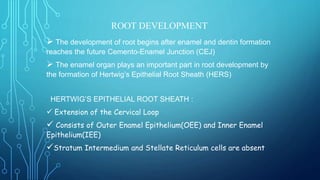
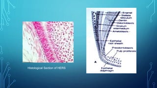
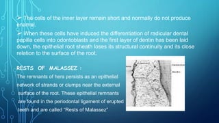
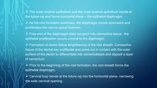
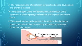
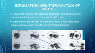
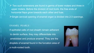
SEMINAR PRESENTATION
SUBJECT – DENTAL ANATOMY AND HISTOLOGY
TOPIC : FORMATION OF ROOT
PRESENTED BY : ANSHUMAN BAG
B.D.S 1ST YEAR
CONTENTS TO BE DISCUSSED :
 Root formation as a process seen in the Advanced Bell Stage
 Hertwig’s Epithelial Root Sheath (HERS)
 Rests of Malassez
 Bifurcation and Trifurcation of Roots
 Enamel Pearls
ROOT DEVELOPMENT
 The development of root begins after enamel and dentin formation
reaches the future Cemento-Enamel Junction (CEJ)
 The enamel organ plays an important part in root development by
the formation of Hertwig’s Epithelial Root Sheath (HERS)
HERTWIG’S EPITHELIAL ROOT SHEATH :
 Extension of the Cervical Loop
 Consists of Outer Enamel Epithelium(OEE) and Inner Enamel
Epithelium(IEE)
Stratum Intermedium and Stellate Reticulum cells are absent
Histological Section of HERS
 The cells of the inner layer remain short and normally do not produce
enamel.
 When these cells have induced the differentiation of radicular dental
papilla cells into odontoblasts and the first layer of dentin has been laid
down, the epithelial root sheath loses its structural continuity and its close
relation to the surface of the root.
RESTS OF MALASSEZ :
The remnants of hers persists as an epithelial
network of strands or clumps near the external
surface of the root. These epithelial remnants
are found in the periodontal ligament of erupted
teeth and are called “Rests of Malassez”
 The outer enamel epithelium and the inner enamel epithelium bends at
the future cej and forms horizontal plane – the epithelial diaphragm.
 As the root formation continues, the diaphragm moves downward and
proliferates the narrow apical foramen.
 Free end of the diaphragm does not grow into connective tissue , the
epithelial proliferation occurs coronal to the diaphragm.
 Formation of dentin follow lenghthening of the root sheath. Connective
tissue of the dental sac proliferate and come out in contact with the outer
surface of the dentin to differentiate into cementoblasts and deposit a layer
of cementum.
 Prior to the beginning of the root formation, the root sheath forms the
epithelial diaphragm.
 Cervical loop bends at the future cej into the horizontal plane, narrowing
the wide cervical opening.
 The horizontal plane of diaphragm remains fixed during development
and growth of the root.
 In the last stages of the root development, proliferation of the
epithelium in diaphragm lags behind than that of pulpal connective
tissue.
Wide apical foramen reduces first to the width of the diaphragm
opening and later further narrowed by apposition of dentin and
cementum at root apex.
BIFURCATION AND TRIFURCATION OF
ROOTS
 Differential growth of the epithelial diaphragm in multi-rooted teth
causes the division of the root trunk into 2-3 roots.
 Expansion of the cervical loop occurs in such a way that a long
tongue with extension develop in horizontal diaphragm.
 Two such extensions are found in germs of lower molars and three in
upper molars. Before the division of root trunk, the free ends of
horizontal flaps grow towards each other and fuse.
Single cervical opening of enamel organ is divided into 2-3 openings.
ENAMEL PEARLS :
If epithelial cells of root sheath remain adherent
to dentin surface, they may differentiate into
ameloblast and produce enamel. They are the
droplets of enamel found in the furcation area of
a multi-rooted tooth.
REFERENCES
 ORBAN’S ORAL HISTOLOGY AND EMBRYOLOGY
 PICTURES FROM GOOGLE IMAGES
THANK YOU

Development of Root

  • 1.
    SEMINAR PRESENTATION SUBJECT –DENTAL ANATOMY AND HISTOLOGY TOPIC : FORMATION OF ROOT PRESENTED BY : ANSHUMAN BAG B.D.S 1ST YEAR
  • 2.
    CONTENTS TO BEDISCUSSED :  Root formation as a process seen in the Advanced Bell Stage  Hertwig’s Epithelial Root Sheath (HERS)  Rests of Malassez  Bifurcation and Trifurcation of Roots  Enamel Pearls
  • 3.
    ROOT DEVELOPMENT  Thedevelopment of root begins after enamel and dentin formation reaches the future Cemento-Enamel Junction (CEJ)  The enamel organ plays an important part in root development by the formation of Hertwig’s Epithelial Root Sheath (HERS) HERTWIG’S EPITHELIAL ROOT SHEATH :  Extension of the Cervical Loop  Consists of Outer Enamel Epithelium(OEE) and Inner Enamel Epithelium(IEE) Stratum Intermedium and Stellate Reticulum cells are absent
  • 4.
  • 5.
     The cellsof the inner layer remain short and normally do not produce enamel.  When these cells have induced the differentiation of radicular dental papilla cells into odontoblasts and the first layer of dentin has been laid down, the epithelial root sheath loses its structural continuity and its close relation to the surface of the root. RESTS OF MALASSEZ : The remnants of hers persists as an epithelial network of strands or clumps near the external surface of the root. These epithelial remnants are found in the periodontal ligament of erupted teeth and are called “Rests of Malassez”
  • 6.
     The outerenamel epithelium and the inner enamel epithelium bends at the future cej and forms horizontal plane – the epithelial diaphragm.  As the root formation continues, the diaphragm moves downward and proliferates the narrow apical foramen.  Free end of the diaphragm does not grow into connective tissue , the epithelial proliferation occurs coronal to the diaphragm.  Formation of dentin follow lenghthening of the root sheath. Connective tissue of the dental sac proliferate and come out in contact with the outer surface of the dentin to differentiate into cementoblasts and deposit a layer of cementum.  Prior to the beginning of the root formation, the root sheath forms the epithelial diaphragm.  Cervical loop bends at the future cej into the horizontal plane, narrowing the wide cervical opening.
  • 7.
     The horizontalplane of diaphragm remains fixed during development and growth of the root.  In the last stages of the root development, proliferation of the epithelium in diaphragm lags behind than that of pulpal connective tissue. Wide apical foramen reduces first to the width of the diaphragm opening and later further narrowed by apposition of dentin and cementum at root apex.
  • 8.
    BIFURCATION AND TRIFURCATIONOF ROOTS  Differential growth of the epithelial diaphragm in multi-rooted teth causes the division of the root trunk into 2-3 roots.  Expansion of the cervical loop occurs in such a way that a long tongue with extension develop in horizontal diaphragm.
  • 9.
     Two suchextensions are found in germs of lower molars and three in upper molars. Before the division of root trunk, the free ends of horizontal flaps grow towards each other and fuse. Single cervical opening of enamel organ is divided into 2-3 openings. ENAMEL PEARLS : If epithelial cells of root sheath remain adherent to dentin surface, they may differentiate into ameloblast and produce enamel. They are the droplets of enamel found in the furcation area of a multi-rooted tooth.
  • 10.
    REFERENCES  ORBAN’S ORALHISTOLOGY AND EMBRYOLOGY  PICTURES FROM GOOGLE IMAGES
  • 11.