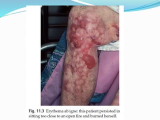
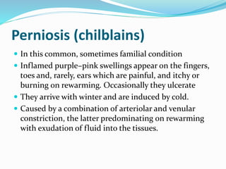
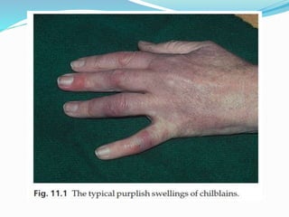
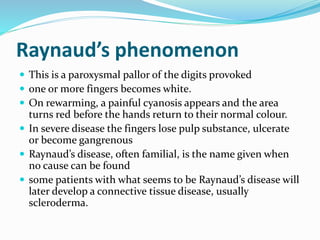
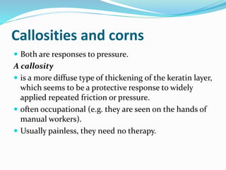
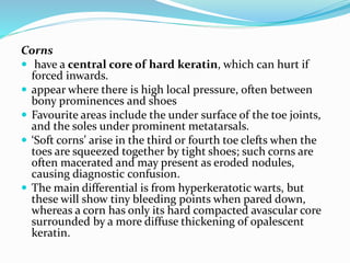
Miliaria
 result of plugging or rupture of sweat ducts.
 It occurs in hot humid climates, at any age and is common
in over-clothed infants in hot nurseries.
 The physical signs depend on where the ducts are blocked.
1. Miliaria crystallina presents as tiny clear noninflamed
vesicles that look like dew. This is the most superficial
type.
2. Miliaria rubra (prickly heat) Tiny erythematous and
very itchy papules.
3. Miliaria profunda These consist of larger erythematous
papules or pustules. This is the deepest type.
4. Miliaria Pustulosa
Treatment
 The best treatment is to move to a cooler climate or
into air conditioning.
 Clothing that prevents the evaporation of sweat (e.g.
nylon shirts) should be avoided; cotton is best.
 Salicylic acid 2% in isopropyl alcohol applied daily to
prone areas for prevention.
 Topical steroids reduce irritation but should only be
used briefly
 Calamine lotion cools and soothes
Erythema ab igne
 Its reticulate pigmented erythema, with variable
scaling, is caused by damage from long-term exposure
to local heat – usually from an open fire, hot water
bottle or heating pad.
 The condition has become less common with the
advent of central heating.
Perniosis (chilblains)
 In this common, sometimes familial condition
 Inflamed purple–pink swellings appear on the fingers,
toes and, rarely, ears which are painful, and itchy or
burning on rewarming. Occasionally they ulcerate
 They arrive with winter and are induced by cold.
 Caused by a combination of arteriolar and venular
constriction, the latter predominating on rewarming
with exudation of fluid into the tissues.
Treatment
 Warm housing and clothing help.
 Oral calcium channel blocker nifedipine may be
useful
 The blood pressure should be monitored at the start of
treatment and at return visits.
 Nicotinamide (500 mg three times daily) alone or in
addition to calcium-channel blockers
 Sympathectomy may be advised in severe cases.
Raynaud’s phenomenon
 This is a paroxysmal pallor of the digits provoked
 one or more fingers becomes white.
 On rewarming, a painful cyanosis appears and the area
turns red before the hands return to their normal colour.
 In severe disease the fingers lose pulp substance, ulcerate
or become gangrenous
 Raynaud’s disease, often familial, is the name given when
no cause can be found
 some patients with what seems to be Raynaud’s disease will
later develop a connective tissue disease, usually
scleroderma.
Treatment
 The main treatment is to protect the vulnerable digits from
cold.
 Smoking should be abandoned.
 Calcium-channel blockers (e.g. nifedipine 10–30 mg three
times daily) are the most effective agents although they
work best in patients with primary Raynaud’s disease.
 Patients should be warned about dizziness caused by postural hypotension
 Diltiazem (30–60 mg three times daily) is less effective
than nifedipine but has fewer side-effects.
 The systemic vasodilator inositol nicotinate may help
 A combination of low-dose acetylsalicylic acid and the
antiplatelet drug dipyridamole is also worth trying.
SunburnCause
 UVB penetrates the epidermis and superficial dermis,
stimulating the production and release of prostaglandins,
leukotrienes, histamine, interleukin 1 (IL-1) and tumour
necrosis factor a (TNF-a).
 These cause pain and stimulate the production of the
inducible nitric oxide synthase (iNOS) enzyme. This
generates high concentrations of nitric oxide which cause
the characteristic dermal vasodilatation and redness.
Presentation and course
 Skin exposed to too much UVB becomes red (redness is
maximal after 1 day) several hours, painful and may blister.
 settles over the next 2–3 days, leaving sheet-like
desquamation, diffuse pigmentation (a ‘tan’) and,
sometimes, discrete lentigines.
Differential diagnosis
 Phototoxic reactions
Treatment
 Symptomatic
 Baths may be cooling and oily shake lotions (e.g. oily
calamine lotion), oil in-water lotions or creams are
comforting.
 Potent topical corticosteroids help if used early and briefly.
 Oral aspirin (a prostaglandin synthesis inhibitor) relieves
the pain.
 Sprays containing benzocaine also relieve pain, but
occasionally sensitize.
Phototoxicity
 A drug that absorb UVR to cause a non
immunological reaction.
 Most blamed drugs window glass, protective against
sunburn, does not protect against most phototoxic
drug reactions.
Presentation and course
 Tenderness and redness occur only in areas exposed
both to sufficient drug and to sufficient UVR
 The signs and symptoms are those of sunburn.
 The skin may later develop a deep tan.
Differential diagnosis
 Photoallergic reactions
Investigations
 photopatch testing
Treatment
 This is the same as for sunburn.
 Drugs should be stopped if further exposure to
ultraviolet light is likely.
Photoallergy
 Drugs, topical or systemic, and chemicals on the skin
can interact with UVR and cause immunological
reactions.
Cause
 UVR converts an immunologically inactive form of a
drug into an antigenic molecule inducing an
immunological reaction, analogous to allergic contact
dermatitis, is induced if the antigen remains in the
skin or is formed there on subsequent exposure to the
drug and UVR. Many of the same drugs that cause
phototoxic reactions can also cause photoallergic ones.
Presentation
 similar to phototoxicity, but the reaction usually becomes
eczematous, appears later and lasts longer.
 The eruption will be on exposed areas such as the hands,
the V of the neck, the nose, the chin and the forehead.
There is also a tendency to spare the upper lip under the
nose, the eyelids and the submental region
 Often, the eruption does not occur on the first exposure
to ultraviolet, but only after a second or further exposures.
A lag phase of one or more weeks is needed to induce an
immune response.
Course
 They tend to resolve when either the drug or the exposure to UVR is
stopped, but this may take several weeks.
Complications
 Some drugs, such as the sulphonamides, can cause chronic actinic
dermatitis.
Investigations
 Photopatch testing can confirm the diagnosis. The chemical is applied
for 24 h and the skin is then irradiated with UVA. An acute
photoallergic contact dermatitis is then elicited.
 A control patch, not irradiated, rules out ordinary allergic contact
dermatitis.
Treatment
 The drug should be stopped and the patient protected from further
ultraviolet exposure
 Potent topical corticosteroids or a short course of a systemic
corticosteroid will hasten resolution and provide symptomatic relief.
Polymorphic light eruption
 This is the most frequent cause of a so-called ‘sun allergy’.
Cause
 It is speculated that UVR causes a natural body chemical to change into
an allergen.
 Mechanisms are similar to those in drug photoallergy.
 Some people seem genetically predisposed, because other family
members may also be affected.
Presentation
 Small itchy red papules, papulovesicles or eczematous plaques arise
from 2 h to 5 days, most commonly at 24 h, after exposure to UVR.
 The eruption is itchy and usually confined to sun-exposed areas,
remembering that some UVR passes through thin clothing.
 Not all exposed skin develops disease so there are papules and plaques
rather than generalized redness.
Treatment
 sunscreens
 Protective clothing, such as wide-brimmed hats, long-
sleeved shirts and long trousers
 In some patients, a 4-week course of psoralen with UVA
(PUVA) in the late spring can create enough tan to confer
protection for the rest of the season.
 Moderately potent topical steroids usually improve the
eruption.
 A tapering course of systemic steroids for severe or early
spring outbreaks.
 Hydroxychloroquine may be effective when used over the
sunny season.
Callosities and corns
 Both are responses to pressure.
A callosity
 is a more diffuse type of thickening of the keratin layer,
which seems to be a protective response to widely
applied repeated friction or pressure.
 often occupational (e.g. they are seen on the hands of
manual workers).
 Usually painless, they need no therapy.
Corns
 have a central core of hard keratin, which can hurt if
forced inwards.
 appear where there is high local pressure, often between
bony prominences and shoes
 Favourite areas include the under surface of the toe joints,
and the soles under prominent metatarsals.
 ‘Soft corns’ arise in the third or fourth toe clefts when the
toes are squeezed together by tight shoes; such corns are
often macerated and may present as eroded nodules,
causing diagnostic confusion.
 The main differential is from hyperkeratotic warts, but
these will show tiny bleeding points when pared down,
whereas a corn has only its hard compacted avascular core
surrounded by a more diffuse thickening of opalescent
keratin.
Treatment
 The right treatment for corns is to eliminate the pressure
that caused them, but patients may be slow to accept this.
While regular paring reduces the symptoms temporarily
 well-fitting shoes are essential
 Corns under the metatarsals can be helped by soft spongy
soles, but sometimes need orthopaedic surgery to alter
weight bearing.
 Special care is needed with corns on ischaemic or diabetic
feet, which are at greater risk of infection and ulceration.
Dermatosis to physical stimuli

Dermatosis to physical stimuli

  • 2.
    Miliaria  result ofplugging or rupture of sweat ducts.  It occurs in hot humid climates, at any age and is common in over-clothed infants in hot nurseries.  The physical signs depend on where the ducts are blocked. 1. Miliaria crystallina presents as tiny clear noninflamed vesicles that look like dew. This is the most superficial type. 2. Miliaria rubra (prickly heat) Tiny erythematous and very itchy papules. 3. Miliaria profunda These consist of larger erythematous papules or pustules. This is the deepest type. 4. Miliaria Pustulosa
  • 3.
    Treatment  The besttreatment is to move to a cooler climate or into air conditioning.  Clothing that prevents the evaporation of sweat (e.g. nylon shirts) should be avoided; cotton is best.  Salicylic acid 2% in isopropyl alcohol applied daily to prone areas for prevention.  Topical steroids reduce irritation but should only be used briefly  Calamine lotion cools and soothes
  • 4.
    Erythema ab igne Its reticulate pigmented erythema, with variable scaling, is caused by damage from long-term exposure to local heat – usually from an open fire, hot water bottle or heating pad.  The condition has become less common with the advent of central heating.
  • 6.
    Perniosis (chilblains)  Inthis common, sometimes familial condition  Inflamed purple–pink swellings appear on the fingers, toes and, rarely, ears which are painful, and itchy or burning on rewarming. Occasionally they ulcerate  They arrive with winter and are induced by cold.  Caused by a combination of arteriolar and venular constriction, the latter predominating on rewarming with exudation of fluid into the tissues.
  • 7.
    Treatment  Warm housingand clothing help.  Oral calcium channel blocker nifedipine may be useful  The blood pressure should be monitored at the start of treatment and at return visits.  Nicotinamide (500 mg three times daily) alone or in addition to calcium-channel blockers  Sympathectomy may be advised in severe cases.
  • 9.
    Raynaud’s phenomenon  Thisis a paroxysmal pallor of the digits provoked  one or more fingers becomes white.  On rewarming, a painful cyanosis appears and the area turns red before the hands return to their normal colour.  In severe disease the fingers lose pulp substance, ulcerate or become gangrenous  Raynaud’s disease, often familial, is the name given when no cause can be found  some patients with what seems to be Raynaud’s disease will later develop a connective tissue disease, usually scleroderma.
  • 10.
    Treatment  The maintreatment is to protect the vulnerable digits from cold.  Smoking should be abandoned.  Calcium-channel blockers (e.g. nifedipine 10–30 mg three times daily) are the most effective agents although they work best in patients with primary Raynaud’s disease.  Patients should be warned about dizziness caused by postural hypotension  Diltiazem (30–60 mg three times daily) is less effective than nifedipine but has fewer side-effects.  The systemic vasodilator inositol nicotinate may help  A combination of low-dose acetylsalicylic acid and the antiplatelet drug dipyridamole is also worth trying.
  • 11.
    SunburnCause  UVB penetratesthe epidermis and superficial dermis, stimulating the production and release of prostaglandins, leukotrienes, histamine, interleukin 1 (IL-1) and tumour necrosis factor a (TNF-a).  These cause pain and stimulate the production of the inducible nitric oxide synthase (iNOS) enzyme. This generates high concentrations of nitric oxide which cause the characteristic dermal vasodilatation and redness. Presentation and course  Skin exposed to too much UVB becomes red (redness is maximal after 1 day) several hours, painful and may blister.  settles over the next 2–3 days, leaving sheet-like desquamation, diffuse pigmentation (a ‘tan’) and, sometimes, discrete lentigines.
  • 12.
    Differential diagnosis  Phototoxicreactions Treatment  Symptomatic  Baths may be cooling and oily shake lotions (e.g. oily calamine lotion), oil in-water lotions or creams are comforting.  Potent topical corticosteroids help if used early and briefly.  Oral aspirin (a prostaglandin synthesis inhibitor) relieves the pain.  Sprays containing benzocaine also relieve pain, but occasionally sensitize.
  • 14.
    Phototoxicity  A drugthat absorb UVR to cause a non immunological reaction.  Most blamed drugs window glass, protective against sunburn, does not protect against most phototoxic drug reactions. Presentation and course  Tenderness and redness occur only in areas exposed both to sufficient drug and to sufficient UVR  The signs and symptoms are those of sunburn.  The skin may later develop a deep tan.
  • 15.
    Differential diagnosis  Photoallergicreactions Investigations  photopatch testing Treatment  This is the same as for sunburn.  Drugs should be stopped if further exposure to ultraviolet light is likely.
  • 17.
    Photoallergy  Drugs, topicalor systemic, and chemicals on the skin can interact with UVR and cause immunological reactions. Cause  UVR converts an immunologically inactive form of a drug into an antigenic molecule inducing an immunological reaction, analogous to allergic contact dermatitis, is induced if the antigen remains in the skin or is formed there on subsequent exposure to the drug and UVR. Many of the same drugs that cause phototoxic reactions can also cause photoallergic ones.
  • 18.
    Presentation  similar tophototoxicity, but the reaction usually becomes eczematous, appears later and lasts longer.  The eruption will be on exposed areas such as the hands, the V of the neck, the nose, the chin and the forehead. There is also a tendency to spare the upper lip under the nose, the eyelids and the submental region  Often, the eruption does not occur on the first exposure to ultraviolet, but only after a second or further exposures. A lag phase of one or more weeks is needed to induce an immune response.
  • 20.
    Course  They tendto resolve when either the drug or the exposure to UVR is stopped, but this may take several weeks. Complications  Some drugs, such as the sulphonamides, can cause chronic actinic dermatitis. Investigations  Photopatch testing can confirm the diagnosis. The chemical is applied for 24 h and the skin is then irradiated with UVA. An acute photoallergic contact dermatitis is then elicited.  A control patch, not irradiated, rules out ordinary allergic contact dermatitis. Treatment  The drug should be stopped and the patient protected from further ultraviolet exposure  Potent topical corticosteroids or a short course of a systemic corticosteroid will hasten resolution and provide symptomatic relief.
  • 22.
    Polymorphic light eruption This is the most frequent cause of a so-called ‘sun allergy’. Cause  It is speculated that UVR causes a natural body chemical to change into an allergen.  Mechanisms are similar to those in drug photoallergy.  Some people seem genetically predisposed, because other family members may also be affected. Presentation  Small itchy red papules, papulovesicles or eczematous plaques arise from 2 h to 5 days, most commonly at 24 h, after exposure to UVR.  The eruption is itchy and usually confined to sun-exposed areas, remembering that some UVR passes through thin clothing.  Not all exposed skin develops disease so there are papules and plaques rather than generalized redness.
  • 24.
    Treatment  sunscreens  Protectiveclothing, such as wide-brimmed hats, long- sleeved shirts and long trousers  In some patients, a 4-week course of psoralen with UVA (PUVA) in the late spring can create enough tan to confer protection for the rest of the season.  Moderately potent topical steroids usually improve the eruption.  A tapering course of systemic steroids for severe or early spring outbreaks.  Hydroxychloroquine may be effective when used over the sunny season.
  • 25.
    Callosities and corns Both are responses to pressure. A callosity  is a more diffuse type of thickening of the keratin layer, which seems to be a protective response to widely applied repeated friction or pressure.  often occupational (e.g. they are seen on the hands of manual workers).  Usually painless, they need no therapy.
  • 26.
    Corns  have acentral core of hard keratin, which can hurt if forced inwards.  appear where there is high local pressure, often between bony prominences and shoes  Favourite areas include the under surface of the toe joints, and the soles under prominent metatarsals.  ‘Soft corns’ arise in the third or fourth toe clefts when the toes are squeezed together by tight shoes; such corns are often macerated and may present as eroded nodules, causing diagnostic confusion.  The main differential is from hyperkeratotic warts, but these will show tiny bleeding points when pared down, whereas a corn has only its hard compacted avascular core surrounded by a more diffuse thickening of opalescent keratin.
  • 27.
    Treatment  The righttreatment for corns is to eliminate the pressure that caused them, but patients may be slow to accept this. While regular paring reduces the symptoms temporarily  well-fitting shoes are essential  Corns under the metatarsals can be helped by soft spongy soles, but sometimes need orthopaedic surgery to alter weight bearing.  Special care is needed with corns on ischaemic or diabetic feet, which are at greater risk of infection and ulceration.