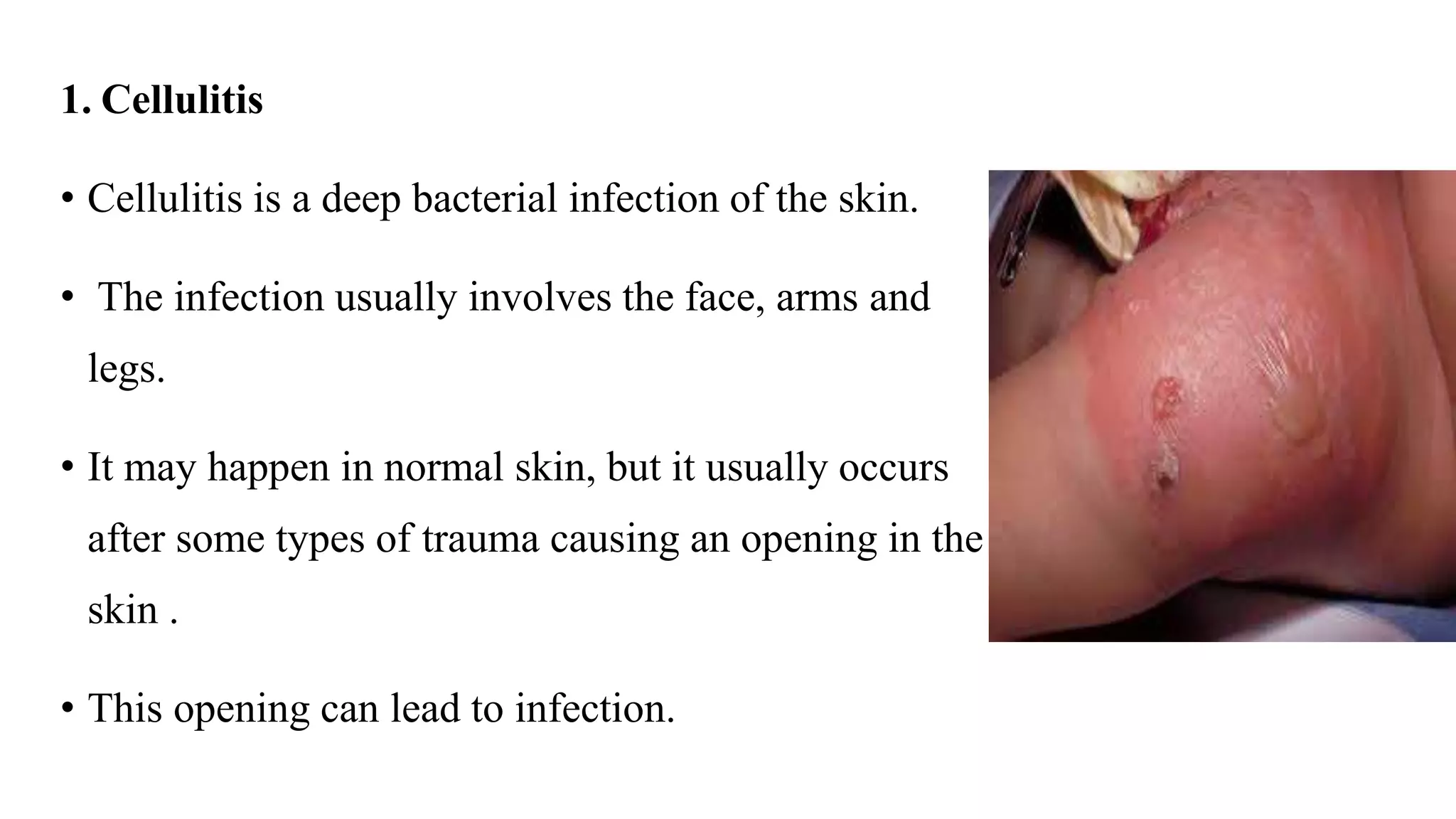
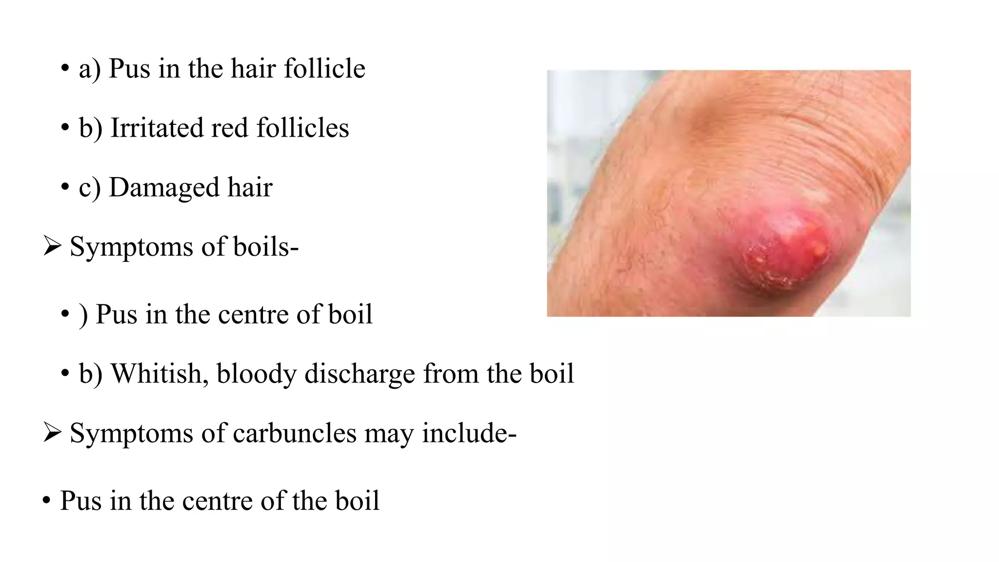
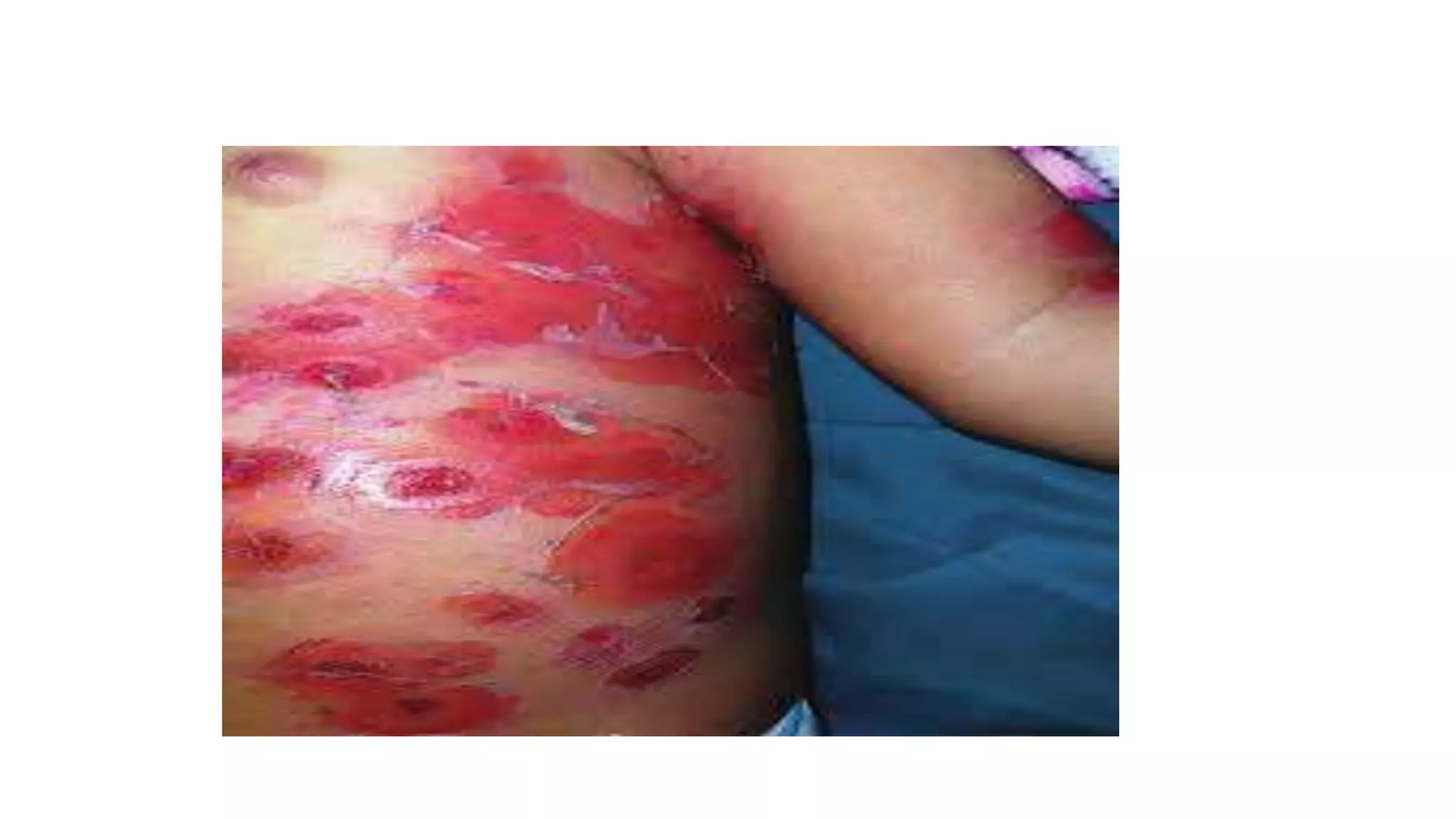
2. PYODERMAS/ BACTERIAL SKIN INFECTIONS
• Skin is largest organ of body.
• It maintains homeostasis, protects underlying tissues and organs, and protects
body from mechanical injury, damaging substances, and ultraviolet rays of
sun.
5. 1. Impetigo
• Impetigo is a superficial infection of the skin, caused by bacteria.
• The lesions are often grouped and have a red base.
• These lesions open and become crusty and have honey- color which is typical
of impetigo.
• It is contagious and can spread to all the members of the family and also
leads to re-infection of children.
6.
7. • Etiology
• Common bacterias that are found on skin normally causes impetigo.
When bacteria enters an open area in the skin, infection occur.
The most common causative organisms are- Group A hemolytic
Streptococcus & Staphylococcus aureus.
Impetigo is most common in children, but adults may also have it due to
poor hygiene and warm temperatures.
8. • Clinical features
Impetigo usually occurs on face, neck, arms and limbs. But the lesions can
be observed on any part of the body.
Impetigo starts as a small vesicle or fluid filled lesion.
The lesion then ruptures and fluid drains leaving areas that are covered with
honey colored crusts.
The lesions may all look different, with different sizes and shapes.
Child may also presents with swollen lymph nodes.
9. • Diagnostic evaluation
Diagnosed based on complete history
Physical examination
The lesions of impetigo are unique and are clearly diagnosable.
Culture of lesion can be done to confirm the diagnosis and the type f
bacteria.
10. • Management
• Specific treatment is given based on –
1. Child’s age, over all health and medical history
2. Extent of condition
3. Child’s tolerance to specific medications
• Treatment:
For child with many lesions oral antibiotics are given
11. If child has only few lesions, topical antibiotics are applied directly on the affected
area.
Bathe the child daily with an antibacterial soap to help decrease the chance of
spreading the infection.
Proper hand washing technique by everyone in the house hold is very important to
help diagnosis the chance of spreading the infection.
Keep the child’s finger nails short to decrease the chances of scratching and spreading
the infection.
Avoid sharing of garments, towels and other house hold items to prevent the spreading
of infection.
12. 1. Cellulitis
• Cellulitis is a deep bacterial infection of the skin.
• The infection usually involves the face, arms and
legs.
• It may happen in normal skin, but it usually occurs
after some types of trauma causing an opening in the
skin .
• This opening can lead to infection.
13. • Etiology
• Cellulitis is caused by a bacterial infection of a wound area of skin that is no
longer intact. The most common bacterial cause of Cellulitis include the
following-
Group A – Hemolytic streptococcus
Streptococcus pneumoniae
Staphylococcus aureus.
14. • Clinical features
Swelling of the skin , Tenderness
Warm skin , Pain
Bruising , Blisters
Fever , Headache
Chills
Feeling weak
Red streaks
15. Very large area of red inflamed skin
Fever
affected area has- 1. Numbness 2. Tingling
skin appears black
16. • Diagnostic evaluation
Diagnosis is usually based on medical history and physical examination of
the child.
Blood and skin samples may be taken to confirm the diagnosis and the type
of bacteria that is present.
• Management
Oral or iv antibiotics
Warm, wet dressings on infection site
17. Surgical intervention
Elevate the effected part and reduce activity of that part.
• Complications
o Meningitis
o Septic arthritis
o Glomerulonephritis
18. • Folliculitis
• Folliculitis is the inflammation of hair follicles due to an infection, injury or
irritation.
• It is characterized by tender, swollen areas that form around the follicles,
often on the neck, breast, buttocks and face.
19. • Boils
• Boils are pus filled lesions that are painful and usually firm. Boils are
usually located the waist area, groins, buttocks and under the arm.
20. • Carbuncles
• Carbuncles are clusters of boils. These are usually found in the back of the
neck or thigh.
• Clinical features
• The following are the most common symptoms of folliculitis, boils and
carbuncles. However each child may experience symptoms differently.
Symptoms of folliculitis-
21. • a) Pus in the hair follicle
• b) Irritated red follicles
• c) Damaged hair
Symptoms of boils-
• ) Pus in the centre of boil
• b) Whitish, bloody discharge from the boil
Symptoms of carbuncles may include-
• Pus in the centre of the boil
22. • Whitish, bloody discharge from the boils
• Fever
• Fatigue.
• Diagnostic evaluation
Thorough medical history and physical examination
Culture of wound
• Management
• Topical antibiotics
23. • For carbuncles and boils,
a warm compress to promote drainage of lesion
Oral and IV antibiotics
Possible removal of boils and carbuncles.
24. STAPHYLOCOCCAL SCALDED SKIN SYNDROME(Ritter Disease)
• Staphylococcal scalded skin syndrome (SSSS) is a response to a
Staphylococcus infection.
• It causes a reddening and blistering of the skin that gives it a scalded or
burned look.
• SSSS is most common in infants and children, but can also affect adults with
a depressed immune system or problems with kidney function. The condition
can range from mild to life-threatening, though it is usually not life-
threatening in children.
25.
26. • Etiology
caused predominantly Staphylococcus aureus strains producing Exfoliative
Toxins (ETs) are the causative organisms.
found in naso-pharynx and, less commonly, the umbilicus, urinary tract, a
superficial abrasion, conjunctivae, and blood
spreads hematogenously
27. Associated Disease
• Renal failure (as Exfoliative Toxins eliminated through the kidneys),
• Malignancy,
• Immunosuppression
28. • Signs and symptoms
The condition often begins with a low-grade fever and general redness of the
skin.
The skin may feel like sandpaper and look wrinkled.
The rash usually spreads quickly and may especially affect the area around the
mouth as well as areas of skin that are creased, such as the arms, groin, legs and
neck.
This is followed by the formation of fluid-filled blisters that rupture easily.
The top layer of skin may start to peel away off in sheets, revealing reddish, moist
skin below.
29. weakness
joint and muscle pain
chills
not feeling well
• Pathophysiology
Caused by an exfoliative toxin
The toxins likely act as proteases(an enzyme which breaks down proteins
and peptides.) that target the protein desmoglein-1 (DG-1)
Exotoxin causes separation of the epidermis beneath the granular cell layer.
30. • Diagnosis
History, physical examination
Gram stain or Culture
Skin biopsy - the surgeon removes a portion of the tissue mass which is then
sent to pathology to make the diagnosis.
PCR
31. • Management
A.Pharmacologic
Systemic therapy, either orally, in cases of localized involvement, or
parenterally, with a semisynthetic penicillinase-resistant penicillin, should be
prescribed because the staphylococci are usually penicillin resistant
Clindamycin may be added to inhibit bacterial protein (toxin) synthesis
Hydroxyzine 2mg/ml, 2.5 ml every 6 hours for pruritus
32. Mupirocin ointment
Erythromycin eye ointment, to both lower lids twice a day
Cloxacillin 250mg/ml, 2ml every 6 hours on an empty stomach, 1 hour prior
to meals
A.Non-pharmacologic
The skin should be gently moistened and cleansed.
33. Application of an emollient provides lubrication and decreases discomfort.
• Prognosis
• Recovery is usually rapid, but complications such as excessive fluid loss,
electrolyte imbalance, faulty temperature regulation, pneumonia, septicemia,
and cellulitis may cause increased morbidity.
34. • NURSING MANAGEMENT
1. Nursing Diagnosis
•Impaired Skin Integrity related to environmental agents, somatic factors,
immunologic deficit
• Nursing Interventions and Rationales
• Carry out therapeutic regimens as prescribed, or support and assist parents in
carrying out treatment plan, to promote skin healing.
• Provide moist environment (dressing or ointment) for optimum wound healing.
35. • Administer topical treatments and applications
• Administer systemic medications, if ordered.
• Prevent secondary infection and autoinoculation, since these delay healing.
• Reduce external stimuli that aggravate condition, causing delay in healing.
• Encourage rest to support body's natural defenses.
• Encourage well-balanced diet to support body's natural defenses.
• Administer skin care and general hygiene measures to promote skin healing.
36. • Risk for Infection related to presence of infective organisms
• Nursing Interventions and Rationales
• Implement Standard Precautions to prevent spread of infection.
• Isolate affected child from susceptible individuals if indicated to prevent
spread of infection.
• Maintain careful hand washing after caring for child to remove infective
organisms.
37. • Avoid unnecessary close contact with affected child during infective stage of
disease.
• Use correct technique for disposal of dressings, solutions, and other fomites
in contact with lesion(s) to safely dispose of infective organisms.
• Teach and reinforce positive habits of hygienic care to decrease risk of
infection.
38. • Pain related to skin lesions, pruritus
• Nursing Interventions and Rationales
• Avoid or reduce external stimuli that aggravate discomfort, such as clothing
and bed linen.
• Implement other appropriate nonpharmacologic pain reduction techniques.
• Administer medications to relieve discomfort, restlessness, or irritability.
• Advocate for child regarding appropriate topical anesthesia or sedation-
analgesia for wound suturing or cleansing to prevent unnecessary pain and
emotional trauma.
39. • Apply soothing treatments and topical applications as ordered to relieve pain
or pruritus.