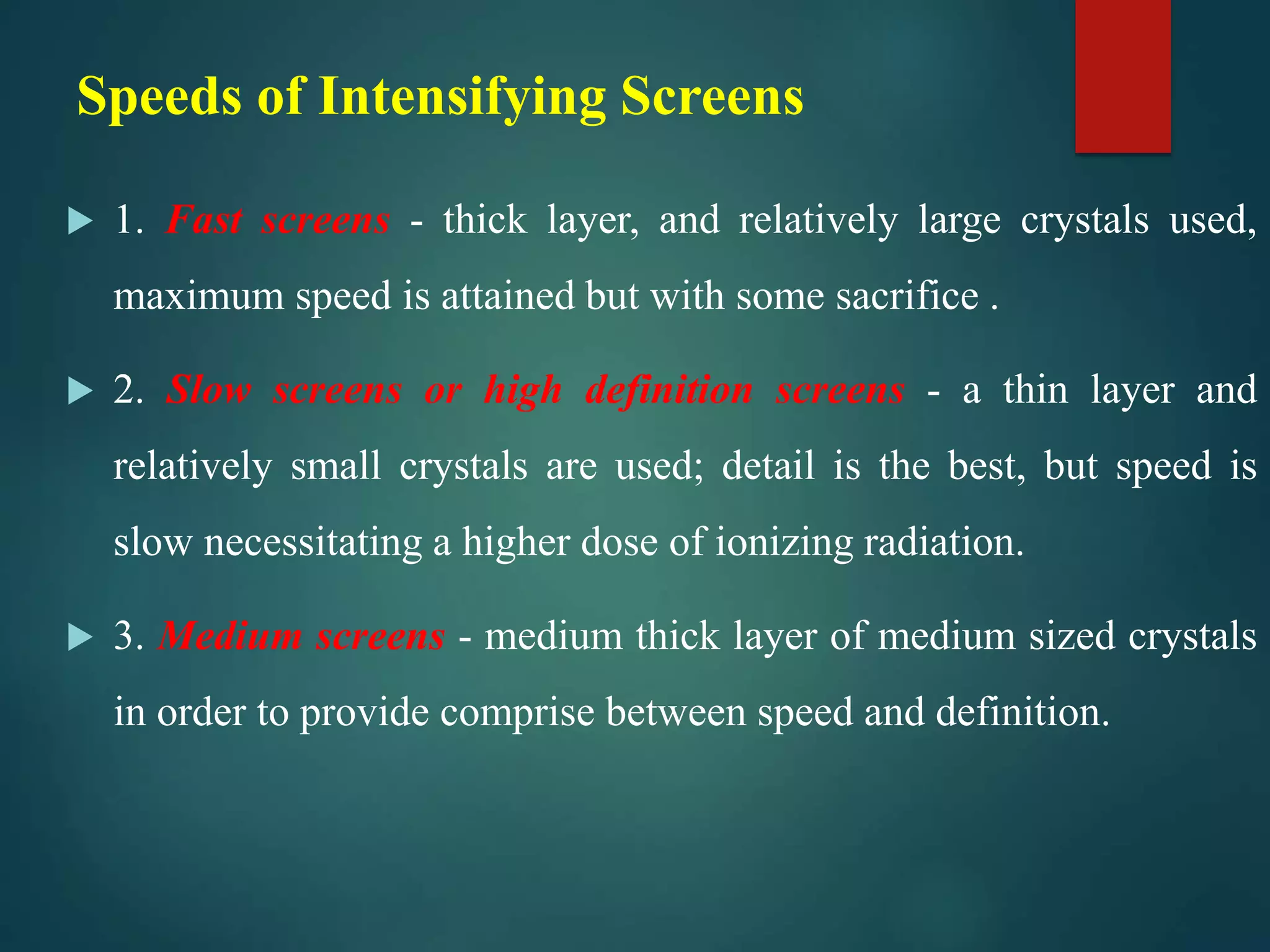
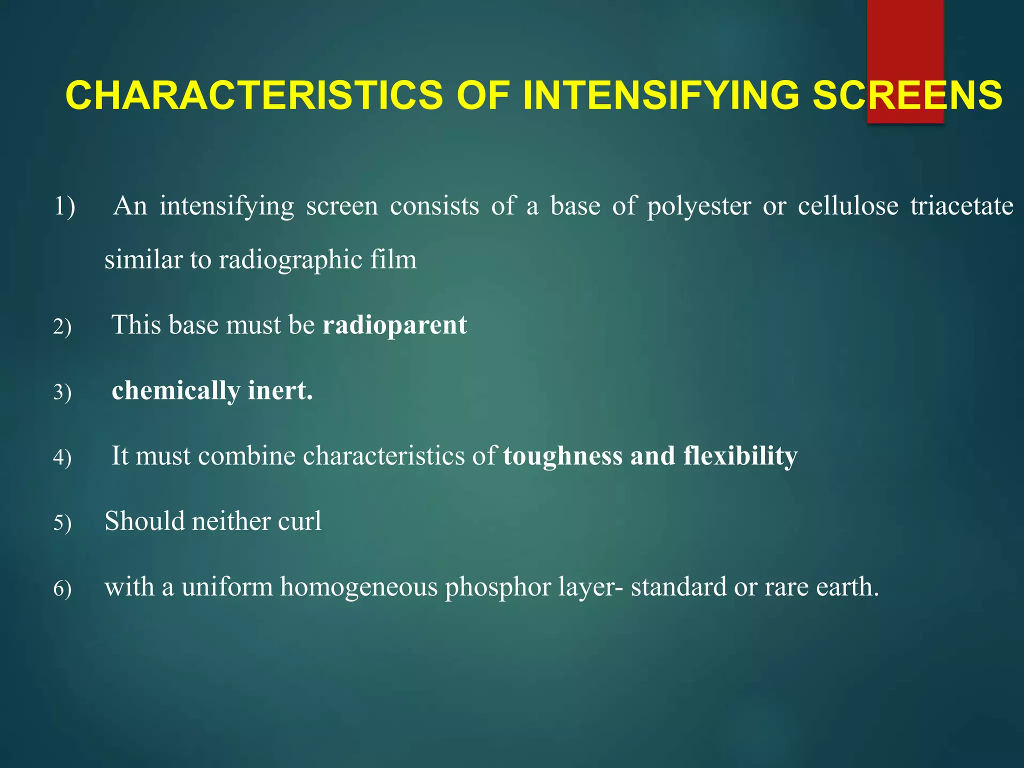
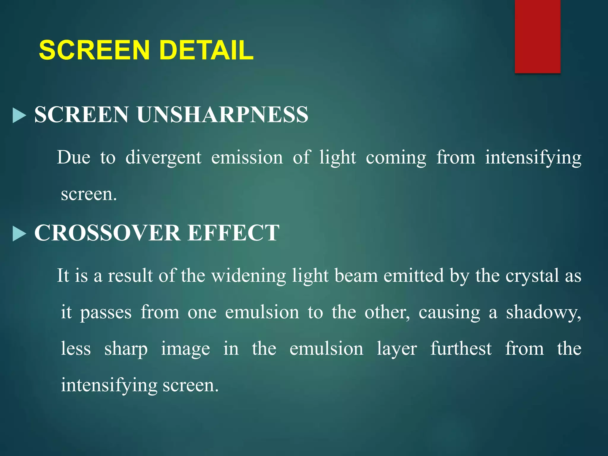
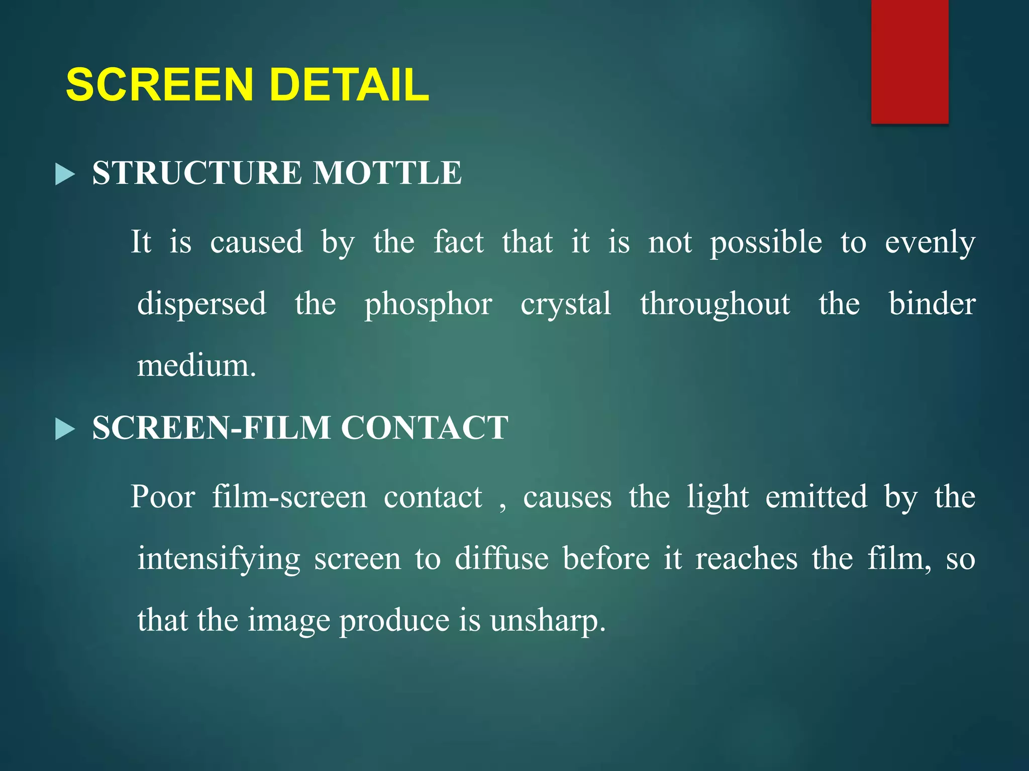
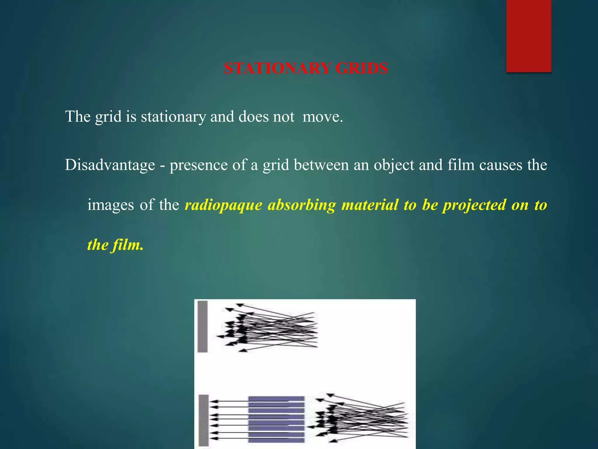
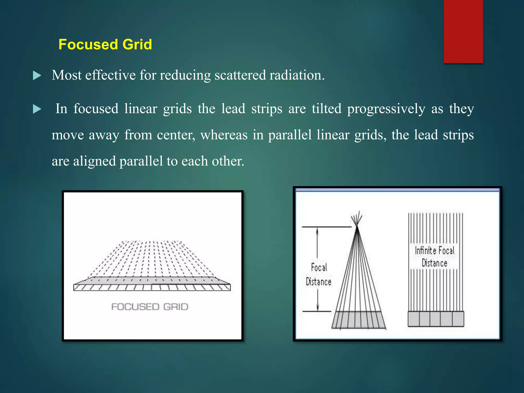
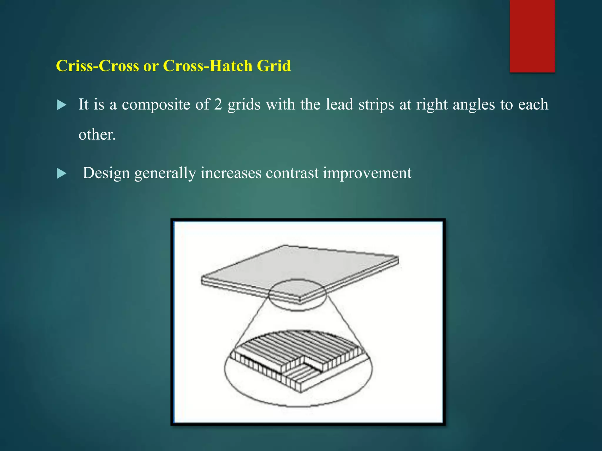
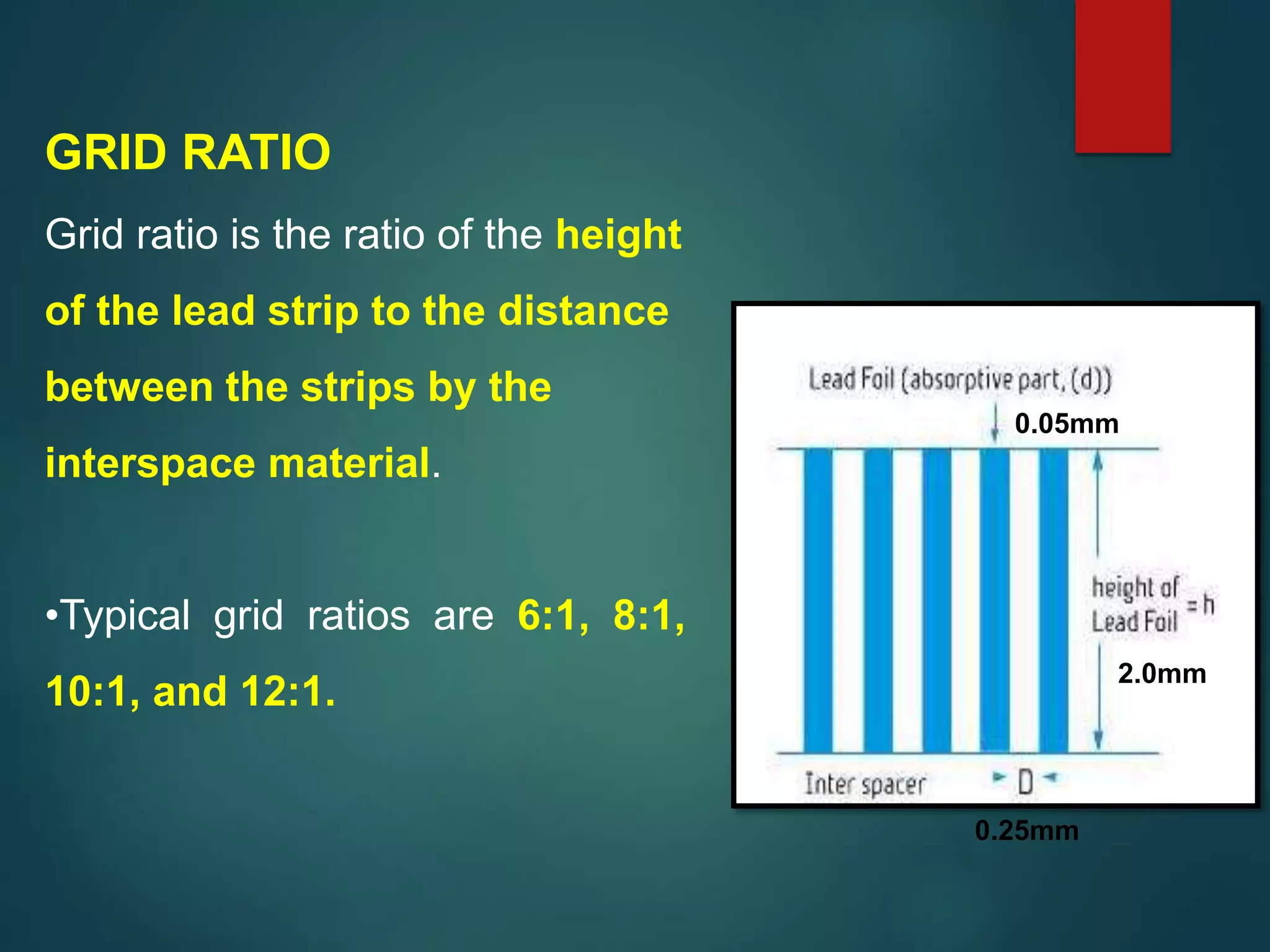
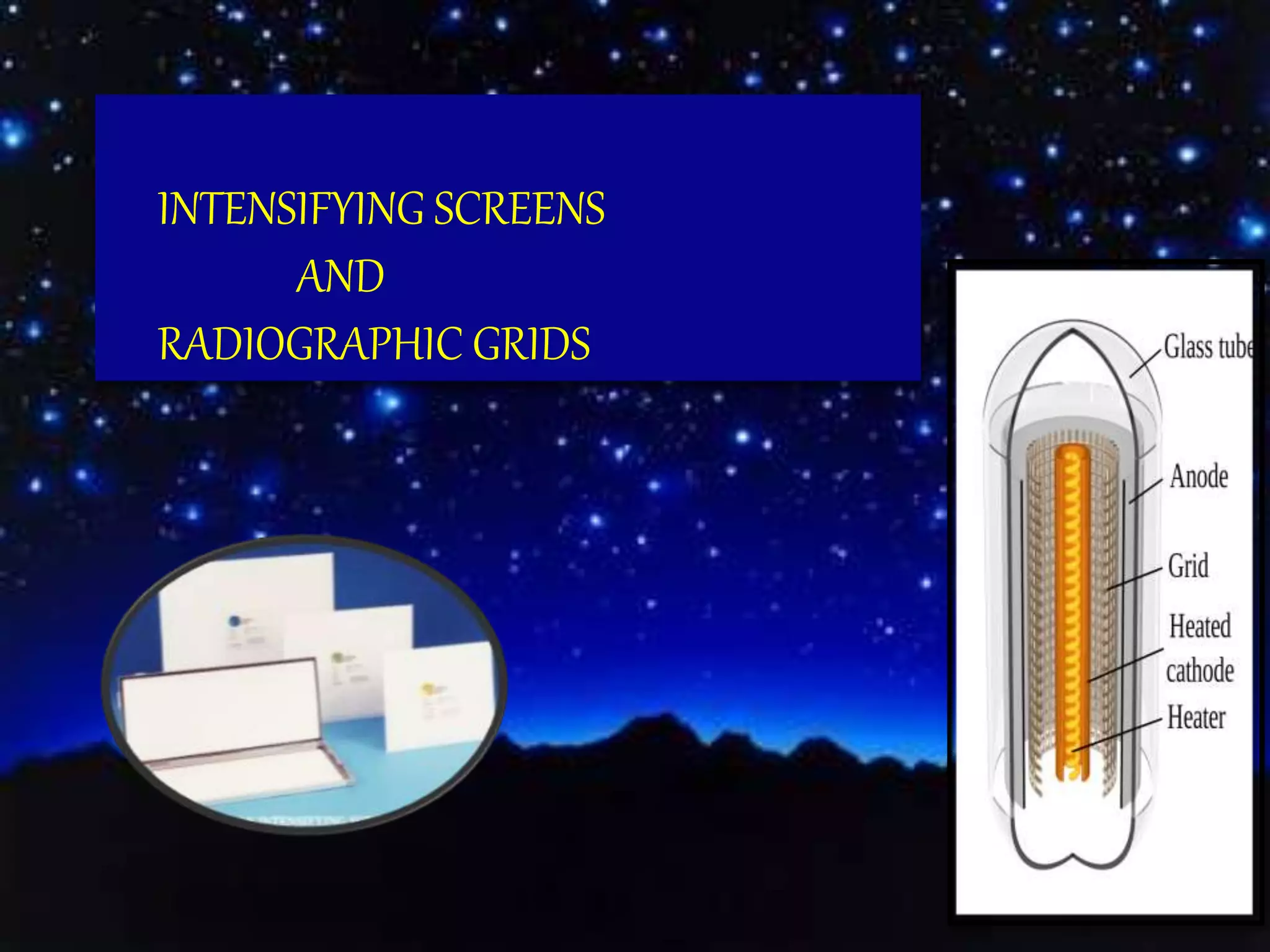
This document provides information on intensifying screens and radiographic grids. It discusses the history, construction, and functions of intensifying screens, including the types of phosphors used. It also covers screen speed, detail, and care. For radiographic grids, it outlines the history and development of grids as well as grid design, patterns, specifications and factors such as ratio and frequency. Research studies evaluating different screen-film combinations and their effects on image quality and radiation dose are also summarized.
brief description about CONTENTS Introduction Principles of panoramic imaging Image layer Panoramic machines Panoramic film Patient positioning Interpreting the panoramic imaging INDICATION Advantages Disadvantages Conclusion References
3. INTRODUCTION • Panoramic imaging also called pantomography is a technique for producing a single tomographic image of facial structures that includes both the maxillary and mandibular dental arches and their supporting structures . • This is a curvilinear variant of conventional tomography.
4. PRINCIPLES OF PANORAMIC IMAGE FORMATION • Patero and Numata - describe the principles of panoramic radiography • based on the principle of reciprocal movement of x-ray source and an image receptor around a central point or plane called the image layer, in which the OBJECT of image is located. • OBJECT in front or behind this image are not clearly captured because of their movement relative to the centre of rotation of the receptor and the x-ray source.
5. The film and x-ray tubehead move around the patient in opposite directions in panoramic radiography
6. ROTATION CENTER The pivotal point or axis around which the cassette carrier and tube head rotate is termed rotation center Three basic rotation center used in panoramic radiography Double centre rotation Triple centre rotation moving centre rotation The location and number of rotational centers INFLUENCE size and shape of focal trough
7. IMAGE LAYER • Also known as focal trough • It is a three dimensional curved zone where the structures lying within this layer are reasonably well defined on final panoramic image. • The structures seen on a panoramic image are primarily those located within image layer. • OBJECTSoutside the image layer are blurred magnified are reduced in size. Even distorted to the extent of not being recognizable. • This shape of image layer varies with the brand of equipment used.
8. FOCAL TROUGH
9. FACTORS AFFECTING SIZE OF IMAGE LAYER: Arc path Velocity of receptor and X-ray tube head Alignment of x-ray beam Collimator width The location of image layer change with extensive machine used so recalibration may be necessary if consistently suboptimal images are produced. As a position of object is moved within the image layer size and shape of image layer change.
10. PANORAMIC UNIT
11. A, Orthophos XG Plus extraoral x-ray machine. B, Orthoralix 8500 extraoral x-ray machine. C, Example of a digital panoramic system
12. PARTS OF PANORAMIC UNITS a. x-ray tube head b. head positioner: chin rest notched bite block forehead rest lateral head support c. exposure controls
13. X-RAY TUBE HEAD: • Similar to intraoral x-ray tube head • Each has a filament to produce electrons and a target to produce x-rays • Collimator is a lead plate with narrow vertical slit • Narrow x-ray beam emerges from collimator minimize patient exposure to radiation
1
IDEAL IMAGE CHARACTERISTICS
FACTORS RELATED TO THE RADIATION BEAM
FACTORS RELATED TO THE OBJECT
FACTORS RELATED TO THE TECHNIQUE
FACTORS RELATED TO RECORDING OF THE ROENTGEN IMAGE OF THE OBJECT
DARK/ LIGHT IMAGE IDEAL IMAGE
IDEAL QUALITY CRIETRIA
The Indian Dental Academy is the Leader in continuing dental education , training dentists in all aspects of dentistry and
offering a wide range of dental certified courses in different formats.
Radiology in orthodontics /certified fixed orthodontic courses by Indian dent...Indian dental academy
The Indian Dental Academy is the Leader in continuing dental education , training dentists in all aspects of dentistry and offering a wide range of dental certified courses in different formats.
Indian dental academy provides dental crown & Bridge,rotary endodontics,fixed orthodontics,
Dental implants courses.for details pls visit www.indiandentalacademy.com ,or call
0091-9248678078
Radiology in orthodontics dr.kavitha /certified fixed orthodontic courses by ...Indian dental academy
The Indian Dental Academy is the Leader in continuing dental education , training dentists in all aspects of dentistry and offering a wide range of dental certified courses in different formats.
Indian dental academy provides dental crown & Bridge,rotary endodontics,fixed orthodontics,
Dental implants courses.for details pls visit www.indiandentalacademy.com ,or call
0091-9248678078
The Indian Dental Academy is the Leader in continuing dental education , training dentists in all aspects of dentistry and
offering a wide range of dental certified courses in different formats.for more details please visit
www.indiandentalacademy.com
Image receptors & accessories/certified fixed orthodontic courses by Indian d...Indian dental academy
The Indian Dental Academy is the Leader in continuing dental education , training dentists in all aspects of dentistry and offering a wide range of dental certified courses in different formats.
Indian dental academy provides dental crown & Bridge,rotary endodontics,fixed orthodontics,
Dental implants courses.for details pls visit www.indiandentalacademy.com ,or call
0091-9248678078
Intensifying screens are major component of the image receptor used in conventional radiography.Its function is to convert the X-rays into visible light through the process of fluorescence.
Pulmonary Thromboembolism - etilogy, types, medical- Surgical and nursing man...VarunMahajani
Disruption of blood supply to lung alveoli due to blockage of one or more pulmonary blood vessels is called as Pulmonary thromboembolism. In this presentation we will discuss its causes, types and its management in depth.
Tom Selleck Health: A Comprehensive Look at the Iconic Actor’s Wellness Journeygreendigital
Tom Selleck, an enduring figure in Hollywood. has captivated audiences for decades with his rugged charm, iconic moustache. and memorable roles in television and film. From his breakout role as Thomas Magnum in Magnum P.I. to his current portrayal of Frank Reagan in Blue Bloods. Selleck's career has spanned over 50 years. But beyond his professional achievements. fans have often been curious about Tom Selleck Health. especially as he has aged in the public eye.
Follow us on: Pinterest
Introduction
Many have been interested in Tom Selleck health. not only because of his enduring presence on screen but also because of the challenges. and lifestyle choices he has faced and made over the years. This article delves into the various aspects of Tom Selleck health. exploring his fitness regimen, diet, mental health. and the challenges he has encountered as he ages. We'll look at how he maintains his well-being. the health issues he has faced, and his approach to ageing .
Early Life and Career
Childhood and Athletic Beginnings
Tom Selleck was born on January 29, 1945, in Detroit, Michigan, and grew up in Sherman Oaks, California. From an early age, he was involved in sports, particularly basketball. which played a significant role in his physical development. His athletic pursuits continued into college. where he attended the University of Southern California (USC) on a basketball scholarship. This early involvement in sports laid a strong foundation for his physical health and disciplined lifestyle.
Transition to Acting
Selleck's transition from an athlete to an actor came with its physical demands. His first significant role in "Magnum P.I." required him to perform various stunts and maintain a fit appearance. This role, which he played from 1980 to 1988. necessitated a rigorous fitness routine to meet the show's demands. setting the stage for his long-term commitment to health and wellness.
Fitness Regimen
Workout Routine
Tom Selleck health and fitness regimen has evolved. adapting to his changing roles and age. During his "Magnum, P.I." days. Selleck's workouts were intense and focused on building and maintaining muscle mass. His routine included weightlifting, cardiovascular exercises. and specific training for the stunts he performed on the show.
Selleck adjusted his fitness routine as he aged to suit his body's needs. Today, his workouts focus on maintaining flexibility, strength, and cardiovascular health. He incorporates low-impact exercises such as swimming, walking, and light weightlifting. This balanced approach helps him stay fit without putting undue strain on his joints and muscles.
Importance of Flexibility and Mobility
In recent years, Selleck has emphasized the importance of flexibility and mobility in his fitness regimen. Understanding the natural decline in muscle mass and joint flexibility with age. he includes stretching and yoga in his routine. These practices help prevent injuries, improve posture, and maintain mobilit
Explore natural remedies for syphilis treatment in Singapore. Discover alternative therapies, herbal remedies, and lifestyle changes that may complement conventional treatments. Learn about holistic approaches to managing syphilis symptoms and supporting overall health.
Prix Galien International 2024 Forum ProgramLevi Shapiro
June 20, 2024, Prix Galien International and Jerusalem Ethics Forum in ROME. Detailed agenda including panels:
- ADVANCES IN CARDIOLOGY: A NEW PARADIGM IS COMING
- WOMEN’S HEALTH: FERTILITY PRESERVATION
- WHAT’S NEW IN THE TREATMENT OF INFECTIOUS,
ONCOLOGICAL AND INFLAMMATORY SKIN DISEASES?
- ARTIFICIAL INTELLIGENCE AND ETHICS
- GENE THERAPY
- BEYOND BORDERS: GLOBAL INITIATIVES FOR DEMOCRATIZING LIFE SCIENCE TECHNOLOGIES AND PROMOTING ACCESS TO HEALTHCARE
- ETHICAL CHALLENGES IN LIFE SCIENCES
- Prix Galien International Awards Ceremony
MANAGEMENT OF ATRIOVENTRICULAR CONDUCTION BLOCK.pdfJim Jacob Roy
Cardiac conduction defects can occur due to various causes.
Atrioventricular conduction blocks ( AV blocks ) are classified into 3 types.
This document describes the acute management of AV block.
The prostate is an exocrine gland of the male mammalian reproductive system
It is a walnut-sized gland that forms part of the male reproductive system and is located in front of the rectum and just below the urinary bladder
Function is to store and secrete a clear, slightly alkaline fluid that constitutes 10-30% of the volume of the seminal fluid that along with the spermatozoa, constitutes semen
A healthy human prostate measures (4cm-vertical, by 3cm-horizontal, 2cm ant-post ).
It surrounds the urethra just below the urinary bladder. It has anterior, median, posterior and two lateral lobes
It’s work is regulated by androgens which are responsible for male sex characteristics
Generalised disease of the prostate due to hormonal derangement which leads to non malignant enlargement of the gland (increase in the number of epithelial cells and stromal tissue)to cause compression of the urethra leading to symptoms (LUTS
Flu Vaccine Alert in Bangalore Karnatakaaddon Scans
As flu season approaches, health officials in Bangalore, Karnataka, are urging residents to get their flu vaccinations. The seasonal flu, while common, can lead to severe health complications, particularly for vulnerable populations such as young children, the elderly, and those with underlying health conditions.
Dr. Vidisha Kumari, a leading epidemiologist in Bangalore, emphasizes the importance of getting vaccinated. "The flu vaccine is our best defense against the influenza virus. It not only protects individuals but also helps prevent the spread of the virus in our communities," he says.
This year, the flu season is expected to coincide with a potential increase in other respiratory illnesses. The Karnataka Health Department has launched an awareness campaign highlighting the significance of flu vaccinations. They have set up multiple vaccination centers across Bangalore, making it convenient for residents to receive their shots.
To encourage widespread vaccination, the government is also collaborating with local schools, workplaces, and community centers to facilitate vaccination drives. Special attention is being given to ensuring that the vaccine is accessible to all, including marginalized communities who may have limited access to healthcare.
Residents are reminded that the flu vaccine is safe and effective. Common side effects are mild and may include soreness at the injection site, mild fever, or muscle aches. These side effects are generally short-lived and far less severe than the flu itself.
Healthcare providers are also stressing the importance of continuing COVID-19 precautions. Wearing masks, practicing good hand hygiene, and maintaining social distancing are still crucial, especially in crowded places.
Protect yourself and your loved ones by getting vaccinated. Together, we can help keep Bangalore healthy and safe this flu season. For more information on vaccination centers and schedules, residents can visit the Karnataka Health Department’s official website or follow their social media pages.
Stay informed, stay safe, and get your flu shot today!
Anti ulcer drugs and their Advance pharmacology ||
Anti-ulcer drugs are medications used to prevent and treat ulcers in the stomach and upper part of the small intestine (duodenal ulcers). These ulcers are often caused by an imbalance between stomach acid and the mucosal lining, which protects the stomach lining.
||Scope: Overview of various classes of anti-ulcer drugs, their mechanisms of action, indications, side effects, and clinical considerations.
Title: Sense of Smell
Presenter: Dr. Faiza, Assistant Professor of Physiology
Qualifications:
MBBS (Best Graduate, AIMC Lahore)
FCPS Physiology
ICMT, CHPE, DHPE (STMU)
MPH (GC University, Faisalabad)
MBA (Virtual University of Pakistan)
Learning Objectives:
Describe the primary categories of smells and the concept of odor blindness.
Explain the structure and location of the olfactory membrane and mucosa, including the types and roles of cells involved in olfaction.
Describe the pathway and mechanisms of olfactory signal transmission from the olfactory receptors to the brain.
Illustrate the biochemical cascade triggered by odorant binding to olfactory receptors, including the role of G-proteins and second messengers in generating an action potential.
Identify different types of olfactory disorders such as anosmia, hyposmia, hyperosmia, and dysosmia, including their potential causes.
Key Topics:
Olfactory Genes:
3% of the human genome accounts for olfactory genes.
400 genes for odorant receptors.
Olfactory Membrane:
Located in the superior part of the nasal cavity.
Medially: Folds downward along the superior septum.
Laterally: Folds over the superior turbinate and upper surface of the middle turbinate.
Total surface area: 5-10 square centimeters.
Olfactory Mucosa:
Olfactory Cells: Bipolar nerve cells derived from the CNS (100 million), with 4-25 olfactory cilia per cell.
Sustentacular Cells: Produce mucus and maintain ionic and molecular environment.
Basal Cells: Replace worn-out olfactory cells with an average lifespan of 1-2 months.
Bowman’s Gland: Secretes mucus.
Stimulation of Olfactory Cells:
Odorant dissolves in mucus and attaches to receptors on olfactory cilia.
Involves a cascade effect through G-proteins and second messengers, leading to depolarization and action potential generation in the olfactory nerve.
Quality of a Good Odorant:
Small (3-20 Carbon atoms), volatile, water-soluble, and lipid-soluble.
Facilitated by odorant-binding proteins in mucus.
Membrane Potential and Action Potential:
Resting membrane potential: -55mV.
Action potential frequency in the olfactory nerve increases with odorant strength.
Adaptation Towards the Sense of Smell:
Rapid adaptation within the first second, with further slow adaptation.
Psychological adaptation greater than receptor adaptation, involving feedback inhibition from the central nervous system.
Primary Sensations of Smell:
Camphoraceous, Musky, Floral, Pepperminty, Ethereal, Pungent, Putrid.
Odor Detection Threshold:
Examples: Hydrogen sulfide (0.0005 ppm), Methyl-mercaptan (0.002 ppm).
Some toxic substances are odorless at lethal concentrations.
Characteristics of Smell:
Odor blindness for single substances due to lack of appropriate receptor protein.
Behavioral and emotional influences of smell.
Transmission of Olfactory Signals:
From olfactory cells to glomeruli in the olfactory bulb, involving lateral inhibition.
Primitive, less old, and new olfactory systems with different path
TEST BANK for Operations Management, 14th Edition by William J. Stevenson, Ve...kevinkariuki227
TEST BANK for Operations Management, 14th Edition by William J. Stevenson, Verified Chapters 1 - 19, Complete Newest Version.pdf
3. INTRODUCTION
X-rays were discovered by W.C. Roentgen because of their
ability to cause fluorescence.
X-ray photons cannot be seen. The image produced by X-rays
may be captured on a film, may be viewed directly (fluoroscopy)
or on a monitor with digital radiology.
Roentgen initially used a sheet of platinocyanide to view the
fluorescence produced by X-ray photons.
4. Combination of screen films, intensifying screens and
cassettes are used in making extra oral images.
Main function of screens is to reduce radiation to the patient.
5. Currently, there are two groups of X-ray films for dental purposes:
1. Non-screen - Those with emulsions more sensitive to direct exposure of
X - rays.
These are primarily used as intraoral films and provide excellent image
quality.
2. Screen - Those with emulsions more sensitive to blue [standard] OR green
[rareearth] light.
Emitted when X-rays strike the intensifying screens.
The X-ray photons are converted to visible light photons.
6. Word “intensifying” means those screens which intensify the effect
of x-rays on the film
Intensifying screen is a device that transfers X-ray energy into
visible light; visible light in turn exposes screen film.
Total thickness is 380 µm.
6
INTENSIFYING SCREENS
7. Intensifying screen is a plastic sheet coated with
fluorescent material called phosphors.
Phosphors are materials which convert photon
energy to light.
LUMINESCENCE is the emission of light
from a substance bombarded by radiation.
These are two types; fluorescence and
phosphorescence.
INTENSIFYING SCREENS
8. Fluorescence - luminescence is excited only
during the period of irradiation and will terminate
at completion of the X-ray exposure.
Phosphors in intensifying screens produce
fluorescence.
Phosphorescence is afterglow.
Irradiated material continues to emit light for a
time after cessation of exposure to radiation and
will continue to produce an image which is not
wanted.
10. `
There are three types of intensifying screens:
a) Standard - slow screens
b) Rare earth - fast screens
c) Combination
11. Standard screens use calcium tungstate phosphors, while rare
earth screens use gadolinium or lanthanum phosphors.
The commercial name for rare earth screens is Lanex.
More efficient at converting X-rays to visible light thus
reducing the radiation further to the patient.
12. Intensifying screen is placed in a cassette in close contact with
a film. The visible light from its fluorescent image will add to
the latent image on the film.
Its function is to reinforce the action of X-rays by subjecting
the emulsion to the effect of light as well as ionizing radiation.
14. Speeds of Intensifying Screens
1. Fast screens - thick layer, and relatively large crystals used,
maximum speed is attained but with some sacrifice .
2. Slow screens or high definition screens - a thin layer and
relatively small crystals are used; detail is the best, but speed is
slow necessitating a higher dose of ionizing radiation.
3. Medium screens - medium thick layer of medium sized crystals
in order to provide comprise between speed and definition.
15. CHARACTERISTICS OF INTENSIFYING SCREENS
1) An intensifying screen consists of a base of polyester or cellulose triacetate
similar to radiographic film
2) This base must be radioparent
3) chemically inert.
4) It must combine characteristics of toughness and flexibility
5) Should neither curl
6) with a uniform homogeneous phosphor layer- standard or rare earth.
16. SCREEN SPEED AND DETAIL
The relationship between screen speed and detail is a reciprocal one: as the
speed of the screen increases, the amount of detail decreases.
FACTORS AFFECTING SPEED
• Phosphor type
• Phosphor grain size
• Thickness of phosphor layer
• Presence of reflective/absorptive layer
• Dye tint in binder
• Exposure technique
17. TYPES OF PHOSPHORS
CALCIUM TUNGSTATE
RARE EARTHS
• Are soft, malleable metals that can be made to emit light upon stimulation by
X-rays.
• First introduced in 1970’s by Wickersheim, Alves, and Buchanan.
18. SCREEN DETAIL
SCREEN UNSHARPNESS
Due to divergent emission of light coming from intensifying
screen.
CROSSOVER EFFECT
It is a result of the widening light beam emitted by the crystal as
it passes from one emulsion to the other, causing a shadowy,
less sharp image in the emulsion layer furthest from the
intensifying screen.
19. SCREEN DETAIL
STRUCTURE MOTTLE
It is caused by the fact that it is not possible to evenly
dispersed the phosphor crystal throughout the binder
medium.
SCREEN-FILM CONTACT
Poor film-screen contact , causes the light emitted by the
intensifying screen to diffuse before it reaches the film, so
that the image produce is unsharp.
20. Poor Screen Contact
Screen contact is tested using a wire
mesh test tool.
Wire mesh is placed on top of the
cassette.
Radiograph is taken and the film
processed.
Image is viewed from 2 to 3 meters
from the view box.
Poor contact will appear as a cloudy and
blurry area on the film.
Wire-Mesh Test Results
21. Poor Screen Contact
Common reasons for poor contact include:
• Warped cassette frame.
• Sprung or cracked cassette frame.
• Foreign matter in the cassette.
22. SPECTRUM MATCHING
Film-screen combination must be matched so that the emission
characteristics of the screen match the spectral sensitivity of the
film.
This is called spectrum matching.
calcium tungstate emits a
broad blue spectrum.
rare earth emits a green spectrum.
23. PHOTOTIMERS
Phototimer should have capacity to recognise
differences in screen speed caused by variation in
kvp(kilovoltage peak).
Most of the phototimers take 30 milliseconds for
to measure the radiation and terminate exposure
24. ASYMMETRIC SCREENS
Screens in the cassette can be of two types or speeds.
When types of screens are different, they are referred to
as Asymmetric screens.
One side may be high contrast and the other side wide
latitude. The combined image is superior.
Some people use two different speeds in cassette for full
spine radiography.
25. Care of Screens
High quality radiography requires that the screens be clean
and free of artifacts.
Avoid touching the screens with hands.
Do not slide the film in or out when loading the cassette.
26. Care of Screens
Keeping the dark room clean will help reduce dirt or
dust getting into the cassette.
Use only specially formulated screen cleaner with anti
static properties.
Make sure they are dry before reloading with film.
27. ADVANTAGES OF USING SCREENS
• Reduces the dose .
• Short exposure time
• Increases x-ray tube life
DISADVANTAGE OF USING SCREEN
• Screen unsharpness
28. An evaluation of rare-earth imaging systems in
panoramic radiography
Barton M. et al ,to evaluate the efficacy of rare earth imaging systems in
panoramic radiography
Panoramic radiographs were made of ninety-nine consenting adult patients who
had image-analysis test devices placed within their oral cavities.
Calcium tungstate screen-film systems were found to have the highest contrast
but with resolution comparable to rare-earth screen-film systems under clinical
test conditions.
Calcium tungstate systems required up to twice the radiation exposure of the
patient. It was found that some rare-earth screen-film combinations may produce
clinically acceptable panoramic radiographs while reducing the patient's
radiation exposure.
Oral Surgery, Oral Medicine, Oral Pathology ,58, (4), 2000,475–482
29. Clinical comparison of conventional and rare earth
screen-film systems for cephalometric radiographs
To evalaute and compare cephalometric and posteroanterior radiographs
taken with conventional (CaWO4) and rare earth screens.
INFERENCE - The rare earth screen-film system was judged to be roughly
comparable to a conventional system, with the additional advantage of
reduced radiation exposure to the patient.
Oral Surgery, Oral Medicine, Oral Pathology, 53( 3), 2005, 322–32
30. Proximal surface caries detection with direct-
exposure and rare earth screen/film imaging
Aim to compare five imaging systems for their diagnostic accuracy in
detection of proximal surface dental caries.
D-speed film marginally outperformed the other four systems, but the three
screen/film systems matched the diagnostic accuracy of E-speed film.
Radiation reductions between 62% and 92% were achieved with the
screen/film systems when compared to the two conventional dental films.
The feasibility of designing a screen/film bite-wing cassette was shown, but
the poor diagnostic accuracy of the present bite-wing system indicated a need
for a new technology in caries detection.
Oral Surgery, Oral Medicine, Oral Pathology 66(6), 2001, 734–745
31. SIMULATION OF DENTAL INTENSIFYING SCREEN FOR
INTRAORAL RADIOGRAPHIC USING MCNP5 CODE
Materials that can be used to build a screen for intraoral radiology were
evaluated using a Monte Carlo code.
Simulations were performed with fluorescent and non-fluorescent materials, in
order to compare the absorption characteristics of the x-ray beam and electrons
production by photoelectric effect for the following materials: oxysulphide
gadolinium, calcium tungstate
The simulations showed a greater electron production in calcium tungstate
with voltage 70 kV, compared to the other materials tested.
The calcium tungstate material was considered a material with interesting
characteristics for the production of radiology screens, because it presented a
greater flow of electrons produced from a smaller amount of radiation
absorbed.
International Nuclear Atlantic Conference october 2011, 24-28.
33. • Scatter radiation is produced when primary radiation passes
through a subject .
• Radiographic fog is produced on receptor which degrades the
diagnostic quality of the image.
RADIOGRAPHIC GRIDS
34. There are 3 factors that determine the amount of scatter radiation
produced:
Field size of radiographic beam
Patient thickness/size of the area to be radiographed
kVp level
Grids are highly recommended for use when using more
than 70 kVp.
35. History
The first radiographic grid was made in
1913 by the American radiologist Gustav
Bucky.
Consisted of wide strips of lead that were
spaced 2 cm apart and running in 2
directions-along the length of the film and
then across the film.
36. In 1920 Hollis Potter , a Chicago radiologist, improved Dr Bucky's
radiographic grid design.
Dr Potter realigned the lead strips so that they ran in only 1
direction, made the lead strips thinner and therefore less obvious on
the image.
Designed a device known as the Potter-Bucky diaphragm which
allowed the radiographic grid to move during the exposure.
37. By moving the radiographic grid, the lead strips became blurred and
were no longer visible on the film.
All these improvements resulted in a practical grid device for
radiographic image applications.
Anti scatter radiation grid - designed so that 80% to 96% of scatter
radiation is removed prior to image receptor.
Most antiscatter radiographic grids allow transmission of 60% of the
primary beam to the image receptor.
Term "grid clean-up" pertains to the amount of absorption of scatter
radiation by the grid
40. STATIONARY GRIDS
The grid is stationary and does not move.
Disadvantage - presence of a grid between an object and film causes the
images of the radiopaque absorbing material to be projected on to
the film.
41. • Grid is moved side ways across the film during exposure.
• This leads to the blurring out of the shadows of grid strips, thus
they are not visible on the film.
POTTER-BUCKY DIAPHGRAM
42. GRID PATTERNS
Parallel Linear Grid
Lead strips run parallel to each other.
Strips are never aligned with the primary beam because they are all
vertical (except for strips directly under the central ray).
Parallel linear grid allows for tube angulation, minimizing the risk of
grid cutoff.
43. There are 2 linear arrangements,
Short dimension: Lead strips parallel to short dimension, also known
as a "decubitus" grid
Long dimension: Lead strips parallel to long dimension.
44. Focused Grid
Most effective for reducing scattered radiation.
In focused linear grids the lead strips are tilted progressively as they
move away from center, whereas in parallel linear grids, the lead strips
are aligned parallel to each other.
45. Canting
Canting is the process used to tilt the lead strips when forming a
focused linear grid.
They are angled to match the beam divergence, which helps reduce
scatter radiation.
46. Criss-Cross or Cross-Hatch Grid
It is a composite of 2 grids with the lead strips at right angles to each
other.
Design generally increases contrast improvement
47. When Criss-cross grids are used, no tube tilt is permitted because any
angulation would result in grid cutoff because lead strips are running
in both directions.
Criss-cross grid is mainly suitable where a grid with a very high ratio
is required
49. 0.25mm
2.0mm
0.05mm
GRID RATIO
Grid ratio is the ratio of the height
of the lead strip to the distance
between the strips by the
interspace material.
•Typical grid ratios are 6:1, 8:1,
10:1, and 12:1.
50. Higher grid ratios require more precise centering of the X-ray beam
and remove the greatest amount of scatter from the primary X-ray
beam
Higher the grid ratio, greater the tendency to improve contrast.
Grid ratio of 8:1 is recommended when imaging below 90 kVp
Grid ratio of 10:1 or 12:1 is recommended for examinations requiring
kVp greater than 90 kVp.
51. Grid frequency refers to the number of lead strips per inch or
centimeter.
Typical grid frequencies
103 line pairs/in (lpi),
178 lpi or 200 lpi
Grid Frequency
52. Bucky Factor
Bucky factor is the ratio of incident radiation intensity reaching the
radiographic grid to the transmitted radiation intensity passing
through the radiographic grid.
Higher the Bucky factor ,the greater the exposure factor and
radiation dosage to the patient.
B = INCIDENT RADIATION
TRANSMITTED RADIATION
53. CONTRAST IMPROVEMENT FACTOR
Ratio of the contrast of a finished radiograph made with a radiographic grid
compared to the contrast of a radiograph made without the antiscatter
radiographic grid.
Contrast improvement factor in a radiographic grid is represented as the "K"
factor.
K = X-ray contrast with grid
X-ray contrast without grid
Typical K factors range between 1.5 to 3.5.
54. Grid Cutoff
Grid cutoff refers to the uneven density or loss of density on the
resultant image due to undesirable absorption of the primary X-ray
beam by the radiographic grid.
Grid cutoff most commonly occurs when the primary beam is
angled into the lead.
55. causes
The central ray is not perpendicular to the grid
surface (off-level).
The central ray is not aimed at the center of the
grid (off-center).
The distance from the X-ray tube head to the
grid surface is beyond a given tolerance (off-
focus).
The grid is upside down.
56. Advantages of Grid
Reduce film fog
Increase radiographic (film) contrast.
Disadvantages of a Grid
•Exposure required to produce a radiograph when a grid is used
approximately doubles.
•An increased exposure time must be used to expose a film.
Hence a grid should be used only when improved image
quality and high contrast are necessary.
57. Non-metallic grid for radiographic measurement
Krithika et al, aim to suggest an easier, non-metallic radiographic
grid system for measuring the working length and radiographic
size of pathologic areas during endodontic diagnosis and
prognosis determination.
Conventional methods of visually determining the size of a
lesion from a radiograph are not accurate and standardized.
This nonmetallic grid system can also be used for measuring the
size of a lesion.
This method is a simple, effective and accurate way of
measuring objects on a radiograph.
Aust Endod J 2008; 34: 36–38
58. Intraoral Periapical Radiographs with Grids for Implant
Dentistry
Intraoral periapical radiographs (IOPAR) are widely used for the
preoperative planning and evaluation for most minor oral surgical
procedures owing to it simplicity, significantly lower cost, less
radiation exposure and easy availability in a dental clinical set-up.
Using these radiographs with a grid aids in increasing the accuracy
of the linear measurements for the treatment planning
Journal of Maxillofacial and Oral Surgery,December 2014,13(4), 603-605
59. Intraoral Periapical Radiographs with Grids for Implant
Dentistry
Intraoral periapical radiographs (IOPAR) are widely used for the
preoperative planning and evaluation for most minor oral surgical
procedures owing to it simplicity, significantly lower cost, less
radiation exposure and easy availability in a dental clinical set-up.
Using these radiographs with a grid aids in increasing the accuracy
of the linear measurements for the treatment planning
Journal of Maxillofacial and Oral Surgery,December 2014,13(4), 603-605
60. Clinical and radiographic evaluation of factors influencing the
presence or absence of interproximal gingival papillae
Perez et al, aim to evaluated factors that may influence the presence or
absence of interproximal papillae.
Clinical evaluation consisted of visual determination, and quantitative
analyses were made using millimeter grids on radiographs. Patients (n =
45) were divided into three groups according to age.
The distance from the contact point to the bone crest had significant
influence on papilla presence in both anterior and posterior sites (P < .05),
whereas the width between roots of adjacent teeth did not.
The papilla was missing more frequently in anterior sites. The presence of
papillae was not age-dependent.
Int J Periodontics Restorative Dent. 2012 Apr;32(2):e68-74
61. Comparative Evaluation of Two Established Age Estimation
Techniques(Two Histological and Radiological) by Image Analysis
Software Using Single Tooth
M adhu Shrestha et al, aim to standardize an age estimation method using
single tooth in absence of modern methods.
Age estimation in this study was carried out by two radiological methods like
(Tooth Coronal index(TCI) and Kvaal’s method) and two histological
(Kashyaps and Koteswara modification of Gustafson’s method and secondary
dentine estimation method).
A new radiographic technique using an acrylic base and radiographic grid
lines was employed using paralleling technique.
Tooth Coronal Index known as TCI can be used as a reliable parameter for
age estimation using single tooth in forensic investigation of individuals of
unknown data and dental anthropology
The Open Forensic Science Journal, 2015, 8, 1-7
63. References
Curry TS, Dowdey JE, Murry RC.
Christensen's Physics of Diagnostic Radiology. 4th ed. Philadelphia,
PA: Lippincott Williams & Wilkins; 1990. Chapter 8.
Oral radiology Principles and Interpretation,
White and Pharoah – Fifth edition
Textbook of Dental and maxillofacial Radiology,
Freny. R. Karjodkar – 2 edition
64. Essentials of Dental Radiography and Radiology,
Eric Whaites – Fourth edition.
Intensifying Screens
Salford University, Imaging BSC1 N.J. Oldnall
Intensifying Screens.docNick Oldnall
N. Serman & S. Singer
INTENSIFYING SCREENS, CASSETTES AND SCREEN
FILMS
65. Bushong SC. Radiologic Science for Technologists: Physics,
Biology, and Protection. 7th ed. St. Louis, MO: Mosby, Inc;
2001.
Comparative Evaluation of Two Established Age Estimation
Techniques(Two Histological and Radiological) by Image
Analysis Software Using Single Tooth-The Open Forensic
Science Journal, 2015, 8, 1-7.
Clinical and radiographic evaluation of factors influencing the
presence or absence of interproximal gingival papillae-Int J
Periodontics Restorative Dent. 2012 Apr;32(2):e68-74
66. Simulation of dental intensifying screen for intraoral
radiographic using mcnp5 code. INAC ,October 2011, 24-28.
Exposure Standards for Digital and Analogue Dry Skull
Orthopantomography. Beaini et al. J Forensic Res 2011, 2:1
An evaluation of rare-earth imaging systems in panoramic
radiography. Oral Surgery, Oral Medicine, Oral Pathology
Volume 58, Issue 4, October 2000 , Pages 475–482