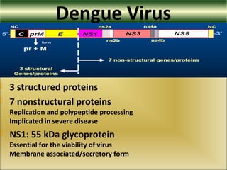
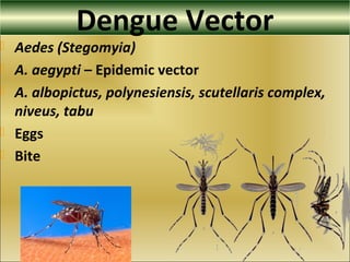
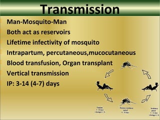
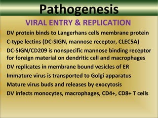
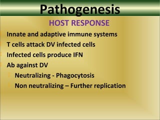
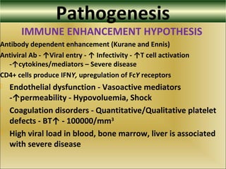
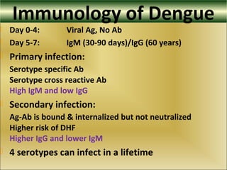
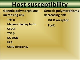
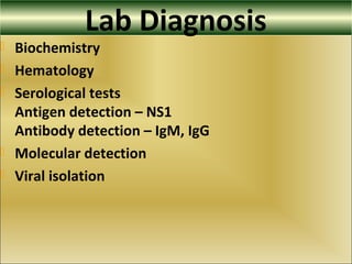
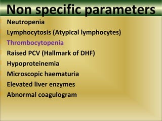
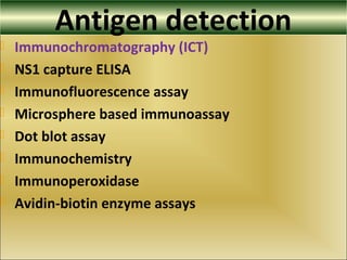
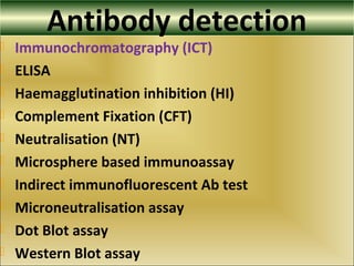
Prologue
 Introduction
 Classification
 Epidemiology
 Virus
 Vector
 Transmission
 Immunology
 Pathogenesis
 Clinical Features
 Lab diagnosis
 Nonspecific
 Immunology
 NAT
 Virus isolation
 Prevention & future
Dengue
 Dengue - Swahili “Ka-Dinga pepo”
Sudden cramp like disease caused by an evil spirit
 Dandy Fever, Breakbone Fever, Breakheart Fever
 Infectious thrombocytopenic purpura
 Mosquito borne Flavivirus infection
 Tropical and subtropical areas
 Earliest record - Chinese encyclopedia of disease
symptoms and remedies 265-420 AD
Classification
 WHO Classification 2009
 Uncomplicated
 Severe
 WHO Classification 1997
 Undifferentiated fever
 Dengue Fever (DF)
 Dengue Hemorrhagic Fever (DHF)
 Gd I – Easy bruising, + Tourniquet test, fever
 Gd II – Spontaneous bleeding
 Gd III – Shock
 Gd IV – Severe Shock (Unrecordable pulse and BP)
Epidemiology
 Second to malaria in tropical areas
 Threatens 2.5 bn (40%) population
 Disease burden – 1600 DALYs/million population
 Sporadic – Endemic – Hyperendemic – Epidemic –
Pandemic ???
 Major public health problem
 No approved vaccines
 Ineffective antiviral therapy
Epidemiology
 1799 – Egypt and Indonesia, 1780 – US
 Fewer epidemics till 1940s
 First epidemic DHF – 1954 Manila
 Increased epidemicity by 1980-90s
worldwide, all serotypes, DEN2
 1998 – Dengue pandemic, DEN3
56 countries worldwide
 2005 – South America, DEN3
 DF 100 m, DHF 50 m, Deaths 50000
 Case Fatality Rate - 0.5-3.5%
Dengue - SEAR
Dengue - SEAR
Dengue - SEAR
 1963 – Kolkata epidemic
80 regular outbreaks in many parts of India except North East
 1967, 70, 82, 88, 92 – DEN1, DEN2
 1996 – Major outbreak, DEN2
 2009 – Delhi and other areas, DEN3
 Ongoing
Dengue - India
Dengue Virus
 Spherical (Diameter 40-50 nm)
 Lipid enveloped
 Positive strand RNA
 Virus - 1940s, serotypes – 1956
 4 serotypes: DEN1 - DEN4
 10.7 kb RNA genome
 10,233 nucleotides and 3411 amino acids
Dengue Virus
 3 structured proteins
 7 nonstructural proteins
Replication and polypeptide processing
Implicated in severe disease
 NS1: 55 kDa glycoprotein
Essential for the viability of virus
Membrane associated/secretory form
Dengue Vector
 Aedes (Stegomyia)
 A. aegypti – Epidemic vector
 A. albopictus, polynesiensis, scutellaris complex,
niveus, tabu
 Eggs
 Bite
Transmission
 Man-Mosquito-Man
 Both act as reservoirs
 Lifetime infectivity of mosquito
 Intrapartum, percutaneous,mucocutaneous
 Blood transfusion, Organ transplant
 Vertical transmission
 IP: 3-14 (4-7) days
Pathogenesis
VIRAL ENTRY & REPLICATION
 DV protein binds to Langerhans cells membrane protein
 C-type lectins (DC-SIGN, mannose receptor, CLEC5A)
 DC-SIGN/CD209 is nonspecific mannose binding receptor
for foreign material on dendritic cell and macrophages
 DV replicates in membrane bound vesicles of ER
 Immature virus is transported to Golgi apparatus
 Mature virus buds and releases by exocytosis
 DV infects monocytes, macrophages, CD4+, CD8+ T cells
Pathogenesis
HOST RESPONSE
 Innate and adaptive immune systems
 T cells attack DV infected cells
 Infected cells produce IFN
 Ab against DV
 Neutralizing - Phagocytosis
 Non neutralizing – Further replication
Pathogenesis
IMMUNE ENHANCEMENT HYPOTHESIS
Antibody dependent enhancement (Kurane and Ennis)
Antiviral Ab - ↑Viral entry - ↑ Infectivity - ↑T cell activation
-↑cytokines/mediators – Severe disease
CD4+ cells produce IFNY, upregulation of FcY receptors
 Endothelial dysfunction - Vasoactive mediators
-↑permeability - Hypovoluemia, Shock
 Coagulation disorders - Quantitative/Qualitative platelet
defects - BT↑ - 100000/mm3
 High viral load in blood, bone marrow, liver is associated
with severe disease
Immunology of Dengue
 Day 0-4: Viral Ag, No Ab
 Day 5-7: IgM (30-90 days)/IgG (60 years)
 Primary infection:
Serotype specific Ab
Serotype cross reactive Ab
High IgM and low IgG
 Secondary infection:
Ag-Ab is bound & internalized but not neutralized
Higher risk of DHF
Higher IgG and lower IgM
 4 serotypes can infect in a lifetime
Host susceptibility
 Genetic polymorphisms
increasing risk
 TNF α
 Mannan binding lectin
 CTLA4
 TGF β
 DC-SIGN
 HLA
 G6PD deficiency
 Genetic polymorphisms
decreasing risk
 Vit D receptor
 FcyR
Incubation peroid
Clinical Features
 Asymptomatic 80%
 Uncomplicated fever
 Severe illness 5%
 Life threatening
 Diabetes
 Asthma
 Febrile phase
 2-7 days
 Fever (Biphasic)
 Headache
 Mucosal bleed
 Muscle ache
 Joint pain
 Vomiting
 Rash (Measles like)
 Diarrhoea
Clinical Features
 Critical phase
 1-2 days
 Hypotension
 Pleural effusion
 Ascites
 Severe bleed (GIT)
 Organ dysfunction
 Recovery phase
 2-3 days
 Altered consciousness
 Seizures
 Itching
 Bradycardia
Clinical Features (DF)
CASE DEFINITION OF DENGUE FEVER (WHO 2006)
 Suspected - Acute febrile illness + >2 of
headache, retro orbital pain, myalgia, arthralgia, rash,
hemorrhagic manifestations and leucopenia
 Probable - Clinical description and serology
reciprocal HI Ab titre > 1280, comparable IgG ELISA or
positive IgM Ab test
 Confirmed
 Reportable - Any probable or confirmed case
Clinical Features (DHF)
CASE DEFINITION OF DHF (WHO 2006)
 Acute onset high fever for 2-7 days
 Hemorrhagic manifestations with at least a positive
tourniquet test
 Platelet <100 x 109
per litre
 Hemoconcentration (rising PCV >20%) or
 Evidence of plasma leakage- ascites, pleural effusion, low
serum protein
Laboratory criteria
LABORATORY CRITERIA FOR DENGUE FEVER (WHO 2006)
 Isolation of the DV from serum or autopsy samples or
 Fourfold or greater rise in reciprocal IgG or IgM Ab titres
to DV Ags in paired sera or
 DV Ag in autopsy tissue, serum or CSF by
immunochemistry, IFA or ELISA or
 Detection of DV genomic sequences in autopsy tissue,
serum or CSF by PCR
Differential diagnosis
 Chickungunya
 West Nile, JE
 Influenza
 Measles
 Rubella
 Typhoid
 Meningococcemia
 Leptospirosis
 Rickettsial infections
 Malaria
Specimens Blood, serum, plasma
 Washed leucocytes
 CSF
 Saliva
 Homogenized/minced autopsy tissues
 Homogenized pooled mosquitoes
 Wet ice - - Thiomersal or sodium azide
 <24 hrs: 4-80
C, -700
C for longer duration
 Acute phase sample
 Convalescent phase sample
Lab Diagnosis
 Biochemistry
 Hematology
 Serological tests
Antigen detection – NS1
Antibody detection – IgM, IgG
 Molecular detection
 Viral isolation
Non specific parameters
 Neutropenia
 Lymphocytosis (Atypical lymphocytes)
 Thrombocytopenia
 Raised PCV (Hallmark of DHF)
 Hypoproteinemia
 Microscopic haematuria
 Elevated liver enzymes
 Abnormal coagulogram
Antigen detection
 Immunochromatography (ICT)
 NS1 capture ELISA
 Immunofluorescence assay
 Microsphere based immunoassay
 Dot blot assay
 Immunochemistry
 Immunoperoxidase
 Avidin-biotin enzyme assays
Antibody detection
 Immunochromatography (ICT)
 ELISA
 Haemagglutination inhibition (HI)
 Complement Fixation (CFT)
 Neutralisation (NT)
 Microsphere based immunoassay
 Indirect immunofluorescent Ab test
 Microneutralisation assay
 Dot Blot assay
 Western Blot assay
ICT
 Lateral flow immunochromatography
 Most frequently used
 Sensitive and specific
 Simple, rapid, easy
 Can be used for mass screening
ELISA
 MAC ELISA
 NS1 serotype specific IgG ELISA
 Pathozyme IgM/Pathozyme IgG
 Platelia Dengue NS1 Ag ELISA – mAb
 IgG avidity ELISA
 Capture ultramicro ELISA
 Dot ELISA
 AuBioDOT IgM capture
MAC ELISA
 Most widely used
 Simple, rapid, requires minimal equipment
 Specificity is not very high
 Unequivocal diagnosis – 4 fold rise
 Not a confirmatory test when single sample used
 Cannot be used to identify serotypes
 Useful tool for surveillance of DF, DHF and DSS
HI
 Frequently used
 Sensitive, easy and reliable
 Detectable by day 5-6 of fever
 Titers >1280 considered significant
 HI Ab persist for at least 48 years
 Suitable for seroepidemiologic studies
 Not suitable for secondary infections
 Not specific
CFT
 Not widely used - Difficult
 Complement is consumed during Ag-Ab reactions
 Detectable earlier than HI Ab
 More specific in primary infections
 Not specific for secondary infections
 Not valuable for seroepidemiologic studies
NT
 Most sensitive and specific
 Serum dilution plaque reduction
 Suitable for primary infections
 NT Ab persist for at least 48 years
 Suitable for seroepidemiologic studies
 Not suitable for secondary infections
 Demanding (time, effort and cost)
Molecular detection
 RT-PCR
 qRT-PCR
 Nested PCR
 Multiplex PCR
 NASBA
 TMA
 bDNA
 RT LAMP
 Nucleic Acid Hybridization
 Self sustained sequence
replication (3SR)
 Strand displacement
amplification (SDA)
Viral isolation
 Mammalian cells - Vero
 Mosquito cell lines – A. albopictus C6/36
Aedes pseudoscutellaris AP61
 Newborn mice (intracerebral)
 Adult mosquitoes (intrathoracic)
A. aegypti, albopictus
Toxorhynchites splendens, amboinensis
 Lengthy, laborious, low sensitivity, expensive
Diagnostic decision
 Choice of test
Rapidity, sensitivity, specificity,
Suitability at a particular stage of disease
Ease of execution, standardization and
automation
 Combination of tests
 Quest for the new Gold Standard
Management & Prevention
 Supportive
 Rehydration
 Antipyretics
 Blood transfusion
 Whole blood
 Packed RBCs
 Platelets
 FFP
 Mosquito reduction
 Habitat reduction
 Prevention of bites
 Anti-dengue drug ?
 New vaccine ?
References1. Chaturvedi UC, Agarwal R, Elbishbishi EA, Mustafa AS. Cytokine Cascade in Dengue Haemorrhagic Fever:
Implications for Pathogenesis. FEMS Immunol Med Microbiol 2000; 28: 183-188.
2. Berry N, Chakravarti A, Gur R, Mathur MD. Serological investigation of a febrile outbreak in Delhi, India, using
a rapid immunochromatographic test. J Clin Microbiol 1998; 36: 2795-6.
3. Tripathi BK, Gupta B, Sinha RS, Prasad S, Sharma DK: Experience in adult population in dengue outbreak in
Delhi. J Assoc Physicians India 1998, 46: 273-76.
4. Kumar M, Pasha S T, Mittal V, Rawat D S, Arya C , Agarwal N, Bhattacharaya D, Lal S, Rai A. Unusual
emergence of guate 98 like molecular subtype of Den -3 during 2003 dengue outbreak in Delhi, dengue
Bull.2004; 28: 101-167.
5. Gubler, DJ 1998. Dengue and dengue hemorrhagic fever. Clin. Microbiol. Rev. 11:480–496.
6. Smith AW, Chen LH, Massad E, Wilson ME. Threat of dengue to blood safety in dengue-endemic countries.
Emerg Infect Dis 2009; 15:8-11.
7. Kurane I, Takasaki T: Dengue fever and dengue hemorrhagic fever: challenges of controlling an enemy still at
large. Rev Med Virol 2001, 11:301-311.
8. Messer WB, Gubler DJ, Harris E, Sivananthan K, de Silva AM: Emergence and global spread of a dengue
serotype 3, subtype III virus. Emerg Infect Dis 2003, 9:800-9.
9. Guzman MG, Kouri G. Dengue: An update. Lancet Infect Dis 2002; 2:33-42.
10. WHO (1997) Dengue hemorrhagic fever: diagnosis, treatment, prevention and control, 2nd edition. Geneva:
World Health Organization.
11. Broor S, Dar L, Sengupta S, Chakaraborty M, Wali JP, Biswas A, Kabra SK, Jain Y, Seth P: Recent dengue
epidemic in Delhi, India. In Factors in the emergence of arbovirus diseases.: Elsevier; 1997: P123-7.
12. Chouhan GS, Rodrigues FM, Shaikh BH, Khangaro SS, Mathur KN, et al. Clinical & virological study of
dengue fever outbreak in Rajasthan.1985. Indian J Med Res 1990; 91: 414-8.
13. Monath TP. Dengue: The risk to developed and developing countries. Proctl Natl Acad Sci USA 1994; 91:
2395-400.
14. Morens DM, Rigau-Perez JG, Lopez-Correa RH,et al.: Dengue in Puerto Rico, 1977: public health response
to characterize and control an epidemic of multiple serotypes. Am J Trop Med Hyg 1986, 35: 197-211.
References15. Halstead SB, Nimmannitya S, Cohen SN: Observations related to pathogenesis of dengue hemorrhagic
fever. Yale J Biol Med 1970, 42: 311-328.
16. WHO: Strengthening implementation of global strategy for dengue fever/ dengue hemorrhagic fever,
prevention and control, report on informal consolation, WHO; 1999.
17. Gubler D J, Sather G E. Laboratory diagnosis of dengue and dengue hemorrhagic fever. In: Homma A,
Cunha J F, editors. Proceedings of the International Symposium on Yellow Fever and Dengue. 1988. pp. 291–
322.
18. World health organization.1997, Dengue hemorrhagic fever: diagnosis, treatment, prevention and control,
second edition. World health organization Geneva, Switzerland.
19. Vaughn DW, Kalayanarooj S, Innis BL, Nimmannitya S et al. Dengue viremia titer, antibody response pattern,
and virus serotype correlate with disease severity. J Infect Dis 2000, 181: 2-9.
20. Groen, J., P. Koraka, J. Velzing and A. D. M. E. Osterhaus. 2000. Evaluation of of dengue virus-specific
immunoglobulin M and G antibodies. Clin. Diagn. Lab. Immunol. 7: 867–871.
21. Mantke, O.D.,K,Lemmer, S.S.Biel, J.et al.2004. Quality control assessment for the serological diagnosis of
dengue virus infection. J.Clin. Virol. 2004; 29: 105-112.
22. Lanciotti, R. S., C. H. Calisher, D. J. Gubler, G. J. Chang, and A. V. Vorndam. 1992. Rapid detection and
typing of dengue viruses from clinical samples by using reverse transcriptase-polymerase chain reaction. J. Clin.
Microbiol. 30: 545–551.
23. Henchal, E. A., J. M. McCown, M. C. Seguin, M. K. Gentry, and W. E.Brandt. 1983. Rapid identification of
dengue virus isolates by using monoclonal antibodies in an indirect immunofluorescence assay. Am. J. Trop.
Med. Hyg. 32: 164–169.
24. Harris, E., T. G. Roberts, L. Smith, J. Selle, L. D. Kramer, S. Valle, E. Sandoval, and A. Balmaseda. 1998.
Typing of dengue viruses in clinical specimens and mosquitoes by single-tube multiplex reverse transcriptase
PCR. J. Clin. Microbiol. 36: 2634–2639.
25. Notomi T et al. Loop-mediated isothermal amplification of DNA. Nucleic Acids Research 2000, 28: E63.
26. Kurane, I., and F. A. Ennis. 1997. Immunopathogenesis of dengue virus infections, p. 273-290. In D. J.
Gubler, and G. Kuno (ed.), Dengue and dengue hemorrhagic fever. CAB International, London, United Kingdom.
Thank You

Dengue

  • 2.
    Prologue  Introduction  Classification Epidemiology  Virus  Vector  Transmission  Immunology  Pathogenesis  Clinical Features  Lab diagnosis  Nonspecific  Immunology  NAT  Virus isolation  Prevention & future
  • 3.
    Dengue  Dengue -Swahili “Ka-Dinga pepo” Sudden cramp like disease caused by an evil spirit  Dandy Fever, Breakbone Fever, Breakheart Fever  Infectious thrombocytopenic purpura  Mosquito borne Flavivirus infection  Tropical and subtropical areas  Earliest record - Chinese encyclopedia of disease symptoms and remedies 265-420 AD
  • 4.
    Classification  WHO Classification2009  Uncomplicated  Severe  WHO Classification 1997  Undifferentiated fever  Dengue Fever (DF)  Dengue Hemorrhagic Fever (DHF)  Gd I – Easy bruising, + Tourniquet test, fever  Gd II – Spontaneous bleeding  Gd III – Shock  Gd IV – Severe Shock (Unrecordable pulse and BP)
  • 5.
    Epidemiology  Second tomalaria in tropical areas  Threatens 2.5 bn (40%) population  Disease burden – 1600 DALYs/million population  Sporadic – Endemic – Hyperendemic – Epidemic – Pandemic ???  Major public health problem  No approved vaccines  Ineffective antiviral therapy
  • 6.
    Epidemiology  1799 –Egypt and Indonesia, 1780 – US  Fewer epidemics till 1940s  First epidemic DHF – 1954 Manila  Increased epidemicity by 1980-90s worldwide, all serotypes, DEN2  1998 – Dengue pandemic, DEN3 56 countries worldwide  2005 – South America, DEN3  DF 100 m, DHF 50 m, Deaths 50000  Case Fatality Rate - 0.5-3.5%
  • 8.
  • 9.
  • 10.
  • 11.
     1963 –Kolkata epidemic 80 regular outbreaks in many parts of India except North East  1967, 70, 82, 88, 92 – DEN1, DEN2  1996 – Major outbreak, DEN2  2009 – Delhi and other areas, DEN3  Ongoing Dengue - India
  • 12.
    Dengue Virus  Spherical(Diameter 40-50 nm)  Lipid enveloped  Positive strand RNA  Virus - 1940s, serotypes – 1956  4 serotypes: DEN1 - DEN4  10.7 kb RNA genome  10,233 nucleotides and 3411 amino acids
  • 13.
    Dengue Virus  3structured proteins  7 nonstructural proteins Replication and polypeptide processing Implicated in severe disease  NS1: 55 kDa glycoprotein Essential for the viability of virus Membrane associated/secretory form
  • 14.
    Dengue Vector  Aedes(Stegomyia)  A. aegypti – Epidemic vector  A. albopictus, polynesiensis, scutellaris complex, niveus, tabu  Eggs  Bite
  • 15.
    Transmission  Man-Mosquito-Man  Bothact as reservoirs  Lifetime infectivity of mosquito  Intrapartum, percutaneous,mucocutaneous  Blood transfusion, Organ transplant  Vertical transmission  IP: 3-14 (4-7) days
  • 16.
    Pathogenesis VIRAL ENTRY &REPLICATION  DV protein binds to Langerhans cells membrane protein  C-type lectins (DC-SIGN, mannose receptor, CLEC5A)  DC-SIGN/CD209 is nonspecific mannose binding receptor for foreign material on dendritic cell and macrophages  DV replicates in membrane bound vesicles of ER  Immature virus is transported to Golgi apparatus  Mature virus buds and releases by exocytosis  DV infects monocytes, macrophages, CD4+, CD8+ T cells
  • 17.
    Pathogenesis HOST RESPONSE  Innateand adaptive immune systems  T cells attack DV infected cells  Infected cells produce IFN  Ab against DV  Neutralizing - Phagocytosis  Non neutralizing – Further replication
  • 18.
    Pathogenesis IMMUNE ENHANCEMENT HYPOTHESIS Antibodydependent enhancement (Kurane and Ennis) Antiviral Ab - ↑Viral entry - ↑ Infectivity - ↑T cell activation -↑cytokines/mediators – Severe disease CD4+ cells produce IFNY, upregulation of FcY receptors  Endothelial dysfunction - Vasoactive mediators -↑permeability - Hypovoluemia, Shock  Coagulation disorders - Quantitative/Qualitative platelet defects - BT↑ - 100000/mm3  High viral load in blood, bone marrow, liver is associated with severe disease
  • 19.
    Immunology of Dengue Day 0-4: Viral Ag, No Ab  Day 5-7: IgM (30-90 days)/IgG (60 years)  Primary infection: Serotype specific Ab Serotype cross reactive Ab High IgM and low IgG  Secondary infection: Ag-Ab is bound & internalized but not neutralized Higher risk of DHF Higher IgG and lower IgM  4 serotypes can infect in a lifetime
  • 20.
    Host susceptibility  Geneticpolymorphisms increasing risk  TNF α  Mannan binding lectin  CTLA4  TGF β  DC-SIGN  HLA  G6PD deficiency  Genetic polymorphisms decreasing risk  Vit D receptor  FcyR
  • 21.
  • 22.
    Clinical Features  Asymptomatic80%  Uncomplicated fever  Severe illness 5%  Life threatening  Diabetes  Asthma  Febrile phase  2-7 days  Fever (Biphasic)  Headache  Mucosal bleed  Muscle ache  Joint pain  Vomiting  Rash (Measles like)  Diarrhoea
  • 23.
    Clinical Features  Criticalphase  1-2 days  Hypotension  Pleural effusion  Ascites  Severe bleed (GIT)  Organ dysfunction  Recovery phase  2-3 days  Altered consciousness  Seizures  Itching  Bradycardia
  • 24.
    Clinical Features (DF) CASEDEFINITION OF DENGUE FEVER (WHO 2006)  Suspected - Acute febrile illness + >2 of headache, retro orbital pain, myalgia, arthralgia, rash, hemorrhagic manifestations and leucopenia  Probable - Clinical description and serology reciprocal HI Ab titre > 1280, comparable IgG ELISA or positive IgM Ab test  Confirmed  Reportable - Any probable or confirmed case
  • 25.
    Clinical Features (DHF) CASEDEFINITION OF DHF (WHO 2006)  Acute onset high fever for 2-7 days  Hemorrhagic manifestations with at least a positive tourniquet test  Platelet <100 x 109 per litre  Hemoconcentration (rising PCV >20%) or  Evidence of plasma leakage- ascites, pleural effusion, low serum protein
  • 26.
    Laboratory criteria LABORATORY CRITERIAFOR DENGUE FEVER (WHO 2006)  Isolation of the DV from serum or autopsy samples or  Fourfold or greater rise in reciprocal IgG or IgM Ab titres to DV Ags in paired sera or  DV Ag in autopsy tissue, serum or CSF by immunochemistry, IFA or ELISA or  Detection of DV genomic sequences in autopsy tissue, serum or CSF by PCR
  • 27.
    Differential diagnosis  Chickungunya West Nile, JE  Influenza  Measles  Rubella  Typhoid  Meningococcemia  Leptospirosis  Rickettsial infections  Malaria
  • 28.
    Specimens Blood, serum,plasma  Washed leucocytes  CSF  Saliva  Homogenized/minced autopsy tissues  Homogenized pooled mosquitoes  Wet ice - - Thiomersal or sodium azide  <24 hrs: 4-80 C, -700 C for longer duration  Acute phase sample  Convalescent phase sample
  • 29.
    Lab Diagnosis  Biochemistry Hematology  Serological tests Antigen detection – NS1 Antibody detection – IgM, IgG  Molecular detection  Viral isolation
  • 30.
    Non specific parameters Neutropenia  Lymphocytosis (Atypical lymphocytes)  Thrombocytopenia  Raised PCV (Hallmark of DHF)  Hypoproteinemia  Microscopic haematuria  Elevated liver enzymes  Abnormal coagulogram
  • 31.
    Antigen detection  Immunochromatography(ICT)  NS1 capture ELISA  Immunofluorescence assay  Microsphere based immunoassay  Dot blot assay  Immunochemistry  Immunoperoxidase  Avidin-biotin enzyme assays
  • 32.
    Antibody detection  Immunochromatography(ICT)  ELISA  Haemagglutination inhibition (HI)  Complement Fixation (CFT)  Neutralisation (NT)  Microsphere based immunoassay  Indirect immunofluorescent Ab test  Microneutralisation assay  Dot Blot assay  Western Blot assay
  • 33.
    ICT  Lateral flowimmunochromatography  Most frequently used  Sensitive and specific  Simple, rapid, easy  Can be used for mass screening
  • 34.
    ELISA  MAC ELISA NS1 serotype specific IgG ELISA  Pathozyme IgM/Pathozyme IgG  Platelia Dengue NS1 Ag ELISA – mAb  IgG avidity ELISA  Capture ultramicro ELISA  Dot ELISA  AuBioDOT IgM capture
  • 35.
    MAC ELISA  Mostwidely used  Simple, rapid, requires minimal equipment  Specificity is not very high  Unequivocal diagnosis – 4 fold rise  Not a confirmatory test when single sample used  Cannot be used to identify serotypes  Useful tool for surveillance of DF, DHF and DSS
  • 36.
    HI  Frequently used Sensitive, easy and reliable  Detectable by day 5-6 of fever  Titers >1280 considered significant  HI Ab persist for at least 48 years  Suitable for seroepidemiologic studies  Not suitable for secondary infections  Not specific
  • 37.
    CFT  Not widelyused - Difficult  Complement is consumed during Ag-Ab reactions  Detectable earlier than HI Ab  More specific in primary infections  Not specific for secondary infections  Not valuable for seroepidemiologic studies
  • 38.
    NT  Most sensitiveand specific  Serum dilution plaque reduction  Suitable for primary infections  NT Ab persist for at least 48 years  Suitable for seroepidemiologic studies  Not suitable for secondary infections  Demanding (time, effort and cost)
  • 39.
    Molecular detection  RT-PCR qRT-PCR  Nested PCR  Multiplex PCR  NASBA  TMA  bDNA  RT LAMP  Nucleic Acid Hybridization  Self sustained sequence replication (3SR)  Strand displacement amplification (SDA)
  • 40.
    Viral isolation  Mammaliancells - Vero  Mosquito cell lines – A. albopictus C6/36 Aedes pseudoscutellaris AP61  Newborn mice (intracerebral)  Adult mosquitoes (intrathoracic) A. aegypti, albopictus Toxorhynchites splendens, amboinensis  Lengthy, laborious, low sensitivity, expensive
  • 41.
    Diagnostic decision  Choiceof test Rapidity, sensitivity, specificity, Suitability at a particular stage of disease Ease of execution, standardization and automation  Combination of tests  Quest for the new Gold Standard
  • 42.
    Management & Prevention Supportive  Rehydration  Antipyretics  Blood transfusion  Whole blood  Packed RBCs  Platelets  FFP  Mosquito reduction  Habitat reduction  Prevention of bites  Anti-dengue drug ?  New vaccine ?
  • 43.
    References1. Chaturvedi UC,Agarwal R, Elbishbishi EA, Mustafa AS. Cytokine Cascade in Dengue Haemorrhagic Fever: Implications for Pathogenesis. FEMS Immunol Med Microbiol 2000; 28: 183-188. 2. Berry N, Chakravarti A, Gur R, Mathur MD. Serological investigation of a febrile outbreak in Delhi, India, using a rapid immunochromatographic test. J Clin Microbiol 1998; 36: 2795-6. 3. Tripathi BK, Gupta B, Sinha RS, Prasad S, Sharma DK: Experience in adult population in dengue outbreak in Delhi. J Assoc Physicians India 1998, 46: 273-76. 4. Kumar M, Pasha S T, Mittal V, Rawat D S, Arya C , Agarwal N, Bhattacharaya D, Lal S, Rai A. Unusual emergence of guate 98 like molecular subtype of Den -3 during 2003 dengue outbreak in Delhi, dengue Bull.2004; 28: 101-167. 5. Gubler, DJ 1998. Dengue and dengue hemorrhagic fever. Clin. Microbiol. Rev. 11:480–496. 6. Smith AW, Chen LH, Massad E, Wilson ME. Threat of dengue to blood safety in dengue-endemic countries. Emerg Infect Dis 2009; 15:8-11. 7. Kurane I, Takasaki T: Dengue fever and dengue hemorrhagic fever: challenges of controlling an enemy still at large. Rev Med Virol 2001, 11:301-311. 8. Messer WB, Gubler DJ, Harris E, Sivananthan K, de Silva AM: Emergence and global spread of a dengue serotype 3, subtype III virus. Emerg Infect Dis 2003, 9:800-9. 9. Guzman MG, Kouri G. Dengue: An update. Lancet Infect Dis 2002; 2:33-42. 10. WHO (1997) Dengue hemorrhagic fever: diagnosis, treatment, prevention and control, 2nd edition. Geneva: World Health Organization. 11. Broor S, Dar L, Sengupta S, Chakaraborty M, Wali JP, Biswas A, Kabra SK, Jain Y, Seth P: Recent dengue epidemic in Delhi, India. In Factors in the emergence of arbovirus diseases.: Elsevier; 1997: P123-7. 12. Chouhan GS, Rodrigues FM, Shaikh BH, Khangaro SS, Mathur KN, et al. Clinical & virological study of dengue fever outbreak in Rajasthan.1985. Indian J Med Res 1990; 91: 414-8. 13. Monath TP. Dengue: The risk to developed and developing countries. Proctl Natl Acad Sci USA 1994; 91: 2395-400. 14. Morens DM, Rigau-Perez JG, Lopez-Correa RH,et al.: Dengue in Puerto Rico, 1977: public health response to characterize and control an epidemic of multiple serotypes. Am J Trop Med Hyg 1986, 35: 197-211.
  • 44.
    References15. Halstead SB,Nimmannitya S, Cohen SN: Observations related to pathogenesis of dengue hemorrhagic fever. Yale J Biol Med 1970, 42: 311-328. 16. WHO: Strengthening implementation of global strategy for dengue fever/ dengue hemorrhagic fever, prevention and control, report on informal consolation, WHO; 1999. 17. Gubler D J, Sather G E. Laboratory diagnosis of dengue and dengue hemorrhagic fever. In: Homma A, Cunha J F, editors. Proceedings of the International Symposium on Yellow Fever and Dengue. 1988. pp. 291– 322. 18. World health organization.1997, Dengue hemorrhagic fever: diagnosis, treatment, prevention and control, second edition. World health organization Geneva, Switzerland. 19. Vaughn DW, Kalayanarooj S, Innis BL, Nimmannitya S et al. Dengue viremia titer, antibody response pattern, and virus serotype correlate with disease severity. J Infect Dis 2000, 181: 2-9. 20. Groen, J., P. Koraka, J. Velzing and A. D. M. E. Osterhaus. 2000. Evaluation of of dengue virus-specific immunoglobulin M and G antibodies. Clin. Diagn. Lab. Immunol. 7: 867–871. 21. Mantke, O.D.,K,Lemmer, S.S.Biel, J.et al.2004. Quality control assessment for the serological diagnosis of dengue virus infection. J.Clin. Virol. 2004; 29: 105-112. 22. Lanciotti, R. S., C. H. Calisher, D. J. Gubler, G. J. Chang, and A. V. Vorndam. 1992. Rapid detection and typing of dengue viruses from clinical samples by using reverse transcriptase-polymerase chain reaction. J. Clin. Microbiol. 30: 545–551. 23. Henchal, E. A., J. M. McCown, M. C. Seguin, M. K. Gentry, and W. E.Brandt. 1983. Rapid identification of dengue virus isolates by using monoclonal antibodies in an indirect immunofluorescence assay. Am. J. Trop. Med. Hyg. 32: 164–169. 24. Harris, E., T. G. Roberts, L. Smith, J. Selle, L. D. Kramer, S. Valle, E. Sandoval, and A. Balmaseda. 1998. Typing of dengue viruses in clinical specimens and mosquitoes by single-tube multiplex reverse transcriptase PCR. J. Clin. Microbiol. 36: 2634–2639. 25. Notomi T et al. Loop-mediated isothermal amplification of DNA. Nucleic Acids Research 2000, 28: E63. 26. Kurane, I., and F. A. Ennis. 1997. Immunopathogenesis of dengue virus infections, p. 273-290. In D. J. Gubler, and G. Kuno (ed.), Dengue and dengue hemorrhagic fever. CAB International, London, United Kingdom.
  • 45.