This document provides information on the structure and functions of various cell organelles found in eukaryotic cells, including the cytoplasm, mitochondria, chloroplasts, endoplasmic reticulum, Golgi complex, lysosomes, ribosomes and vacuoles. It describes their roles in processes like cellular respiration, photosynthesis, protein synthesis, transport of materials within the cell, and storage of molecules.
Cytoplasm is a gel like fluid present between the plasma membrane and the nucleus
Cytoplasm is the semi-fluid substance of a cell that is present within the cellular membrane and surrounds the nuclear membrane
It is sometimes described as the nonnuclear content of the protoplasm
The endoplasmic reticulum (ER) is a network of interconnected membranes found throughout the cytoplasm of eukaryotic cells. It consists of flat sacs and tubules with a single continuous lumen. The ER is involved in protein transport and synthesis, lipid and steroid synthesis, calcium storage, and processing of toxins. It has two types - smooth ER which lacks ribosomes and is involved in lipid synthesis, and rough ER which is studded with ribosomes and is the main site of protein synthesis.
The cytoplasm is the jelly-like substance within cells that surrounds the organelles and nucleus. It is made up mostly of water along with molecules like enzymes, salts, and cytosol. The cytoplasm contains membrane-enclosed organelles that each perform specialized functions, as well as inclusions that store nutrients or waste. It aids many cellular functions like movement of materials, maintaining cell shape, and acting as a site for metabolic reactions like glycolysis.
Mitochondria are double-membrane organelles found in eukaryotic cells that are considered the powerhouses of the cell. They contain their own DNA and machinery for transcription and translation. Evidence suggests that mitochondria originated from bacteria that were engulfed by early eukaryotic cells in an endosymbiotic event. Mitochondria have similarities to bacteria in terms of size, membrane structure, ribosomes, and DNA shape. They produce energy for the cell through oxidative phosphorylation and are involved in processes like apoptosis. Mitochondrial dysfunction can lead to disease by failing to produce sufficient energy for cellular functions.
The endoplasmic reticulum is a network of membranous tubules and sacs found in eukaryotic cells. It consists of two types - smooth ER and rough ER. Smooth ER is involved in lipid and glycogen metabolism, while rough ER contains ribosomes and is abundant in cells that synthesize proteins. The endoplasmic reticulum provides structure, transports molecules, and aids in various metabolic functions through its enzymes and continuity with other organelles. It helps synthesize proteins and lipids, metabolize drugs and toxins, and provides mechanical support to the cell.
The document discusses the key components and structure of the nucleus. It notes that the nucleus was discovered in 1831 and is located at the center of most cells, where it controls cell activities and houses genetic material. The nucleus contains a double-layered nuclear envelope that encloses chromosomes, nucleolus, and nucleoplasm. Chromosomes contain DNA that provides genetic instructions, while the nucleolus produces ribosomes and the nucleoplasm is a liquid found within the nuclear envelope.
Cytosol is a jelly-like material that surrounds organelles inside cells. It contains proteins that control metabolism and transport molecules between sites of production and places they are used. Cytosol also helps transmit signals from the cell membrane to organelles like the nucleus. The structure of organelles does not influence cytosol's functions of transport and signal transmission.
Chloroplasts are organelles found in plant cells and eukaryotic photosynthetic organisms that conduct photosynthesis. They have a double membrane envelope and contain a stroma, thylakoids, and chloroplast DNA. Thylakoids contain light-absorbing pigments and perform the light reactions of photosynthesis, while the stroma is the site of the dark reactions where CO2 is fixed into sugars. Chloroplasts are essential for photosynthesis as they trap solar energy to produce ATP and NADPH via light reactions, and use these products to fix CO2 into carbohydrates via dark reactions, providing energy for plant growth.
The nucleus is an organelle found in eukaryotic cells that houses the cell's genetic material. It was first observed by Anton van Leeuwenhoek in the 17th century and further described by Robert Brown in the 19th century. The nucleus is surrounded by a double membrane called the nuclear envelope and contains chromatin fibers, nucleoplasm, and one or more nucleoli. It controls cell functions and stores the cell's genetic information in the form of DNA.
The plasma membrane is a flexible yet sturdy lipid bilayer that surrounds the cytoplasm of cells. It is described by the fluid mosaic model, where lipids form a fluid sea containing a mosaic of embedded and floating proteins. The basic structure is a phospholipid bilayer containing cholesterol, glycolipids, and integral and peripheral proteins. Transport across the membrane includes passive diffusion and facilitated diffusion down gradients, as well as active transport against gradients using protein carriers and ATP.
Membranes cover the surface of cells and surround organelles within cells. They serve several functions including maintaining cellular integrity by keeping components inside, selectively controlling movement of molecules in and out, and allowing cellular processes to occur separately within organelles. The plasma membrane forms the boundary of the cell and is made of a phospholipid bilayer with various embedded and attached proteins and carbohydrates. It regulates what enters and exits the cell.
Lysosomes are membrane-bound organelles that contain hydrolytic enzymes. They were discovered in 1955 and function to break down biomolecules through intracellular and extracellular digestion. Lysosomes occur in eukaryotic cells and vary in size, shape, and function. They are formed in the Golgi apparatus and contain around 50 enzymes including proteases, lipases, nucleases and more that function best in acidic environments. Lysosomes play several important roles including digesting cellular waste, breaking down pathogens during phagocytosis, and digesting old cell components through autophagy.
The Golgi apparatus is an organelle found in most eukaryotic cells that was discovered in 1898 by Camillo Golgi. It processes and packages macromolecules after their synthesis in the endoplasmic reticulum. The Golgi apparatus is composed of stacked, flattened sacs called cisternae that modify proteins and lipids through enzymes as they progress through the stacks. It packages macromolecules for secretion from the cell or for other intracellular use and plays an important role in glycosylation.
1. The document discusses cells, which are the basic structural and functional units of living organisms.
2. It provides details on the history of cell discovery from Hooke to Virchow and outlines the three main principles of cell theory.
3. The document describes the key components of cells including the plasma membrane, nucleus, cytoplasm, and various organelles as well as their structure and functions. It provides examples of unicellular and multicellular organisms.
General overview of Plasma/ Cell membrane.
Definition of Plasma/ Cell membrane
Structure of Plasma membrane
1. Sandwitch model ORDanielli- Davson Model
2. Fluid mosaic model
Plasma Membrane Proteins
Chemical Composition of Plasma/ Cell Membrane
Movement across the Cell Membrane
Channels through cell membrane
The document summarizes key aspects of the plasma membrane structure and models. It discusses the plasma membrane's role in separating the cell's cytoplasm from the external environment and controlling molecule movement. The plasma membrane is composed primarily of lipids, proteins, and carbohydrates arranged in a fluid mosaic structure according to the fluid mosaic model. This model proposes the plasma membrane has a fluid-like consistency with protein molecules dotted mosaic-style throughout the lipid bilayer.
Peroxisomes are organelles found in the cytoplasm of plant and animal cells that contain enzymes for oxidizing fatty acids and other organic substances. They produce hydrogen peroxide as a byproduct which is immediately broken down by the enzyme catalase. Peroxisomes play important roles in processes like fatty acid breakdown, bile acid and cholesterol synthesis, and the breakdown of toxic peroxides. Defects in peroxisome function can lead to genetic disorders affecting the nervous system, liver, and other organs.
The cytoskeleton consists of three main types of protein filaments - microtubules, microfilaments, and intermediate filaments. Microtubules provide structure to cells and are involved in cell motility and intracellular transport. Microfilaments are the thinnest filaments and are involved in cell movement and cytoplasmic streaming. Intermediate filaments provide structural support and help maintain cell shape.
The cytoskeleton is a network of protein filaments that extends throughout the cytoplasm. It provides structure and organization to the cell, determining shape and positioning organelles. The three main types of filaments are actin filaments, intermediate filaments, and microtubules. Actin filaments are the thinnest filaments and form structures like filopodia, lamellipodia, and stress fibers. Microtubules are hollow cylinders composed of tubulin dimers and originate from the centrosome. They are involved in processes like cell division, organelle transport, and motility. Cilia and flagella project from the cell surface and use microtubule motors for movement.
The plasma membrane is a selectively permeable membrane that surrounds the cell. It is composed of a phospholipid bilayer with embedded proteins. The fluid mosaic model from 1972 describes the plasma membrane as a fluid bilayer with integral proteins embedded within it, peripheral proteins attached to its surface, and lipid-anchored proteins. The plasma membrane regulates what enters and exits the cell through diffusion, osmosis, facilitated diffusion using channel proteins, and active transport using carrier proteins that require ATP. Endocytosis and exocytosis allow bulk transport across the membrane through vesicles.
This document provides an overview of key cell organelles:
- The cell membrane controls movement of substances in and out of cells. It consists of a lipid bilayer and embedded proteins.
- Mitochondria produce ATP through respiration and regulate metabolism. They have an outer and inner membrane.
- The Golgi apparatus packages and modifies proteins and lipids in the cell.
- The endoplasmic reticulum synthesizes lipids and proteins. It has rough and smooth regions.
- Lysosomes contain enzymes for breaking down biomolecules through autophagy and endocytosis.
- Ribosomes are the sites of protein synthesis in the cell.
This document discusses the structure and function of ribosomes. It begins by introducing ribosomes as large, complex molecules found in all living cells that serve as the primary site of protein synthesis. The document then describes the structure of ribosomes, which consist of two subunits that come together during protein synthesis. It explains the three main steps of protein synthesis carried out by ribosomes - initiation, elongation, and termination - and the role of mRNA and tRNA in translating genetic code into proteins.
The Golgi apparatus was discovered in 1873 by Camillo Golgi and serves important functions in eukaryotic cells. It appears as a complex network of flattened sacs called cisternae, tubules, and vesicles. The Golgi apparatus modifies and packages proteins and lipids and directs them to their proper destinations within the cell or for secretion. It plays a key role in sorting cellular components and is often referred to as the "traffic police of the cell". The Golgi apparatus is involved in processes like cell wall secretion in plants and mucus, lactoprotein, and collagen secretion in animals.
The nucleus is a large membrane-bound organelle found at the center of eukaryotic cells that contains the cell's genetic material in the form of chromosomes. It directs the cell's activities and is involved in cellular reproduction. Key features include being visible under a light microscope due to its acidic nature, containing DNA, RNA, and the nucleolus which produces ribosomes. The nuclear envelope encloses the nucleus and contains pores that regulate material passing in and out.
The nucleus houses a cell's genome and controls cellular activities. It is enclosed by a double membrane nuclear envelope punctuated by nuclear pores that regulate transport between the nucleus and cytoplasm. Within the nucleus, DNA is organized into chromatin and various sub-compartments carry out functions like transcription and RNA processing. Chromosomes occupy distinct territories and nuclear pores facilitate macromolecular transport through the nuclear envelope.
Eukaryotic cells have complex internal structures that allow them to be larger and more specialized than prokaryotic cells. They have a nucleus that contains their DNA and organelles like the endoplasmic reticulum, Golgi apparatus, mitochondria, and chloroplasts that perform specialized functions. Eukaryotic cells also have cytoskeletons and can develop external structures like flagella and cilia. This complex internal organization allows eukaryotic cells to form multicellular organisms and carry out complex processes like photosynthesis.
Ribosomes are organelles found in all cells that synthesize proteins. They consist of RNA and proteins and exist as smaller and larger subunits. Ribosomes were discovered in 1950 and can be seen under electron microscopes. They translate genetic information from mRNA into amino acid chains. Recent research has provided new insights into ribosome structure and function and how they play a role in certain diseases.
The document summarizes key organelles and structures found within eukaryotic cells, including:
1) The cytosol is the jelly-like material within the cell cytoplasm with dissolved substances like amino acids.
2) The nucleus contains the cell's DNA and controls the cell. Inside is the nucleolus which makes ribosomes.
3) Mitochondria convert food into ATP for energy in most eukaryotic cells, with more in muscle cells. They have inner and outer membranes.
4) The endoplasmic reticulum synthesizes proteins and lipids, with ribosomes on the rough ER and no ribosomes on the smooth ER.
This provides a high-level overview of
This document provides information on mitochondria, Golgi bodies, and plastids. It describes the key structures and functions of each organelle. Mitochondria are described as the powerhouses of the cell that produce ATP through cellular respiration. Golgi bodies function in transport, packaging, and modification of proteins and lipids. Plastids, such as chloroplasts, chromoplasts, and leucoplasts, play important roles in photosynthesis, pigment storage, and starch/protein storage in plants.
Ch 08 - Cell : The unit of Life || Class 11 ||SAQIB AHMED
- Robert Hooke first observed cells in 1665 when examining a slice of cork under a microscope. He saw small compartments separated by walls, which he called cells.
- The Cell Theory, developed by Schleiden and Schwann, states that the cell is the basic unit of structure and function of living things, new cells arise from existing cells, and all organisms are composed of one or more cells.
- Cells come in two main types - prokaryotic cells which lack a nucleus and membrane-bound organelles, and eukaryotic cells which have a nucleus surrounded by a nuclear membrane and other membrane-bound structures.
This document provides information on cells at both the prokaryotic and eukaryotic levels. It discusses cell theory and key aspects of prokaryotic cells including their shape, structures like plasmids and mesosomes, and cell envelopes. For eukaryotic cells, it describes the membrane-bound organelles like the nucleus, mitochondria, plastids, Golgi apparatus, endoplasmic reticulum, lysosomes and vacuoles. It also discusses cytoskeletal elements and membrane properties.
Aim : to study cell and it's organelle with help of electron microscope.
Cells are the basic building blocks of living things. The human body is composed of trillions of cells, all with their own specialised function.
Cells are the basic structures of all living organisms.
Cells provide structure for the body, take in nutrients from food and carry out important functions.
Cells group together to form tissues?, which in turn group together to form organs?, such as the heart and brain.
Our cells contain a number of functional structures called organelles?.
These organelles carry out tasks such as making proteins?, processing chemicals and generating energy for the cell.
The nucleus? is based at the centre of the cell and is the ‘control room’ for the cell.
The genome? is found within the nucleus.
CELL FEATURES presentation.pptx andrew.pptxkitati1
Cells have several key features that allow them to carry out essential functions. These include a cell membrane, cytoplasm, nucleus, and various organelles. Plant and animal cells share many structures but plant cells also contain chloroplasts. Cells reproduce, metabolize, maintain homeostasis, pass on hereditary information, respond to stimuli, grow and develop, and evolve adaptations. Prokaryotic and eukaryotic cells differ in their structures, with eukaryotes generally being larger and containing membrane-bound organelles.
Cell biology is the study of the structure and function of cells, the fundamental units of life. There are two main types of cells - prokaryotic and eukaryotic. Eukaryotic cells are larger, more complex, and contain organelles like the nucleus, endoplasmic reticulum, Golgi apparatus, mitochondria, chloroplasts, lysosomes, and peroxisomes, each with specialized functions. The animal cell is surrounded by a plasma membrane and contains a large nucleus housing the DNA, as well as various membrane-bound organelles that carry out specific functions necessary for cell survival and homeostasis.
Filipino
200
English
Philippines
AllImagesVideosMapsNewsFlights
More
About 1,430,000,000 resultsDate
Images of Baroque Artist PPT
bing.com/images
Image result for Baroque Artist PPT. Size: 150 x 112. Source: www.slideserve.com
Image result for Baroque Artist PPT. Size: 150 x 112. Source: vdocuments.net
Image result for Baroque Artist PPT. Size: 150 x 112. Source: www.slideserve.com
Image result for Baroque Artist PPT. Size: 150 x 89. Source: prezi.com
Image result for Baroque Artist PPT. Size: 150 x 112. Source: www.teacherspayteachers.com
Image result for Baroque Artist PPT. Size: 150 x 112. Source: www.slideserve.com
Image result for Baroque Artist PPT. Size: 150 x 84. Source: prezentr.com
Image result for Baroque Artist PPT. Size: 150 x 112. Source: www.slideserve.com
Image result for Baroque Artist PPT. Size: 150 x 112. Source: www.teacherspayteachers.com
Image result for Baroque Artist PPT. Size: 150 x 112. Source: www.slideshare.net
Image result for Baroque Artist PPT. Size: 150 x 112. Source: www.teacherspayteachers.com
Image result for Baroque Artist PPT. Size: 150 x 200. Source: www.scribd.com
Image result for Baroque Artist PPT. Size: 150 x 112. Source: www.slideserve.com
Image result for Baroque Artist PPT. Size: 150 x 112. Source: www.powershow.com
Image result for Baroque Artist PPT. Size: 150 x 112. Source: www.slideshare.net
See all images
Global web icon
slideshare.net
· Jun 28, 2012
Baroque art - SlideShare
Occupation: Visual Artist, Art Instructor
Works For: Art Studio
Oil on canvas. 18” x 16”. 37. France • France’s “sun king,” Louis XIV, preferred Classicism, and he created academies and teachers to perpetuate this Baroque style • The French Baroque is a more reserved style, toward Raphael The artists: • Nicholas Poussin - main exponent of Classical style in France.
https://www.slideshare.net/gwfreeman/baroque-art-13484182
Feedback
EXPLORE 6 RELATED PAGES
PPT - BAROQUE ART PowerPoint Presentation, free download - ID:2696612
1
Global web icon
www.slideserve.com
Baroque PowerPoint Template
Free baroque PowerPoint template is a simple free rose template background with a basic frame for PowerPoint presentations. This background template is a free rose template design for presentations that you can download for baroque presentations in PowerPoint. The renaissance PowerPoint background can also be used in rococo presentations, or classical presentatio
This document provides information about cells through a presentation. It discusses the history of cell discovery, the main structures of plant and animal cells including organelles, differences between prokaryotic and eukaryotic cells, and differences between plant and animal cells. Key figures in cell history discussed include Robert Hooke, Antonie van Leeuwenhoek, Matthias Schleiden, Theodor Schwann, and Rudolf Virchow. The presentation includes diagrams labeling structures of typical plant and animal cells.
The document describes a technology-based lesson on cells for 9th standard science students. It covers various topics related to cells including: the basic unit of life, structural organization of cells, different types of cell organelles, differences between plant and animal cells, and differences between prokaryotic and eukaryotic cells. Diagrams are provided to illustrate cell structures like the plasma membrane, nucleus, mitochondria and more. The key aspects of cell theory are also explained.
1. The cell is the fundamental unit of structure and function in all living organisms.
2. Cells come in a variety of shapes and sizes, and have a plasma membrane, cytoplasm, and organelles that allow them to carry out functions necessary for life.
3. Eukaryotic cells contain a nucleus and membrane-bound organelles, while prokaryotic cells like bacteria lack these structures.
1. A cell is the smallest unit capable of performing life functions and all living things are composed of cells.
2. There are two main types of cells - prokaryotic cells which lack a membrane-bound nucleus and eukaryotic cells which have a membrane-bound nucleus and organelles.
3. Key cell organelles include the nucleus which houses genetic material, mitochondria which generate energy, the endoplasmic reticulum and golgi apparatus which aid in protein transport and modification, and plastids and chloroplasts which perform photosynthesis in plant cells. Together, these organelles allow the cell to carry out all functions necessary for life.
This document discusses the different types of plastids found in plant cells. It begins by describing plastids as double membrane-bound organelles that can vary in shape and contain DNA and ribosomes, allowing them to replicate. Their main functions are food synthesis, carbohydrate and lipid storage. There are three main types of plastids: chloroplasts, which contain chlorophyll and carry out photosynthesis; chromoplasts, which contain carotenoids and give color to flowers and fruits; and leucoplasts, which are colorless and store carbohydrates, proteins or lipids in different plant tissues. The document focuses on describing the structure, components and functions of chloroplasts in detail.
The document discusses the discovery and structure of the Golgi apparatus. It was discovered in 1898 by Italian physician Camillo Golgi and has since been observed under light and electron microscopes. Under electron microscopy, it appears as a stack of flattened sacs called cisternae, along with peripheral tubules and vesicles. The cisternae vary in number between cell types and the Golgi complex plays an important role in processing and transporting materials within the cell.
1. The document discusses the structure and function of cells. It states that cells are the basic unit of life and all organisms are made of one or more cells.
2. The cell theory developed by Schleiden and Schwann states that all organisms are composed of cells, cells come from pre-existing cells, and their functions occur within cells.
3. There are two main types of cells - eukaryotic and prokaryotic. Eukaryotic cells have a nucleus and organelles, while prokaryotic cells like bacteria do not have a nucleus or organelles.
4. Key organelles in eukaryotic cells and their functions are discussed in detail, including the nucleus,
The document discusses the key characteristics and components of cells. It states that the cell is the fundamental unit of life and is the structural and functional basic unit that makes up the whole body. Cells come in both unicellular and multicellular forms. The cell consists of various organelles that allow it to carry out its functions, such as the nucleus, mitochondria, lysosomes, ribosomes, endoplasmic reticulum, and Golgi apparatus. Cells can also be categorized as prokaryotic or eukaryotic based on their structure. The document further describes the roles and characteristics of various cell organelles and components like the nucleus, cell membrane, mitochondria, chloroplasts, and cell wall. It also explains
- Cells are the basic structural and functional units of all living organisms. Cells come in different shapes, sizes, and specialized functions.
- The main structures of a cell include the cell membrane, cytoplasm, organelles, nucleus, and in plant cells, a cell wall. Organelles such as the mitochondria, endoplasmic reticulum, Golgi apparatus, lysosomes, vacuoles, and chloroplasts are contained within the cytoplasm and perform specific functions for the cell.
- The nucleus is the largest organelle and contains genetic material in the form of chromatin fibers and chromosomes. It directs cellular activities and protein synthesis.
Similar to Cytoplasm & cell organelles By Manoj Dhital (M.Sc Medical Microbiology)) (20)
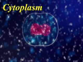
Local Advanced Lung Cancer: Artificial Intelligence, Synergetics, Complex Sys...Oleg Kshivets
Overall life span (LS) was 1671.7±1721.6 days and cumulative 5YS reached 62.4%, 10 years – 50.4%, 20 years – 44.6%. 94 LCP lived more than 5 years without cancer (LS=2958.6±1723.6 days), 22 – more than 10 years (LS=5571±1841.8 days). 67 LCP died because of LC (LS=471.9±344 days). AT significantly improved 5YS (68% vs. 53.7%) (P=0.028 by log-rank test). Cox modeling displayed that 5YS of LCP significantly depended on: N0-N12, T3-4, blood cell circuit, cell ratio factors (ratio between cancer cells-CC and blood cells subpopulations), LC cell dynamics, recalcification time, heparin tolerance, prothrombin index, protein, AT, procedure type (P=0.000-0.031). Neural networks, genetic algorithm selection and bootstrap simulation revealed relationships between 5YS and N0-12 (rank=1), thrombocytes/CC (rank=2), segmented neutrophils/CC (3), eosinophils/CC (4), erythrocytes/CC (5), healthy cells/CC (6), lymphocytes/CC (7), stick neutrophils/CC (8), leucocytes/CC (9), monocytes/CC (10). Correct prediction of 5YS was 100% by neural networks computing (error=0.000; area under ROC curve=1.0).
Title: Sense of Taste
Presenter: Dr. Faiza, Assistant Professor of Physiology
Qualifications:
MBBS (Best Graduate, AIMC Lahore)
FCPS Physiology
ICMT, CHPE, DHPE (STMU)
MPH (GC University, Faisalabad)
MBA (Virtual University of Pakistan)
Learning Objectives:
Describe the structure and function of taste buds.
Describe the relationship between the taste threshold and taste index of common substances.
Explain the chemical basis and signal transduction of taste perception for each type of primary taste sensation.
Recognize different abnormalities of taste perception and their causes.
Key Topics:
Significance of Taste Sensation:
Differentiation between pleasant and harmful food
Influence on behavior
Selection of food based on metabolic needs
Receptors of Taste:
Taste buds on the tongue
Influence of sense of smell, texture of food, and pain stimulation (e.g., by pepper)
Primary and Secondary Taste Sensations:
Primary taste sensations: Sweet, Sour, Salty, Bitter, Umami
Chemical basis and signal transduction mechanisms for each taste
Taste Threshold and Index:
Taste threshold values for Sweet (sucrose), Salty (NaCl), Sour (HCl), and Bitter (Quinine)
Taste index relationship: Inversely proportional to taste threshold
Taste Blindness:
Inability to taste certain substances, particularly thiourea compounds
Example: Phenylthiocarbamide
Structure and Function of Taste Buds:
Composition: Epithelial cells, Sustentacular/Supporting cells, Taste cells, Basal cells
Features: Taste pores, Taste hairs/microvilli, and Taste nerve fibers
Location of Taste Buds:
Found in papillae of the tongue (Fungiform, Circumvallate, Foliate)
Also present on the palate, tonsillar pillars, epiglottis, and proximal esophagus
Mechanism of Taste Stimulation:
Interaction of taste substances with receptors on microvilli
Signal transduction pathways for Umami, Sweet, Bitter, Sour, and Salty tastes
Taste Sensitivity and Adaptation:
Decrease in sensitivity with age
Rapid adaptation of taste sensation
Role of Saliva in Taste:
Dissolution of tastants to reach receptors
Washing away the stimulus
Taste Preferences and Aversions:
Mechanisms behind taste preference and aversion
Influence of receptors and neural pathways
Impact of Sensory Nerve Damage:
Degeneration of taste buds if the sensory nerve fiber is cut
Abnormalities of Taste Detection:
Conditions: Ageusia, Hypogeusia, Dysgeusia (parageusia)
Causes: Nerve damage, neurological disorders, infections, poor oral hygiene, adverse drug effects, deficiencies, aging, tobacco use, altered neurotransmitter levels
Neurotransmitters and Taste Threshold:
Effects of serotonin (5-HT) and norepinephrine (NE) on taste sensitivity
Supertasters:
25% of the population with heightened sensitivity to taste, especially bitterness
Increased number of fungiform papillae
Integrating Ayurveda into Parkinson’s Management: A Holistic ApproachAyurveda ForAll
Explore the benefits of combining Ayurveda with conventional Parkinson's treatments. Learn how a holistic approach can manage symptoms, enhance well-being, and balance body energies. Discover the steps to safely integrate Ayurvedic practices into your Parkinson’s care plan, including expert guidance on diet, herbal remedies, and lifestyle modifications.
Muktapishti is a traditional Ayurvedic preparation made from Shoditha Mukta (Purified Pearl), is believed to help regulate thyroid function and reduce symptoms of hyperthyroidism due to its cooling and balancing properties. Clinical evidence on its efficacy remains limited, necessitating further research to validate its therapeutic benefits.
These simplified slides by Dr. Sidra Arshad present an overview of the non-respiratory functions of the respiratory tract.
Learning objectives:
1. Enlist the non-respiratory functions of the respiratory tract
2. Briefly explain how these functions are carried out
3. Discuss the significance of dead space
4. Differentiate between minute ventilation and alveolar ventilation
5. Describe the cough and sneeze reflexes
Study Resources:
1. Chapter 39, Guyton and Hall Textbook of Medical Physiology, 14th edition
2. Chapter 34, Ganong’s Review of Medical Physiology, 26th edition
3. Chapter 17, Human Physiology by Lauralee Sherwood, 9th edition
4. Non-respiratory functions of the lungs https://academic.oup.com/bjaed/article/13/3/98/278874
Basavarajeeyam is a Sreshta Sangraha grantha (Compiled book ), written by Neelkanta kotturu Basavaraja Virachita. It contains 25 Prakaranas, First 24 Chapters related to Rogas& 25th to Rasadravyas.
- Video recording of this lecture in English language: https://youtu.be/kqbnxVAZs-0
- Video recording of this lecture in Arabic language: https://youtu.be/SINlygW1Mpc
- Link to download the book free: https://nephrotube.blogspot.com/p/nephrotube-nephrology-books.html
- Link to NephroTube website: www.NephroTube.com
- Link to NephroTube social media accounts: https://nephrotube.blogspot.com/p/join-nephrotube-on-social-media.html
3. Cytosol/Matrix/ Hyaloplasm
• Composition : Inorganic compounds – water(75- 85%), salts of
Na, K & other metals.
Organic compounds – carbohydrates, lipids,
proteins, nucleoproteins, nucleic acids, enzymes etc.
• Pheripheral (outer, near plasma membrane) layer is non-granular,
viscous, clear & rigid so called plasma gel or ectoplasm or cortex.
• Inner layer is granular, less viscous called plasma sol or endoplasm
or medulla.
• Functions : provides raw material for cell organelles, site of
biosynthesis of lipid, nucleotides, proteins etc., site of glycolysis
(glucose pyruvic acid i.e. glycolosis).
4. Cyclosis
• It is an active movement of cytoplasm around vacuole or the
movement of food vacuole in cytoplasm(In Paramecium).
• Also called cytoplasmic streaming.
• It helps in,
Movement of cell organelles e.g. food vacuole in Paramecium.
Distribution of materials inside cells
Formation of pseudopodia in Amoeba.
5. Mitochondria (GK mito: thread, chondrion: granule)
• Discovered by Koliker in 1880 AD, later on 1890 AD Altman named it as
Bioplast.
• The term Mitochondria was given by C. Benda in 1898 AD.
• Commonly called “power house of cell”.
• Absent in prokaryotes, present in all eukaryotic cells except mature
mammalian RBC & sieve tube of phloem in vascular plants.
• Mitochondria contains enzymes, protein(70 %) , lipids(25-30%), 0.5% of
RNA & traces of DNA(1%).
• Shape : Rod shaped, Filamentous, Spherical, Oval, Cylindrical or can
change its shape depending upon the condition of cell.
• Size : Size varies from cell to cell.
Yeast cell =1µm3 (smallest) , Toad =20-40µm3(largest).
• Number : variable, found more on growing, dividing & metabolically active
cells. E.g. In Chlamydomonas = 1, Muscles of insects = 500,000 per cell.
• Location : It is located in metabolically active area where energy is required
continuously.
7. • Mitochondria is bounded by a double layered membrane i.e. Inner &
outer membrane.
• Space between inner and outer membrane is called outer chamber,
filled with watery fluid.
• Inner membrane is filled with a matrix which contains inorganic salts,
ribosomes & DNA.
• Outer membrane is smooth. Inner membrane is folded into a
number of folds called cristae, which increases the surface area for
cellular respiration.
• Cavity inside cristae is called Inter cristae space.
• On the matrix, there are numerous tennis racket like structure known
as Oxysomes (F1 particles or elementary particles). In the head of
these particles ATP synthetase is present which controls the synthesis
of ATP, so these are also called ATP particle.
8. Functions of mitochondria
• They are “power house of cell” as they store energy in the form of
ATP in oxysomes.
• It helps in cellular respiration.
• It is the site of Krebs cycle or TCA cycle i.e. conversion of pyruvic
acid into ATP.
• They can synthesize some amino acids.
• Regulate the Ca+ ion concentration inside the cell.
• Helps in yolk formation in ovum.
• It also forms middle part of sperm.
• It is the site of synthesis of haeme of haemoglobin & myoglobin.
(a protein found in the muscle tissue of vertebrates)
9. Plastid
• They are double membrane bound cytoplasmic organelles found in plant
and some protozoans such as Euglena.
• They are absent in bacteria, blue green algae(cyanobacteria), fungi but may
contain chromatophore. Plastid may be coloured or colourless.
• The term plastid was given by Haeckel (1886 AD).
• Types, there are 3 types of plastid, Chloroplast
Chromoplast &
Leucoplast.
Chloroplast (GK, chloros=green, plastos=formed)
• Most common plastid, they have green pigment called chlorophyll.
chloroplast was first observed by Anton Von leeuwenoek in 1697.
• The term chloroplast was given by Schimper in 1883 AD.
• It is found in all photosynthetic cells of plant.
• Shape : Biconvex in higher plants, however it may be filamentous, saucer
shaped, ovoid, discoid, spheroid, star, spiral etc.
• Size : Usually measures 2-3µm in thickness & 50-10µm in diameter but size
may vary.
• Number : Chlamydomonas = 1, Spirogyra = 1-16, In higher plants may
present 20-40 per cell or may be up to 500 per cell.
11. • Bounded by double layered membrane, outer & inner lipo-protenious
membrane with an inter membrane space between them.
• Inner membrane contains two parts, grana & stroma (matrix).
• In stroma or Matrix, dark reaction of photosynthesis takes place. It
contains proteins(> 50%), mRNA, circular DNA, tRNA, ribosomes,
water, Mn2+
, Fe2+, Mg2+, ATP particles & enzymes.
• Grana are embedded in stroma. Light reaction of photosynthesis
takes place in grana. Each granus contains disk shaped membranous
sac called thylakoid. Each grana are connected by a network of
membranous tubules called Intergrana or Stroma lamellae or Frets.
• Grana or thylakoids contains all types of enzymatic components
required for photosynthesis.
12. Functions of Chloroplast
• Chloroplast helps in photosynthesis as it contains chlorophyll.
Chloroplast are the “kitchen of cells”.
• They evolve oxygen during photosynthesis, balance O2 & Co2 in
biosphere, prevent global warming by reducing Co2, they maintain
nature greenery.
• They store starch in protein body called Pyrenoids in
algae.(Spirogyra)
13. Chromoplast (GK, chroma=colour, plastos=formed)
• They are coloured plastids which contains various pigments other than
green.
• They make fruit & flowers attractive to attract insects for pollination.
• Types,
Phaeoplast(dark brown pigments) – E.g. xanthophyll & Fucoxanthin,
found in diatoms, dinoflagelates & brown algae.
Rhodoplast(red pigments) – E.g. R-phycoerythrin & R-phycocyanin,
found in red algae.
Chromatophore in blue green algae. E.g. C-phycoerythrin, C-
phycocyanin & chlorophyll-a.
Chromatophore of photosynthetic bacteria. E.g. carotenoid.
14. Leucoplast(GK, leuos=white, plastios=formed)
• They are colorless plastid. They are found in parts which are not
exposed in sunlight.
• They store & reserve food(starch).
• Types,
Amyloplast : they store starch, found in potato tubers, wheat & rice
grains.
Elaioplast or Oleosome or Lipoplast: they store lipids, found in
sunflower, mustard, ground nut, olive etc.
Proteinoplast or Aleuroplasts : they store protein, found in seeds.
15. Semi-autonomous nature of Mitochondria & Chloroplast
• Mitochondria & chloroplast contains all requirement for protein
synthesis i.e. ribosomes, DNA molecule which can transcript into
RNA (Transcription : DNA mRNA) & ATP molecule & it can
replicate or make its copy of itself during cell division. Thus they are
semi-autonomous cell organelles.
16. Endoplasmic reticulum (ER)
• ER was first observed by Garnier (1897 AD), its ultrastructure was given by Porter,
Claude & Fulham (1945 AD), the term Endoplasmic reticulum was given by Porter
(1953AD).
• ER is present in all cells except germinal cell & mature mammalian RBC. Absent in
prokaryotic cell.
• Types,
Smooth or agranular endoplasmic reticulum(SER) : ribosomes absent.
Rough or granular endoplasmic reticulum(RER) : ribosomes present.
• Structure : composed of three types of structure, Cisternae, Vesicle & Tubules.
Cisternae : long , flattened, sac like , narrow,
two layered, un-branched tubules near nucleus,
40-50µm in diameter & contains ribosomes on
the membrane.
Vesicle : oval, membrane bound structure
having diameter about 25-500µm. They
remains often scattered on cytoplasm.
Tubules : wider, tubular & branched forming
reticular system around Cisternae & Vesicle.
They are about 50-190µm in diameter.
17. • Functions,
ER acts as cell circulatory system. It helps in the transport
of material.
It also acts as cytoskeleton providing mechanical support.
It helps in the storage of glycogen, protein synthesis, lipid
synthesis, synthesis of hormones.
Helps in the formation of primary lysosomes.
Helps is the synthesis of nuclear membrane during cell
division.
Helps in detoxification of harmful drugs.
18. Golgi Complex
• Discovered by Camillo Golgi in 1898 AD in the nerve cell of cat & barn owl.
• It is also called Lipochondrion or Idiosome or Dalton complex or Dictyosomes.
• Absent in prokaryotic cell. Occurs in all eukaryotic cell except mature mammalian
RBC, antherozoids of bryophyta & pteridophyta & sieve tubes of phloem of
angiosperm.
• Shape & size varies according to the cell, present in large numbers in plant cells.
• Structure, Under electron microscope golgi body consist of three smooth
membranous compartments such as; Cisternae or flattened sac, Vacuoles &
Vesicles.
Cisternae or flattened sac : they are elongated,
double layered, flat & curve parallel sac with
swollen ends. They are about 180-230A0 in size.
They are about 3-12 in animal cell & 10-20 in
plant cell.
Vacuoles : They are spherical & lies infornt of
cisternae.
Vesicle : small structure associated with
cisternae & vacuoles. They may be smooth &
coated types.
19. Functions of golgi body
• Helps in cell excretion.
• Balances the fluid inside cells.
• Helps in cytokinesis(division of cytoplasm) during cell division.
• Helps is the formation of primary lysosome.
• Helps is the formation of hormones in endocrine cells.
• Golgi body of intestine helps in the absorption of lipid.
20. Lysosomes(GK, lysis=digestive, soma=body)
• Discovered by Christain de Duve in 1955 AD in the liver cells of rat.
• They are involved in intra cellular digestive activities. They contains digestive or
hydrolysing enzymes capable of lysis or digestion . So they are called suicidial
bags.
• Present in all eukaryoic cells except mammalian RBC, some fungi (Yeast), Euglena
& meristematic cells.
• They are spherical or irregular. Size ranges from 0.2-0.8µm. Average size is 0.5µm.
• Lysosome contains 40 types of enzymes divided into six categories, protease,
nuclease, glycosidase, phosphatase, sulphatase & esterase.
• Lysosomes are of 4 types; Primary, secondary, Autophagosome (autolysosomes)
& Residual bodies (tertiary lysosome or telolysosome).
Primary lysosome; are small sac like bodies. They have digestive enzymes in
inactive forms.
Secondary lysosomes; made by fusion of primary lysosome with other extracellular
or intracellular material. It contains ingested food & digestive enzymes.
Autophagosome; formed when cells feed upon intracellular organelles such as
mitochondria & ER.
Residual bodies; are lysosomes with undigested food, they are generally thrown out
of cell by exocytosis.
21. Functions of lysosomes,
• Autophagy; digestion of reserve food or cell organelles.
• Heterophagy; digestion of foreign food particles.
• Autolysis; self destruction of cell by the release of digesting enzymes.
Because of autolysis lysosomes are called suicidal bags.
• Defense against pathogens.
• Can engulf carcinogens (cancer causing agents).
22. Ribosomes
• First isolated by Claude in 1943AD & named “Microsomes”. These
were first reported by Robinson & Brown (1953AD) in plant cells &
were called “ribosomes” by G. Palade in 1955 AD.
• Found in all cells(prokaryotic & eukaryotic) except mature sperm cell
& RBC.
• They are found freely(in prokaryotes) or remains attached on ER.
They are “The site of protein synthesis”.
• In Eukaryotic cells ribosomes are found in two forms, in cytoplasm
(cytoplasmic ribosomes) i.e. free form and bound form(RER & outer
nuclear membrane).
• They are also found inside some cell organelles like mitochondria &
plastids.
• Shape : sphere, dome or cap like shape.
• Size : 70s=200-290A0 × 170-210A0 ; 80s=300-340A0 × 200-240A0
• Number : depends upon the RNA content of cell, ribosomes are more
on plasma cells, liver cells, meristematic cells, cancer cells, endocrine
cell. Escherichia coli contains 20,000-30,000 ribosomes.
23. • Types & structure,
70s ribosomes : smaller in size , found in prokaryotic cell, they have sedimentation
coefficient 70s and molecular weight 2.7 ×10⁶ daltons. (70s = 30s & 50s) .
80s ribosomes : larger in size, found in cytoplasm of Eukaryotic cell, they have
sedimentation coefficient 80s & molecular weight 4.5 ×10⁶ - 5.0 ×10⁶ daltons.
(80s=40s & 60s). 40s & 60s
[1 dalton = 1.65 ×10⁻²⁴ gm]
Larger subunit 50s & 60s are dome shaped & attached to ER by glycoprotein.
Smaller subunit 30s & 40s are oval shaped & fits into the cap on flat side of larger
subunit. It is the binding site of mRNA.
When many ribosomes are attached to mRNA it is called polysome or
polyribosome.
Functions,
Site of protein synthesis,
protein factories of cells, stores
proteins.
Synthesize enzymes for intracellular
& extracellular use.
24. Vacuoles (latin, vaccus=empty sac)
• Single membrane cell organelles bounded by a tonoplast, fluid filled
sac.
• Plant cell contains larger vacuoles while in animal cell small vacuoles
are present.
• Structure,
Vacuole has two parts, Tonoplast & cell-sap.
Tonoplast is outer layer, lipoprotenious, selectively permeable
membrane about 40A0 in thickness.
Cell sap is a fluid present inside vacuole, it contains water, minerals,
sugar, amino acids, proteins, waste products etc.
• Functions,
Stores water, minerals & organic compounds
Helps in digestion(food vacuoles), osmoregulation(contractile
vacuoles) in protozoans.
Gas vacuoles provides buoyancy in prokaryotes.
25. Centrioles, Basal bodies & Centrosome
• Centrioles are small hollow structure present in animal cell, absent in
plant cells.
• Centrosomes are formed by two darkly stained granules of centrioles
surrounded by a transparent centrosphere or cytocentrum. As
centriole are found in pairs so called diplosome.
• They are found in the base of cilia & flagella called basal bodies.
• They are found in protozoans, algae & all animal cells except mature
mammalian RBC & prokaryotes.
• Functions, controls the movement of cilia & flagella, helps in the
formation of spindle fibres.
26. • Microbodies;
- Sphaerosomes : small spherical bodies, helps in lipid metabolism.
- Peroxisomes : circular shaped, protects cell organelles from toxic
substances, involve in oxidation of fatty acids.
- Glyoxysomes : spherical shaped, helps in carbohydrate metabolism
• Microtubules (Neuro-tubules) : They are slender protein tubes or
threads, found in cilia, flagella , nerve cells, meristematic cells etc.
They provide mechanical support to cell, helps in spindle fibre
formation, locomotion, feeding, distribution of coloured pigments in
cells, carry nerve impulses.
• Microfilaments :They are long, thin & fine protein filaments present
in muscle fibres. They help in cyclosis, locomotion, movement,
absorption, endocytosis, helps in cleavage during cell division.
• Cilia & flagella : They helps in movement(flagella in bacteria, cilia in
Paramecium), food capturing(Paramecium), cilia of kidney(nephrons)
moves the filtrate, in respiratory tract cilia helps in elimination of
solid particles, cilia of larva helps in their dispersal, eggs of amphibian
& mammals are driven out from oviduct by cilia.
27. Nucleus
• Nucleus is the “Brain of cell”, it is the most significant cell
organelles which controls all the cellular activities of cell &
carries the heriditary information.
• It was first described by Robert brown in 1831 AD in orchid cell
(Nepali name : Sunakhari or Sungava).
• It was first observed by Anton Von Leeuwenhoek.
• Occurrence :
- found in all eukaryotic cell except mature mammalian RBC,
sieve tube of phloem, tracheids & vessels of xylem contains true
nucleus.
- all prokaryotic cell contains incipient nucleus or nucleoid or
prokaryon or genophore i.e nucleus without nuclear
membrane & nucleolus.
28. • Number :
- Uninucleate (monokaryotic cell) : single nucleus e.g.
oesteoblast cell, simple plant & animal cell, some fungal
cell etc.
- Binucleate cell : two nuclei e.g. Paramecium(macronucleus
& micronucleus), cancer cells, liver cells, cartilage cells etc.
- Polynucleate/ multinucleate cells : many nuclei e.g.
Rhizopus (fungi), Vaucheria (green algae).
• Shape & Size :
- varies from cell to cell. It may be spherical, cuboidal,
ellipsoidal(depressed sphere), discoid or even irregular.
- In young cell it occupies about 25% & in mature cell it
occupies about 10% of cell volume.
29. Ultra-structure of Nucleus
Ribosomes
ER
Outer membrane
&
Inner membrane
Peri-Nuclear Space
Nucleolus or Plasmasome
Chromatin Network or reticulum
Heterochromatin
Nucleoplasm/Nuclear sap
Nuclear Pore(Annulus)
Euchromatin
Fig : Ultra-structure of Nucleus
Nuclear membrane or Karyotheca
30. • Ultra structure of Nucleus shows following parts,
- Nuclear membrane/ karyotheca
- Nuclear sap/ nucleoplasma
- Chromatin fibre/ nuclear reticulum
- Nucleolus/plasmasome
• Nuclear membrane :
It is a thin, transparent membrane, discovered by Erclab in 1845 AD.
Composed of two membranes inner & outer membrane. Space
between inner & outer membrane is called peri-nuclear space filled
with fluid.
Outer membrane is continuous with RER.
It contains nuclear pore, it helps in the exchange of material between
nucleus & cytoplasm. Pore is fitted with a cylindrical structure called
Annulus & forms pore complex or pore basket.
Function : It provides definite shape & size to nucleus, helps in
passage of organic & inorganic nutrients, helps in phagocytosis &
pinocytosis.
31. • Nuclear Sap :
It is a homogeneous, semi fluid & transparent gel.
Composed of water, nucleotides, sugars, minerals, proteins,
ribosomes, lipids, mRNA, tRNA etc.
It also contains basic proteins, which take basic stain(dye)
e.g. nucleoprotamines, nucleohistones and acidic
proteins(non histones proteins) e.g. phosphoprotein.
Functions : It maintains definite shape & size of nucleus,
provides nutrients, supports nucleolus & chromatin fibers, it
is the site of enzyme activities.
32. • Chromatin fibers: (GK, chroma=colour)
It is a thread like, coiled & elongated structure.
It forms network called chromatin reticulum.
It was first observed by W. Fleming (1882 AD).
It is of two types,
Heterochromatin : darkly stained, occurs around nucleolus &
periphery, it is metabolically innert, it contains large amount of
RNA & less amount of DNA.
Euchromatin : light in colour, contains large amount of DNA, it
is metabolically & genetically active.
Chromatin threads shows beaded structure due to dense region
of DNA & proteins called Chromomeres.
During cell division chromatin fibre condense by dehydration &
spirilization into short rods like structure called chromosomes.
33. • The term chromosome was given by Waldayer(1888 AD).
• It is a thread like filamentous body, heriditary in structure, it
store replicate & transmit cooded information of biological
importances.
• Chromosome is composed of 50% protein & 50% DNA.
• Humans= 23 paris(2n or diploid) of chromosomes, 22 paris are
similar in male & female called Autosomes or Homologous
chromosomes.
• 23rd pair is sex chromosome, in female=XX & in male=XY.
• “X” chromosome is rod shaped while “Y” chromosome is “J”
shaped.
• Pea(Pisum sativum)=14 pairs
• Carrot(Daucus carota)=18 pairs
• Maize(Zea mays)=20 pairs
• Onion(Allium sepa)=16 pairs
• Nematode worms= 2 pairs
• Protozoans=300 in each cell
34. • Structure & function of
chromosomes
Chromosome is covered externally by a
pellicle which contains matrix inside.
In matrix chromonema is present which is
composed of two sub chromatids called
chromonemata.
Tips of chromosome is called telomere, it
prevents the end of chromosome from
sticking together.
Secondary constriction II is the site of
breakage & subsequent fusion.
Secondary constriction I (nucleolar
organizer)is necessary for the formation of
nucleolus.
Primary constriction or centromere is
functional during cell division.
Satellite is the part below secondary
constriction I. It is sphere shaped & short. It
contains SAT- chromosome(sine acid
thymo nucleinico). It serves as identifying
markers for secondary constriction.
Telomere
Secondary constriction II
Secondary constriction I
(nucleolar organizer)
Primary constriction(Centromere)
Pellicle
Matrix
Chromonema
Satellite
(Contains two
Chromonemata)
35. • According to the location of centromere chromosome are of
following types,
• Functions of chromosome
It contains DNA which acts as genetic material & transfer it
to offspring.
Controls the synthesis of proteins.
Helps to produce variation through DNA.
36. • Nucleolus :
It is a spherical, dark coloured granule. It has three
regions, granular, fibrillar & amorphous regions.
It is composed of RNA & non-histones acidic proteins.
Functions : It stores & synthesize RNA, helps in spindle
fibre formation, helps in the synthesis of nuclear
protein.
• Functions of Nucleus :
Controls cellular activities.
Helps in cell division.
Controls the synthesis of proteins.
Helps in ribosome formation.
Transfer hereditary character.
Helps in genetic variation.
37. Cell inclusions
• As a result of metabollic activities, several kinds of non-
living substances are produced within the cell, called cell
inclusions or deutoplasmic bodies or Egrastic bodies. E.g.
proteins, starch, cellulose, organic acids, Nectar, Resins,
oils, plant pigments etc.
• 3 types, Reserve materials, Excretory material &
Secretory materials.
• Reserve materials : stored product inside cells.
E.g. – carbohydrates : glucose, fructose, sucrose, Starch
grains, inulin(soluble polysaccharide), pectin, cellulose,
glycogen.
- Nitrogenous matter : proteins & amino acids
- Fats & oils
38. • Excretory materials : waste products of plants, not useful
for plants but valuable for human.
E.g. – Tannins(bitter compound found in bark , leaves, fruit ,
seeds. Used to manufacture ink & for tanning of leather).
- Resins (found in stem of pine, used in paints & varnishing).
- Latex(white milky substance produced by lactiferous cells
of Rubber plant).
- Gums(produced by cellulose )
- Alkaloids (nitrogenous substance found in root, seed , bark
& leaf, used in medicines).
- Essential oils(found in leaf, used to manufacture oil, soaps,
perfume etc.)
- Organic acids(found in citrus fruits).
39. • Secretory materials : secreted by protoplasm.
E.g. – Nectar
- Plant pigments(E.g. Anthocyanins, anthoxanthins,
chlorophyll-a, b, c ).
- Hormones & enzymes(promotes growth & development).