Downloaded 224 times
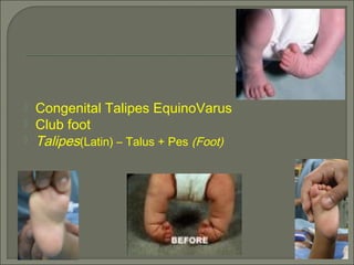
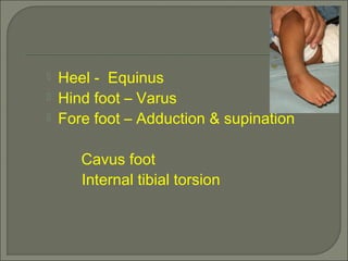
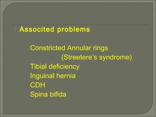
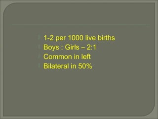


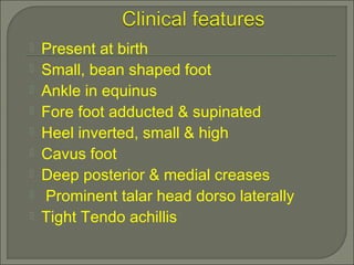


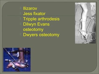

This document summarizes congenital talipes equino varus, or club foot, which occurs in approximately 1-2 per 1000 live births. Club foot is characterized by an inverted heel, hindfoot varus, and forefoot adduction and supination. It is caused by unknown factors that may include genetic defects or abnormal positioning in the womb. Treatment involves manipulation, casting, and sometimes surgery to correct contractures and produce a plantigrade foot.


