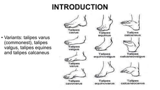
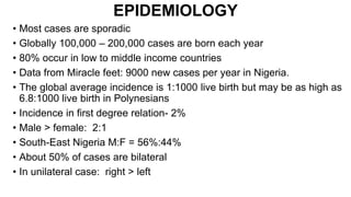
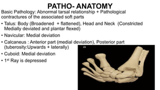
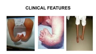
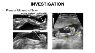
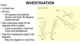
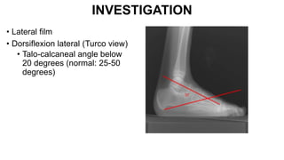
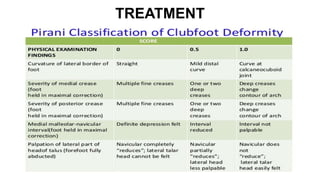
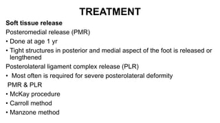
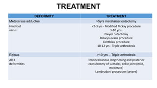
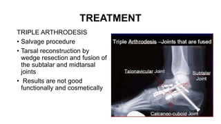
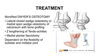
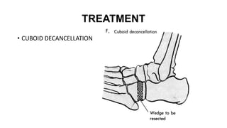
RESIDENTS’ PRESENTATION
ORTHOPEDIC DEPARTMENT
FMC LOKOJA
TOPIC: PATHOLOGY AND MANAGEMENT
OF CONGENITAL TALIPES EQUINOVARUS
DEFORMITY
BY
DR ENEJO JOSEPH
OUTLINE
• INTRODUCTION
• EPIDEMIOLOGY
• ANATOMY
• ETIOLOGY
• CLASSIFICATION
• CLINICAL FEATURES
• MANAGEMENT
• COMPLICATIONS
• FOLLOW UP
• PROGNOSIS
• CONCLUSION
INTRODUCTION
• The term ‘talipes’ is derived from talus(
Latin=ankle) and pes(Latin=foot).
• Equino & varus (turned inward)
• Congenital talipes equino varus also known as
Club foot
• Diagnosis is clinical
• It is a congenital deformity of the foot and ankle
characterized by equinus deformity at the
ankle, inversion at the subtalar, adduction at
the midtarsal joint, cavus and internal tibial
torsion
INTRODUCTION
• Variants: talipes varus
(commonest), talipes
valgus, talipes equines
and talipes calcaneus
EPIDEMIOLOGY
• Most cases are sporadic
• Globally 100,000 – 200,000 cases are born each year
• 80% occur in low to middle income countries
• Data from Miracle feet: 9000 new cases per year in Nigeria.
• The global average incidence is 1:1000 live birth but may be as high as
6.8:1000 live birth in Polynesians
• Incidence in first degree relation- 2%
• Male > female: 2:1
• South-East Nigeria M:F = 56%:44%
• About 50% of cases are bilateral
• In unilateral case: right > left
PATHO- ANATOMY
2,3,1,0,4
PATHO- ANATOMY
PATHO- ANATOMY
Basic Pathology: Abnormal tarsal relationship + Pathological
contractures of the associated soft parts
• Talus: Body (Broadened + flattened), Head and Neck (Constricted
Medially deviated and plantar flexed)
• Navicular: Medial deviation
• Calcaneus : Anterior part (medial deviation), Posterior part
(tuberosity:Upwards + laterally)
• Cuboid: Medial deviation
• 1st Ray is depressed
PATHO- ANATOMY
PATHO- ANATOMY
Soft Tissue Contractures:-
• Posterior: Tendo Achilles, Talocalcaneal Capsule,Calcanoefibular lig,
Posterior talofibular lig., Posterior Capsule of the ankle joint)
• Medial –Plantar: (Tibialis posterior, Spring ligament, Talonavicular
capsule, Deltoid ligaments)
• Subtalar: (interosseous ligaments and Y-ligament)
• Plantar: Abductor halluces,Plantar aponeurosis,Quadratus plantae,
Masters knot of Henry)
• Lateral: Tendon sheeth of peroneus muscle, Posterior displacement of
distal fibula
AETIOPATHOGENESIS
The exact cause is unknown
• Various hypothesis has been postulated
• Primary germ cell defect of talus  Irani
• Abnormal mechanical intra-uterine forces  Hippocrates
• Early intra-uterine growth arrest of the foot  Bohm
• Hereditary  Wynne- Davies
• Contractile myofibroblastic tissue in the Musculotendinous units
Ippolito and Ponseti
• Vascular insufficiency  Keith
• Muscle imbalance from neurological deficit
• Genetic: PITX1, TBX5
AETIOPATHOGENESIS
PATHOLOGY
• Club foot
• Midfoot cavus (tight intrinsics, FHL, FDL)
• Forefoot adductus (tight tibialis posterior)
• Hindfoot varus (tight tendoachilles, tibialis posterior)
• Hindfoot equinus (tight tendoachilles )
• The ankle, subtalar and midtarsal joints are involved
• Abnormal gait
• Pain
• Callosities
• Osteoarthritis
AETIOPATHOGENESIS
Associated findings
• Absent or hypoplastic anterior tibial artery
• Atrophy of muscles around the calf
• Abnormal foot is smaller
CLASSIFICATION
According To Cause
• Idiopathic
• Secondary:
• Neurogenic
• syndromic (Streeters dysplasia, arthrogryposis, edwards syndrome)
• Postural / Positional
CLASSIFICATION
According to stage of treatment (CUMMIN)
• Delayed
• Untreated
• Treated
• Resistant
• Recurrent
• Neglected
• Complex
Harold and walker
Grade I = correctable beyond neutral
Grade II = correctable to neutral or < 20 degree heel varus
Grade III = fixed equinous or >20 degree heel varus
CLASSIFICATION
According to severity of deformity
CLASSIFICATION
• Others
• Browns classification
• Pirani classification
CLINICAL FEATURES
MANAGEMENT
HISTORY
• Biodata: Age, sex,
• Associated conditions
• Pregnancy, antenatal and delivery history
• Family history
• Previous treatment
• General examination: facie,
• MSS: Spine, muscle tone
INVESTIGATION
• Prenatal Ultrasound Scan
INVESTIGATION
Xrays
• Limited use
• AP film
• 30 degrees foot plantar
flexion and tube 30 degrees
perpendicular
• Talo-calcaneal angle 20-40
degrees (kite’s angle)
• Less or parallel in club foot
(hind foot parallelism)
• Talo-first metatarsal angle 0-20
degrees
• May be negative in club foot
INVESTIGATION
• Lateral film
• Dorsiflexion lateral (Turco view)
• Talo-calcaneal angle below
20 degrees (normal: 25-50
degrees)
INVESTIGATION
• CT- Scan :
• FBC
• Genotype
• Genetic studies
• E/U/CR
• GXM
TREATMENT
Treatment: Multidisciplinary
The role of parents/guardian
AIMS
• Plantigrade
• Pliable
• Pain-free
• Cosmetically acceptable
TREATMENT
• Non-operative
• Operative
TREATMENT
Non operative
• Ignacio Ponseti technique (1940s)
• Joseph Hiram Kite technique (1930s)
• Turco’s Method
• French technique
Surgical
• Soft Tissue Operations: ETA; PMR; TATT;
• Bony Operations: Calcaneo-cuboid; Osteotomy of Calcaneus; Talectomy;
Triple Arthrodesis
• Combined Bony & Soft Tissue Operation
• Ilizarov / Joshi’s External Stabilization System (JESS)
TREATMENT
Ponseti technique
• Goal is to rotate foot laterally around a fixed talus
Treatment phase
• Weekly Serial manipulation and casting (long leg cast)
Order of correction (cave)
• Midfoot cavus
• Forefoot adductus
• Hindfoot varus
• Hindfoot equinus (TAL)
TREATMENT
TREATMENT
• After the last cast Tendo-achelles lengthening
Maintenance phase
• FAB for 23 hrs a day for 3 months and night splint till 2-3 yrs of
age
• Chance of recurrence up to 4 or 5 yrs of age
TREATMENT
French technique
• Goal is to reduce talonavicular joint, stretch out medial tissues
• Sequentially correct forefoot adduction, hindfoot varus and
equinus of calcaneum.
• Electrical stimulation of peroneal muscles done
• Reduction maintained by adhesive taping
TREATMENT
Kite’s technique
• Foot manipulated with Calcaneo-cuboid joint as fulcrum
• Correction in a sequential order
• Foot adduction
• Heel varus
• Equinus
• Advised change of cast every 3 weeks till correction is achieved.
• After correction Denis Browne splint applied
TREATMENT
SURGICAL PROCEDURES
A SOFT TISSUE OPERATIONS: Posterior Release,
Posteromedial Release, Posterolateral Release, Circumferential
Release (Cincinati), Steindler Operation, Tendon transfer, Illizarov
technique
B BONY OPERATIONS: Calcaneo-cuboid orthrodesis, Osteotomy of
the calcaneus, Talectomy, Tripple Arthodesis,Tibial Osteotomy
C COMBINED SOFT TISSUES + BONY OPERATIONS
D OTHERS
- Transposed Skin Graft
- Fascio-Cutaneous Flap
TREATMENT
Soft tissue release
Posteromedial release (PMR)
• Done at age 1 yr
• Tight structures in posterior and medial aspect of the foot is released or
lengthened
Posterolateral ligament complex release (PLR)
• Most often is required for severe posterolateral deformity
PMR & PLR
• McKay procedure
• Carroll method
• Manzone method
TREATMENT
DEFORMITY TREATMENT
Metatarsus adductus >5yrs metatarsal osteotomy
Hindfoot
varus
<2-3 yrs - Modified Mckay procedure
3-10 yrs -
Dwyer osteotomy
Dillwyn evans procedure
Lichtblau procedure
10-12 yrs - Triple arthrodesis
Eqinus >10 yrs – Triple arthodesis
All 3
deformities
Tendocalcaneus lengthening and posterior
capsulotomy of subtalar, ankle joint (mild,
moderate)
Lambrudoni procedure (severe)
TREATMENT
TRIPLE ARTHRODESIS
• Salvage procedure
• Tarsal reconstruction by
wedge resection and fusion of
the subtalar and midtarsal
joints
• Results are not good
functionally and cosmetically
TREATMENT
Mordified DWYER’S OSTEOTOMY
• Lateral closed wedge osteotomy or
medial open wedge osteotomy of
calcaneum with bone grafting
• Z lengthening of Tendo-achilles
• Medial plantar fasciotomy
Dependent on the flexibility of
subtalar and midtalar joint
TREATMENT
Dillwyns and Evans procedure
• Indicated for a shortened medial column
• wedge resection of calcaneocuboid joint
with arthrodesis
• Age : 4-8 yrs
TREATMENT
LICHTBLAU
• Resection of anterior
end of calcaneum of
1 cm
• Indicated in a long
lateral column
• This can be
combined with
Medial release
TREATMENT
• CUBOID DECANCELLATION
TREATMENT
TALECTOMY
• Age< 4 years
• Rigid paralytic deformities of the foot
• Principle is that by excision, sufficient laxity of soft tissues is
provided to correct equinus and varus deformities without soft
tissue tension
TREATMENT
• Ilizarov and Joshi’s External Stabilization System (JESS) are for older
children with recurrence or residual deformity
COMPLICATIONS
NOM
• Pressure sores
• Compartment syndrome
• Fractures
• Limb loss
• Burns
• Foot anomaly: Rocker bottom foot, bean shaped foot, flat top talus
• Failure of correction
• Recurrence
COMPLICATIONS
Oprative care
• Infection
• Non-union
• Compartment syndrome
• Neurovascular injuries
• Recurrence
FOLLOW UP
Follow-up of patient is essential to:
• Monitor treatment
• Prevent recurrence
PROGNOSIS
• Prognosis is good
• It however depends on
• Age at presentation
• Severity of the disease
• Associated anomalies
• Availability of experience care giver
CONCLUSION
• Club foot deformity as been with mankind for ages
• The etiology is unknown
• Genetic and environmental factors
• Diagnosis is clinical
• Options of care depend on the age at presentation, severity of
deformity and pathology involved
• Surgical correction of resistant clubfoot includes both soft tissue
release & bony osteotomies
• Follow up is essential to prevent recurrence
THANK YOU
REFERENCES
• Tochukwu NU, Nwakamma JI, KachisichonTA, Faith WC. (2020).
Epidemiology and pattern of Clubfoot in Enugu, South-East, Nigeria.
Amr J BSRB, Vol. 11, Issue 2,
• MiracleFeet. Accessed @ https://www.miraclefeet.org>nigeria
• Tope Omogbolagun (2022). Punch News paper 10th June 2022.
• Manjappa CN.Joshi’s External Stabilization System(JESS) Application
for correction of Resistant Club-Foot. The Internet Journal of
Orthopedic Surgery. 2009, Vol. 18, Numb. 1
• ArchampongEQ, NaaederSB, UgwuB.(2015 ). Baja’s principles and
practice of surgery including pathologies in the tropics: Orthopedic
surgery. 5thEd. Vol2. Repro India Ltd. Chapt.54, page 1202-1207.
• Campbell’s operative orthopaedics, 13th Ed (2017),
• Apley’s system of orthopaedics and fractures, 9th Ed (2010),
REFERENCES
• Campbells operative orthopaedics volume 2; 12th edition
• Tachdijian’s pediatric orthopedics volume 2; 4th edition
• Al-falloujiM.A.R (1998). Postgraduate Surgery; The Candidate’s Guide:
Surgical Oncology. 2ndEd. The Bath Press Plc Britain. Pg. 208-236.

CTEV DEFORMITY.pptx

  • 1.
    RESIDENTS’ PRESENTATION ORTHOPEDIC DEPARTMENT FMCLOKOJA TOPIC: PATHOLOGY AND MANAGEMENT OF CONGENITAL TALIPES EQUINOVARUS DEFORMITY BY DR ENEJO JOSEPH
  • 2.
    OUTLINE • INTRODUCTION • EPIDEMIOLOGY •ANATOMY • ETIOLOGY • CLASSIFICATION • CLINICAL FEATURES • MANAGEMENT • COMPLICATIONS • FOLLOW UP • PROGNOSIS • CONCLUSION
  • 3.
    INTRODUCTION • The term‘talipes’ is derived from talus( Latin=ankle) and pes(Latin=foot). • Equino & varus (turned inward) • Congenital talipes equino varus also known as Club foot • Diagnosis is clinical • It is a congenital deformity of the foot and ankle characterized by equinus deformity at the ankle, inversion at the subtalar, adduction at the midtarsal joint, cavus and internal tibial torsion
  • 4.
    INTRODUCTION • Variants: talipesvarus (commonest), talipes valgus, talipes equines and talipes calcaneus
  • 5.
    EPIDEMIOLOGY • Most casesare sporadic • Globally 100,000 – 200,000 cases are born each year • 80% occur in low to middle income countries • Data from Miracle feet: 9000 new cases per year in Nigeria. • The global average incidence is 1:1000 live birth but may be as high as 6.8:1000 live birth in Polynesians • Incidence in first degree relation- 2% • Male > female: 2:1 • South-East Nigeria M:F = 56%:44% • About 50% of cases are bilateral • In unilateral case: right > left
  • 6.
  • 7.
  • 8.
    PATHO- ANATOMY Basic Pathology:Abnormal tarsal relationship + Pathological contractures of the associated soft parts • Talus: Body (Broadened + flattened), Head and Neck (Constricted Medially deviated and plantar flexed) • Navicular: Medial deviation • Calcaneus : Anterior part (medial deviation), Posterior part (tuberosity:Upwards + laterally) • Cuboid: Medial deviation • 1st Ray is depressed
  • 9.
  • 10.
    PATHO- ANATOMY Soft TissueContractures:- • Posterior: Tendo Achilles, Talocalcaneal Capsule,Calcanoefibular lig, Posterior talofibular lig., Posterior Capsule of the ankle joint) • Medial –Plantar: (Tibialis posterior, Spring ligament, Talonavicular capsule, Deltoid ligaments) • Subtalar: (interosseous ligaments and Y-ligament) • Plantar: Abductor halluces,Plantar aponeurosis,Quadratus plantae, Masters knot of Henry) • Lateral: Tendon sheeth of peroneus muscle, Posterior displacement of distal fibula
  • 11.
    AETIOPATHOGENESIS The exact causeis unknown • Various hypothesis has been postulated • Primary germ cell defect of talus  Irani • Abnormal mechanical intra-uterine forces  Hippocrates • Early intra-uterine growth arrest of the foot  Bohm • Hereditary  Wynne- Davies • Contractile myofibroblastic tissue in the Musculotendinous units Ippolito and Ponseti • Vascular insufficiency  Keith • Muscle imbalance from neurological deficit • Genetic: PITX1, TBX5
  • 12.
    AETIOPATHOGENESIS PATHOLOGY • Club foot •Midfoot cavus (tight intrinsics, FHL, FDL) • Forefoot adductus (tight tibialis posterior) • Hindfoot varus (tight tendoachilles, tibialis posterior) • Hindfoot equinus (tight tendoachilles ) • The ankle, subtalar and midtarsal joints are involved • Abnormal gait • Pain • Callosities • Osteoarthritis
  • 13.
    AETIOPATHOGENESIS Associated findings • Absentor hypoplastic anterior tibial artery • Atrophy of muscles around the calf • Abnormal foot is smaller
  • 14.
    CLASSIFICATION According To Cause •Idiopathic • Secondary: • Neurogenic • syndromic (Streeters dysplasia, arthrogryposis, edwards syndrome) • Postural / Positional
  • 15.
    CLASSIFICATION According to stageof treatment (CUMMIN) • Delayed • Untreated • Treated • Resistant • Recurrent • Neglected • Complex Harold and walker Grade I = correctable beyond neutral Grade II = correctable to neutral or < 20 degree heel varus Grade III = fixed equinous or >20 degree heel varus
  • 16.
  • 17.
    CLASSIFICATION • Others • Brownsclassification • Pirani classification
  • 18.
  • 19.
    MANAGEMENT HISTORY • Biodata: Age,sex, • Associated conditions • Pregnancy, antenatal and delivery history • Family history • Previous treatment • General examination: facie, • MSS: Spine, muscle tone
  • 20.
  • 21.
    INVESTIGATION Xrays • Limited use •AP film • 30 degrees foot plantar flexion and tube 30 degrees perpendicular • Talo-calcaneal angle 20-40 degrees (kite’s angle) • Less or parallel in club foot (hind foot parallelism) • Talo-first metatarsal angle 0-20 degrees • May be negative in club foot
  • 22.
    INVESTIGATION • Lateral film •Dorsiflexion lateral (Turco view) • Talo-calcaneal angle below 20 degrees (normal: 25-50 degrees)
  • 23.
    INVESTIGATION • CT- Scan: • FBC • Genotype • Genetic studies • E/U/CR • GXM
  • 24.
    TREATMENT Treatment: Multidisciplinary The roleof parents/guardian AIMS • Plantigrade • Pliable • Pain-free • Cosmetically acceptable TREATMENT • Non-operative • Operative
  • 25.
    TREATMENT Non operative • IgnacioPonseti technique (1940s) • Joseph Hiram Kite technique (1930s) • Turco’s Method • French technique Surgical • Soft Tissue Operations: ETA; PMR; TATT; • Bony Operations: Calcaneo-cuboid; Osteotomy of Calcaneus; Talectomy; Triple Arthrodesis • Combined Bony & Soft Tissue Operation • Ilizarov / Joshi’s External Stabilization System (JESS)
  • 26.
    TREATMENT Ponseti technique • Goalis to rotate foot laterally around a fixed talus Treatment phase • Weekly Serial manipulation and casting (long leg cast) Order of correction (cave) • Midfoot cavus • Forefoot adductus • Hindfoot varus • Hindfoot equinus (TAL)
  • 27.
  • 28.
    TREATMENT • After thelast cast Tendo-achelles lengthening Maintenance phase • FAB for 23 hrs a day for 3 months and night splint till 2-3 yrs of age • Chance of recurrence up to 4 or 5 yrs of age
  • 29.
    TREATMENT French technique • Goalis to reduce talonavicular joint, stretch out medial tissues • Sequentially correct forefoot adduction, hindfoot varus and equinus of calcaneum. • Electrical stimulation of peroneal muscles done • Reduction maintained by adhesive taping
  • 30.
    TREATMENT Kite’s technique • Footmanipulated with Calcaneo-cuboid joint as fulcrum • Correction in a sequential order • Foot adduction • Heel varus • Equinus • Advised change of cast every 3 weeks till correction is achieved. • After correction Denis Browne splint applied
  • 31.
    TREATMENT SURGICAL PROCEDURES A SOFTTISSUE OPERATIONS: Posterior Release, Posteromedial Release, Posterolateral Release, Circumferential Release (Cincinati), Steindler Operation, Tendon transfer, Illizarov technique B BONY OPERATIONS: Calcaneo-cuboid orthrodesis, Osteotomy of the calcaneus, Talectomy, Tripple Arthodesis,Tibial Osteotomy C COMBINED SOFT TISSUES + BONY OPERATIONS D OTHERS - Transposed Skin Graft - Fascio-Cutaneous Flap
  • 32.
    TREATMENT Soft tissue release Posteromedialrelease (PMR) • Done at age 1 yr • Tight structures in posterior and medial aspect of the foot is released or lengthened Posterolateral ligament complex release (PLR) • Most often is required for severe posterolateral deformity PMR & PLR • McKay procedure • Carroll method • Manzone method
  • 33.
    TREATMENT DEFORMITY TREATMENT Metatarsus adductus>5yrs metatarsal osteotomy Hindfoot varus <2-3 yrs - Modified Mckay procedure 3-10 yrs - Dwyer osteotomy Dillwyn evans procedure Lichtblau procedure 10-12 yrs - Triple arthrodesis Eqinus >10 yrs – Triple arthodesis All 3 deformities Tendocalcaneus lengthening and posterior capsulotomy of subtalar, ankle joint (mild, moderate) Lambrudoni procedure (severe)
  • 34.
    TREATMENT TRIPLE ARTHRODESIS • Salvageprocedure • Tarsal reconstruction by wedge resection and fusion of the subtalar and midtarsal joints • Results are not good functionally and cosmetically
  • 35.
    TREATMENT Mordified DWYER’S OSTEOTOMY •Lateral closed wedge osteotomy or medial open wedge osteotomy of calcaneum with bone grafting • Z lengthening of Tendo-achilles • Medial plantar fasciotomy Dependent on the flexibility of subtalar and midtalar joint
  • 36.
    TREATMENT Dillwyns and Evansprocedure • Indicated for a shortened medial column • wedge resection of calcaneocuboid joint with arthrodesis • Age : 4-8 yrs
  • 37.
    TREATMENT LICHTBLAU • Resection ofanterior end of calcaneum of 1 cm • Indicated in a long lateral column • This can be combined with Medial release
  • 38.
  • 39.
    TREATMENT TALECTOMY • Age< 4years • Rigid paralytic deformities of the foot • Principle is that by excision, sufficient laxity of soft tissues is provided to correct equinus and varus deformities without soft tissue tension
  • 40.
    TREATMENT • Ilizarov andJoshi’s External Stabilization System (JESS) are for older children with recurrence or residual deformity
  • 41.
    COMPLICATIONS NOM • Pressure sores •Compartment syndrome • Fractures • Limb loss • Burns • Foot anomaly: Rocker bottom foot, bean shaped foot, flat top talus • Failure of correction • Recurrence
  • 42.
    COMPLICATIONS Oprative care • Infection •Non-union • Compartment syndrome • Neurovascular injuries • Recurrence
  • 43.
    FOLLOW UP Follow-up ofpatient is essential to: • Monitor treatment • Prevent recurrence
  • 44.
    PROGNOSIS • Prognosis isgood • It however depends on • Age at presentation • Severity of the disease • Associated anomalies • Availability of experience care giver
  • 45.
    CONCLUSION • Club footdeformity as been with mankind for ages • The etiology is unknown • Genetic and environmental factors • Diagnosis is clinical • Options of care depend on the age at presentation, severity of deformity and pathology involved • Surgical correction of resistant clubfoot includes both soft tissue release & bony osteotomies • Follow up is essential to prevent recurrence
  • 46.
  • 47.
    REFERENCES • Tochukwu NU,Nwakamma JI, KachisichonTA, Faith WC. (2020). Epidemiology and pattern of Clubfoot in Enugu, South-East, Nigeria. Amr J BSRB, Vol. 11, Issue 2, • MiracleFeet. Accessed @ https://www.miraclefeet.org>nigeria • Tope Omogbolagun (2022). Punch News paper 10th June 2022. • Manjappa CN.Joshi’s External Stabilization System(JESS) Application for correction of Resistant Club-Foot. The Internet Journal of Orthopedic Surgery. 2009, Vol. 18, Numb. 1 • ArchampongEQ, NaaederSB, UgwuB.(2015 ). Baja’s principles and practice of surgery including pathologies in the tropics: Orthopedic surgery. 5thEd. Vol2. Repro India Ltd. Chapt.54, page 1202-1207. • Campbell’s operative orthopaedics, 13th Ed (2017), • Apley’s system of orthopaedics and fractures, 9th Ed (2010),
  • 48.
    REFERENCES • Campbells operativeorthopaedics volume 2; 12th edition • Tachdijian’s pediatric orthopedics volume 2; 4th edition • Al-falloujiM.A.R (1998). Postgraduate Surgery; The Candidate’s Guide: Surgical Oncology. 2ndEd. The Bath Press Plc Britain. Pg. 208-236.