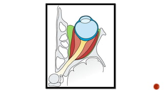
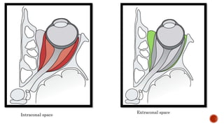
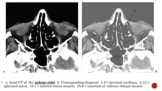
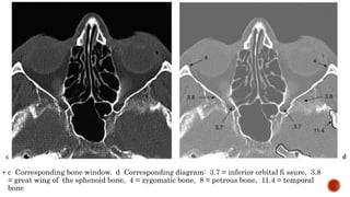
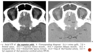
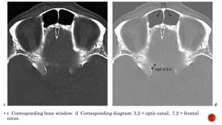
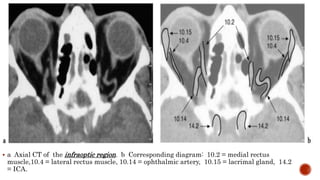
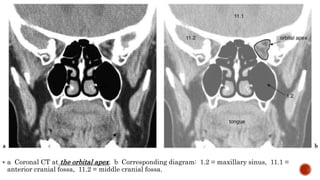
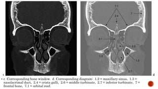
This document provides a summary of CT scans of the orbit and surrounding structures with corresponding diagrams. It includes axial, coronal, and sagittal CT scans at different levels through the orbit, labeled with key anatomical structures visible in each scan such as muscles, bones, sinuses, blood vessels and nerves. Diagrams accompany each scan identifying the structures. The document aims to familiarize readers with cross-sectional anatomy of the orbit and surrounding areas.