The document presents a case report on trigonocephaly, a form of craniosynostosis involving premature fusion of the metopic suture, leading to forehead and orbital deformities. It describes a successful surgical intervention performed on an 11-month-old male patient, which involved a complete fronto-orbital osteotomy to correct the deformities and prevent complications like strabismus and intracranial hypertension. The findings emphasize the importance of early surgical treatment and the positive aesthetic and functional outcomes following the procedure.
![Clinics of Surgery Case Report
Trigonocephalia a Curable Craniosynostosis. Case Report
Luces OAR1*
, Luces OR2
, Millian C3
, Lara G4
, Mejias W5
, Perdomo Y6
and Dumoulins W7
1
Specialist in general surgery and laparoscopy and General urologist of the Venezuelan Institute of Social Security (IVSS), Universidad de
Oriente, Barcelona, Anzoátegui - Venezuela
2
Pediatric surgery specialist, Physician of the State Institute of Sports of Anzoátegui (IDEA), Universidad de Oriente, Barcelona,
Anzoátegui - Venezuela
3
Specialist in Pediatric Neurosurgery or Children, Doctor of the Venezuelan Institute of Social Security (IVSS), Dr. Cesar Rodríguez
Rodríguez Hospital, Puerto la Cruz – Venezuela
4
Specialist in Pediatric Neurosurgery or Children, Dr. J. M. de los Ríos Children's Hospital l, Caracas - Venezuela
5
Specialist in Pediatric Neurosurgery or Children, Dr. Jose Maria Vargas Hospital, Caracas – Venezuela
6
Postgraduate resident of general surgery, Hospital Dr. José Ignacio Baldo, Caracas – Venezuela
7
Specialist in general surgery and laparoscopy, Head of the surgery service, Doctor of the Venezuelan Institute of Social Security (IVSS),
Dr. Cesar Rodríguez Rodríguez Hospital, Puerto la Cruz - Venezuela
8
Medical surgeon - biostatist, Puerto la Cruz - Venezuela
Volume 2 Issue 3- 2019
Received Date: 03 Dec 2019
Accepted Date: 20 Dec 2019
Published Date: 30 Dec 2019
2. Key words
Trigonocephaly treatment;
Craniosynostosis; Metopic
suture; Invasive surgical
technique
1. Trigonocephalia A Curable Craniosynostosis. Case Report
The Trigonocephaly is premature fusion of the metopic suture (part of the frontal suture which joins
the two halves of the frontal bone of the skull) in which a V-shaped abnormality occurs at the front
of the skull. It is characterized by a triangular prominence of the forehead and eyes close together, has
implications for neurological injury secondary variables to the deformity. The reasons for the anterior
fossa surgery are reduced in size and orbits with abnormal spatial arrangement leads secondarily to
disturbances in vision (strabismus) and intracranial hypertension. We want to describe a clinical case
of Trigonocephaly from the Neurosurgery Service of the IVSS Hospital Dr. Cesar Rodríguez, describes
the case of an 11-month-old patient, Male sex with no history of illness, siendo un primer pregnancy,
controlled, simple eutocic delivery without complications, who entered the outpatient service in
November. The physical examination presents good general condition, after having suffered frequent
crying and irritability for 2 months. The purpose of surgery is to perform a complete orbitofrontal
osteotomy in addition to resection of the metopic suture, which includes both orbital roofs and the
pterional region, resulting in remodeling of the front. Trigonocephaly is a type of craniosynostosis,
must have an early surgical treatment. The immediate postoperative changes are satisfactory, in the
immediate postoperative period their appreciation for the aesthetic appearance, about 24 to 48 hours
has a progressively diminishing facial edema, also fixes harmonious and spontaneous intracranial
hypertension.
*Corresponding Author (s): Odionnys Antonio Ramos Luces, Specialist in gen-
eral surgery and laparoscopy and General Urologist of the Venezuelan
Institute of Social Security (IVSS), Universidad de Oriente. AV. University,
Anzoátegui-Venezuela, Tel: (0058) 02812682809; 04265808668, E-mail:
odywan66@yahoo.es
Citation: Luces OAR1
Trigonocephalia a Curable Craniosynostosis. Case Report Clinics of Sugery.
2018;1(5): 1-6
3. Introduction
The pediatrician needs to be able to differentiate true
craniosynostosis that affect the surgical treatment of positional
deformities. The premature fusion of one or several skull sutures
produces well-recognized patterns of deformity, both cranial
and facial, in which, in addition to the child's deformity, serious
functional complications sometimes arise related to breathing,
feeding and vision [1]. Craniosynostosis has variable implications
forneurologicalinjurysecondarytodeformity.Currentstudieshave
shown the presence of alterations in the cerebral cortex underlying
the fused suture by magnetic resonance imaging. However, there
are discrepancies as to whether this is the consequence of the
https://clinicsofsurgery.com/
ISSN 2638-1451](https://image.slidesharecdn.com/cos-v2-1045-240913105412-3c389306/85/Trigonocephalia-a-Curable-Craniosynostosis-Case-Report-1-320.jpg)
![Clinics of Surgery Case Report
Trigonocephalia a Curable Craniosynostosis. Case Report
Luces OAR1*
, Luces OR2
, Millian C3
, Lara G4
, Mejias W5
, Perdomo Y6
and Dumoulins W7
1
Specialist in general surgery and laparoscopy and General urologist of the Venezuelan Institute of Social Security (IVSS), Universidad de
Oriente, Barcelona, Anzoátegui - Venezuela
2
Pediatric surgery specialist, Physician of the State Institute of Sports of Anzoátegui (IDEA), Universidad de Oriente, Barcelona,
Anzoátegui - Venezuela
3
Specialist in Pediatric Neurosurgery or Children, Doctor of the Venezuelan Institute of Social Security (IVSS), Dr. Cesar Rodríguez
Rodríguez Hospital, Puerto la Cruz – Venezuela
4
Specialist in Pediatric Neurosurgery or Children, Dr. J. M. de los Ríos Children's Hospital l, Caracas - Venezuela
5
Specialist in Pediatric Neurosurgery or Children, Dr. Jose Maria Vargas Hospital, Caracas – Venezuela
6
Postgraduate resident of general surgery, Hospital Dr. José Ignacio Baldo, Caracas – Venezuela
7
Specialist in general surgery and laparoscopy, Head of the surgery service, Doctor of the Venezuelan Institute of Social Security (IVSS),
Dr. Cesar Rodríguez Rodríguez Hospital, Puerto la Cruz - Venezuela
8
Medical surgeon - biostatist, Puerto la Cruz - Venezuela
Volume 2 Issue 3- 2019
Received Date: 03 Dec 2019
Accepted Date: 20 Dec 2019
Published Date: 30 Dec 2019
2. Key words
Trigonocephaly treatment;
Craniosynostosis; Metopic
suture; Invasive surgical
technique
1. Trigonocephalia A Curable Craniosynostosis. Case Report
The Trigonocephaly is premature fusion of the metopic suture (part of the frontal suture which joins
the two halves of the frontal bone of the skull) in which a V-shaped abnormality occurs at the front
of the skull. It is characterized by a triangular prominence of the forehead and eyes close together, has
implications for neurological injury secondary variables to the deformity. The reasons for the anterior
fossa surgery are reduced in size and orbits with abnormal spatial arrangement leads secondarily to
disturbances in vision (strabismus) and intracranial hypertension. We want to describe a clinical case
of Trigonocephaly from the Neurosurgery Service of the IVSS Hospital Dr. Cesar Rodríguez, describes
the case of an 11-month-old patient, Male sex with no history of illness, siendo un primer pregnancy,
controlled, simple eutocic delivery without complications, who entered the outpatient service in
November. The physical examination presents good general condition, after having suffered frequent
crying and irritability for 2 months. The purpose of surgery is to perform a complete orbitofrontal
osteotomy in addition to resection of the metopic suture, which includes both orbital roofs and the
pterional region, resulting in remodeling of the front. Trigonocephaly is a type of craniosynostosis,
must have an early surgical treatment. The immediate postoperative changes are satisfactory, in the
immediate postoperative period their appreciation for the aesthetic appearance, about 24 to 48 hours
has a progressively diminishing facial edema, also fixes harmonious and spontaneous intracranial
hypertension.
*Corresponding Author (s): Odionnys Antonio Ramos Luces, Specialist in gen-
eral surgery and laparoscopy and General Urologist of the Venezuelan
Institute of Social Security (IVSS), Universidad de Oriente. AV. University,
Anzoátegui-Venezuela, Tel: (0058) 02812682809; 04265808668, E-mail:
odywan66@yahoo.es
Citation: Luces OAR1
Trigonocephalia a Curable Craniosynostosis. Case Report Clinics of Sugery.
2018;1(5): 1-6
3. Introduction
The pediatrician needs to be able to differentiate true
craniosynostosis that affect the surgical treatment of positional
deformities. The premature fusion of one or several skull sutures
produces well-recognized patterns of deformity, both cranial
and facial, in which, in addition to the child's deformity, serious
functional complications sometimes arise related to breathing,
feeding and vision [1]. Craniosynostosis has variable implications
forneurologicalinjurysecondarytodeformity.Currentstudieshave
shown the presence of alterations in the cerebral cortex underlying
the fused suture by magnetic resonance imaging. However, there
are discrepancies as to whether this is the consequence of the
https://clinicsofsurgery.com/
ISSN 2638-1451](https://image.slidesharecdn.com/cos-v2-1045-240913105412-3c389306/75/Trigonocephalia-a-Curable-Craniosynostosis-Case-Report-1-2048.jpg)
![premature fusion of the suture or simply the cause of the fusion.
Some authors carried out psychological tests in order to know how
the development of patients affected by a simple craniostenosis
was and found that the percentage of patients with a psychomotor
development index below normal was higher in patients with
craniostenosis than in the normal population [2]. It is a relatively
common birth defect with an estimated incidence of 1 / 2,100-1 /
2,500 children [3].
Trigonocephaly within all craniosynostosis occur in approximately
18% of cases, with a clear predominance of scaphocephaly (35%);
Brachycephaly (24.9%) and Plagiocephaly (21.6%) and lastly,
Oxycephaly (11.6%). It occurs in the same proportion between both
sexes. Very little is known about the etiology. So far, the arguments
to prove autosomal recessive inheritance are not convincing.
Although trigonocephaly is a fundamental characteristic of the
9p¬7-9 syndrome, it can also appear in other chromosomopathies
such as 6q +, 7p ¬, 13q +, 14p + and 18p [3]. This consists of the early
closure of the metopic suture. It is a relatively rare craniostenosis.
However, the frontal deformity - keel skull - with the prominence
in the midline corresponding to the sinostotic suture allows it
to be easily identified. The degree of deformity can be variable,
from a purely cosmetic defect caused by the prominence of the
midline, to accentuated defects with retrusion of the superior and
external flanges of the orbit, hypotelorism and axis of the oriented
orbit from inferolateral to superomedial and with Posterolateral
rotation of the orbit [4]. The reasons for the indication for surgical
treatment are decreased anterior fossa and orbits with anomalous
spatial arrangement, which will lead secondarily to vision
disorders (strabismus). In accordance with the above, the surgery
will consist of the remodeling of the frontal shell with elimination
of the prominence of the metopic suture and advancement of the
superoexternal flange of both orbits with placement of a bone graft
at the level of the skull base in order to eliminate the hypotelorism.
4. Case Report
It is presented under the signature of responsibility of the informed
consent of their parents, an 11-month-old male patient, without
a history of illness, whose parents were adolescents of 17 years
in 2010, child product of the first pregnancy, fully controlled
pregnancy with 8 controls prenatal, simple eutocic delivery
without complications, birth weight of 2640 grs and height at
birth of 49 cms. In September, at 7 months of age, he attended Dr.
Luis Ortega de Porlamar hospital, Nueva Esparta, where he was
diagnosed with trigonocephaly type craniosynostosis and was
immediately referred to Dr. Cesar Rodríguez IVSS hospital in
Puerto La Cruz-Anzoátegui, entered the neurosurgery service of
the mentioned hospital in November. In good general condition
after having suffered frequent crying for two months, that after
evaluating the case a possible surgical treatment is suggested. He
was hospitalized in December where he shows up. The neurological
examination shows wakefulness, spontaneous activity, attention to
the environment, reactivity, response to stimuli. Behavior, activity,
affectivity, communication, interest in the environment, neck reflex
and pressure preserved, sensitivity preserved (Figure 1).
The admission laboratory test showed a hematocrit of 31.3%,
hemoglobin at 10.1g / dl, leukocytes 8800, segmented 50%,
lymphocytes 45%, platelets 162,000, glycemia 91 mg / dl, urea
20.4 and creatinine in 02, blood group O, Rh negative factor, with
normal thromboplastin and thrombin time. Skull X-rays were
performed where no fracture traces are observed as well as lytic
or blastic lesions, partial sinostosis is seen that compromises the
sagittal suture in its anterior portion and the coronal suture, with
normal orbital ridges and normal facial massif bone structures
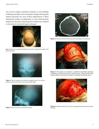
(Figure 2, Figure 3). The simple brain CT (Figure 4) in which
deformity characterized by pointed frontal region was evidenced,
trigonocephaly type craniostenosis is observed as a result of the
early closure of the mitotic suture, prominence of the ventricular
system, with no evidence of hypodense airs that suggest epindymal
transduction of the cerebrospinal fluid, normal anatomic variant in
infants, left temporobasal extracerebral hypodense area compatible
with arachnoid cyst.
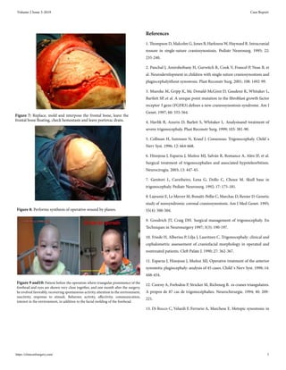
5. Surgical Treatment
Surgical treatment after anesthetic induction, the patient is
intubated orotracheally and placed in a neutral supine position.
The objective of the surgery is to perform a complete fronto orbital
osteotomy in addition to the resection of the metopic suture, which
includes the orbital ceilings and the pterional region, a remodeling
of its forehead originates (Figure 5), the adjustment in two halves
of this, and interposition of a graft taken from the posterior flange
of the craniectomy to annul the keel, through a bicoronal access, a
soft flap is made towards the face, parasagittal and paratemporal
Volume 2 Issue 3-2019 Case Report
Copyright ©2019 Luces OAR1
et al. This is an open access article distributed under the terms of the Creative Commons Attribution License,
which permits unrestricted use, distribution, and build upon your work non-commercially.](https://image.slidesharecdn.com/cos-v2-1045-240913105412-3c389306/85/Trigonocephalia-a-Curable-Craniosynostosis-Case-Report-2-320.jpg)
![holes are made (Figure 6), a frontal bone craniotomy , plus a
classic caudal lateral advance. An attempt is made to raise the
fronto-orbital bar to the level of the lateral margins of the orbit, to
avoid entering the temporal fossa, which is partially achieved. The
advance is also achieved and the frontal bone is replaced, molded
and interposed. Once the advance is achieved, the frontal bone is
left floating (Figure 7), the hemostasis is checked and the portovac
drained, then an operative wound synthesis is performed by planes
(Figure 8). The patient receives a 300 cc globular concentrate
during surgery.
The patient evolved favorably, a febrile, recovering spontaneous
activity, attention to the environment, reactivity conserved,
response to stimuli. Behavior, activity, affectivity, communication,
e interest in the environment adecuad (Figure 9-10), remain
hospitalized for 10 days with medical treatment with cefazidal (30
mg / kg / day).
6. Discussion
The closure of the metopic suture begins in normal conditions
around the end of the first year of life and is the first of the large
sutures to close towards the end of the second year 6. When an
early closure of the same occurs, usually in the intrauterine period,
a characteristic craniofacial malformation called trigonocephaly
occurs [7]. This is considered a relatively frequent type of
craniosynostosis and is considered to be approximately 10% of all
patients treated in a Craniofacial Surgery Unit [5-8]. Although the
trigonocephaly classically has been classified in the group of simple
craniosynostosis, the resulting deformity responds to a broad
phenotypic spectrum depending on the extent and the time at
which the closure of the metopic suture begins. The milder forms of
trigonocephaly have only a slight crest in the midline of the frontal
region, and do not usually require surgical correction, except for
aesthetic reasons. In the most severe cases there is an affectation
of the anterior chondrocranial structures that give rise to the
characteristic “keel” front, with retrusion and obliqueness of both
frontal bones, disappearance of the frontal eminences, hypoplasia
of both supraorbital ridges that are also found destroyed, epicanth
fold and hypoteleorbitism [6, 9].
It is precisely these last deformities that are the most characteristic
and that determine the final aesthetic result in most cases [10,
11]. A wide variety of techniques have been used for the surgical
treatment of trigonocephaly, all of them with very satisfactory
result [5, 9, 12-14]. The classic approach consists of a complete
frontoorbital remodeling by bifrontal scranotomy followed by
orbital osteotomies, with or without disassembly and remodeling
of the frontoorbital bar, and "tongue-in-groove" advancement
or at least expansion of both pterional regions [6, 14]. All these
techniques have achieved very good results in terms of the
immediate correction of the deformities while remaining stable
over time. However, the need for a bicoronal incision, subperiosteal
exposure, wide osteotomies and bone mobilizations involve a
considerable approach, with long surgical times, blood losses that
often require transfusions in the surgical act or in the immediate
postoperative period, and a prolonged hospital stay mainly
due to soft tissue inflammation caused by the wide detachment
of the structures of the operative field. In the current state of
craniofacial surgery, the emphasis of the postoperative results has
been deposited not only in the correction of the deformity and
release of the intracranial organs and systems "incarcerated" by the
sinostosis (cerebral parenchyma, CSF, airway, vision) but mainly
in the aesthetic aspect after the intervention and the maintenance
of the favorable results in the long term. This is more true in so-
called "simple" craniosynostosis or in milder ones, in which there
is a "single" affected suture.
7. Conclusion
Trigonocephaly is a type of craniostenosis that, depending on the
degree of structural compromise, has implications for psychic
developmentaswellasimportantmorphologicaltypeoffrontonasal
type causing hypotelorism, so early surgical treatment is essential
where in addition to fracturing the frontal for remodeling and
rotation it is necessary release the supra orbital bar, advance it
by bone grafts, leave the floating forehead, and thus release the
pressure on the frontal lobes and correct the hypotelorism. The
immediate post-surgical changes are very satisfactory, in the
immediate postoperative period there is a pleasure on the part
of the parents for the aesthetic appearance achieved, it should be
noted that around 24 to 48 hours they will present an important
facial edema that progressively decreases between 4 to 7 days In
addition, this surgical act is allowed to correct, harmonic and
spontaneously, endocranial hypertension. At present we must
think that with the appearance of multidisciplinary teams in
the Craniofacial Surgery Units that have allowed a considerable
reduction in the incidence of morbidity and mortality even in the
Volume 2 Issue 3-2019 Case Report
https://clinicsofsurgery.com/ 3](https://image.slidesharecdn.com/cos-v2-1045-240913105412-3c389306/85/Trigonocephalia-a-Curable-Craniosynostosis-Case-Report-3-320.jpg)


![CTEV [ clubfoot] DR ARUN LAL ,DR MOHAMED ASHRAF travancore medical college k...](https://cdn.slidesharecdn.com/ss_thumbnails/ctevclubfootdrarunlaldrmohamedashraftravancoremedicalcollegekollamkeralaindia-260208063247-18fc466c-thumbnail.jpg?width=640&height=640&fit=bounds)


