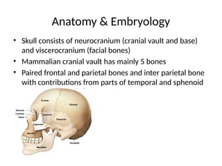
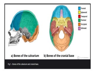
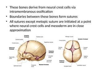
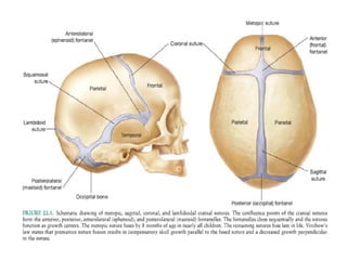
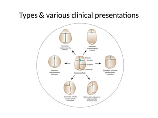
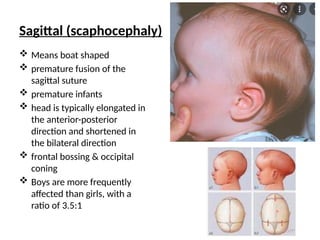
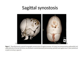
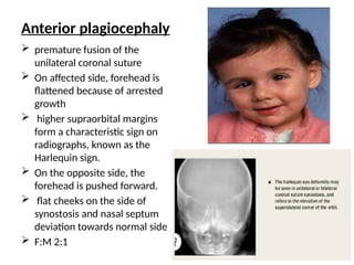
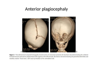
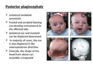
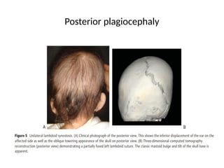
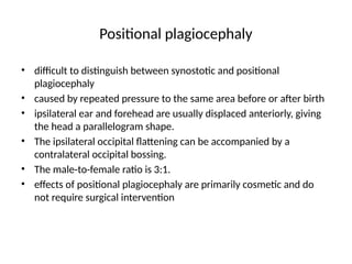
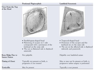
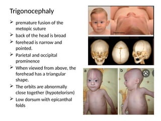
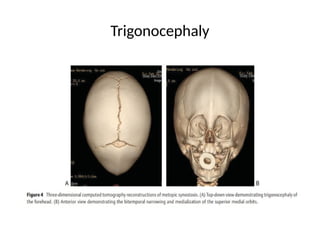
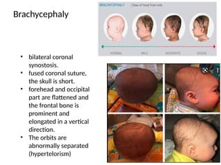
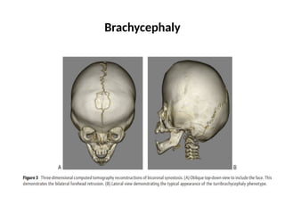
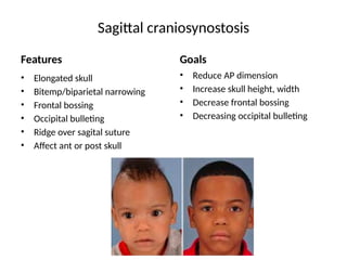
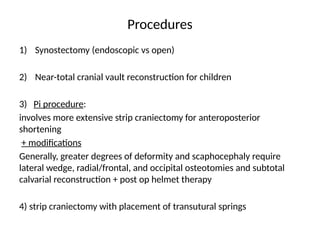
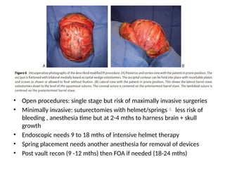
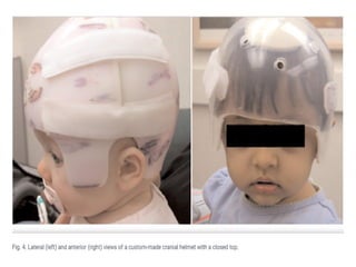
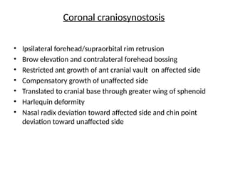
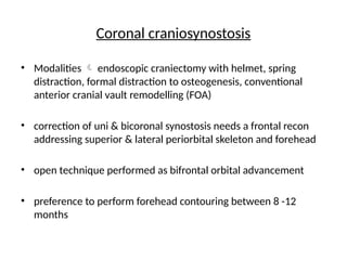
NONSYNDROMIC CRANIOSYNOSTOSIS
PRESENTOR: DR. SHAREEN LAKHANI
RESIDENT PLASTIC SURGERY, PATEL HOSPITAL
Introduction
• Craniosynostosis : premature fusion of cranial sutures
• Calvarial or facial deformities
• Neurocognitive impairment
• Otto was the first to explain pathogenesis in 1830
• Virchow observation: premature suture fusion results in
compensatory growth in other areas of skull
• Recent studies: genetic regulation
• Broadly: 1) single vs multiple suture
2) syndromic vs non syndromic
• Moss and Salentjin (mid 20th
cent) described “functional
matrix theory”
• Theory explains normal cranial vault development
depends on normal neural mass and subsequent
expansion
• Suture itself may not be primary source of deformity
• Complex interplay between cranial base and expanding
brain may be real culprit
• Management involves early diagnosis with interventions
aimed at minimizing constricted brain growth and
craniofacial deformities
Anatomy & Embryology
• Skull consists of neurocranium (cranial vault and base)
and viscerocranium (facial bones)
• Mammalian cranial vault has mainly 5 bones
• Paired frontal and parietal bones and inter parietal bone
with contributions from parts of temporal and sphenoid
• These bones derive from neural crest cells via
intramembranous ossification
• Boundaries between these bones form sutures
• All sutures except metopic suture are initiated at a point
where neural crest cells and mesoderm are in close
approximation
• Growth occurs perpendicular to orientation of sutures
• Driven by rapidly expanding brain during infancy
• When a suture closes prematurely, normal perpendicular
growth doesn’t occur
• Compensatory growth occurs parallel to affected suture
• Importance of cranial sutures:
1) Allows head to deform during parturition
2) Sutures couple rapid brain expansion early in life to
growth of cranium
• Craniosynostosis can impair brain growth and
development
Epidemiology
• Incidence is estimated to be between 1 in 2000 – 2500
live births
• In Pakistan, it has been found as 2 per 2,360 live births
(Craniosynostosis, Craniofacial reconstruction, TC, FOAR. (JPMA 66: 1611; 2016)
• Non syndromic 0.4 to 1 in 1000 births
• Most common suture involved is sagittal (45%), male :
female 4:1
• Unilateral coronal F:M 3:2
• Bilateral coronal, metopic & lambdoid, no gender
predilection
Pathogenesis
• Numerous theories and explanations have
been set forth for development of premature
sutural fusion
• 1. genetic processes
• 2. external environmental causes
Genetics:
• Genetic mutations: FGFR3, FGFR2, TWIST1
• Causing abnormal signaling of growth factors and stem
cells at dura mater-cranial suture interface
• Genetic counseling recommended in all cases, of
particular importance in coronal craniosynostosis or with
multiple suture involvement
• Gene expression in both dura and cranium, with timing
of expression, targeted gene therapy for prevention of
early suture fusion
Other potential causes:
• Plural births
• Fetal constraint upregulates FGFR2 causing change in
cranial base
• Lambdoid type – preterm labor
• Maternal smoking
• Nitrosatable drugs in utero
• Paternal- mechanic, agriculture, forestry sector
• In a nutshell-multifactorial
Types & various clinical presentations
Sagittal (scaphocephaly)
 Means boat shaped
 premature fusion of the
sagittal suture
 premature infants
 head is typically elongated in
the anterior-posterior
direction and shortened in
the bilateral direction
 frontal bossing & occipital
coning
 Boys are more frequently
affected than girls, with a
ratio of 3.5:1
Sagittal synostosis
Anterior plagiocephaly
 premature fusion of the
unilateral coronal suture
 On affected side, forehead is
flattened because of arrested
growth
 higher supraorbital margins
form a characteristic sign on
radiographs, known as the
Harlequin sign.
 On the opposite side, the
forehead is pushed forward.
 flat cheeks on the side of
synostosis and nasal septum
deviation towards normal side
 F:M 2:1
Anterior plagiocephaly
Posterior plagiocephaly
 unilateral lambdoid
synostosis.
 Frontal and occipital bossing
can develop contralateral to
the affected side.
 ipsilateral ear and mastoid
can be displaced downward.
 In majority of cases, the ear
is also displaced in the
anteroposterior direction.
 Clinically, the shape of the
head from above can
resemble a trapezoid
Posterior plagiocephaly
Positional plagiocephaly
• difficult to distinguish between synostotic and positional
plagiocephaly
• caused by repeated pressure to the same area before or after birth
• ipsilateral ear and forehead are usually displaced anteriorly, giving
the head a parallelogram shape.
• The ipsilateral occipital flattening can be accompanied by a
contralateral occipital bossing.
• The male-to-female ratio is 3:1.
• effects of positional plagiocephaly are primarily cosmetic and do
not require surgical intervention
Trigonocephaly
 premature fusion of the
metopic suture
 back of the head is broad
 forehead is narrow and
pointed.
 Parietal and occipital
prominence
 When viewed from above, the
forehead has a triangular
shape.
 The orbits are abnormally
close together (hypotelorism)
 Low dorsum with epicanthal
folds
Trigonocephaly
Brachycephaly
• bilateral coronal
synostosis.
• fused coronal suture,
the skull is short.
• forehead and occipital
part are flattened and
the frontal bone is
prominent and
elongated in a vertical
direction.
• The orbits are
abnormally separated
(hypertelorism)
Brachycephaly
Anatomic & Neurodevelopment consequences
Raised Intra cranial pressure (ICP)
Hydrocephalus (syndromic:Apert)
Mental impairment
Altered morphology of brain
Affected brain growth and development
Raised ICP
• Cranio cerebral disproportion
• venous hypertension resulting from stenosis or complete closure
of the sigmoid/jugular sinus complex.
• The gold standard for detecting elevated ICP is direct monitoring
• Intraparenchymal and intraventricular monitoring is
more accurate than epidural measurements
• Radiographic signs: loss of subdural space, often with
effacement of the basal cisterns and vertex sulci, ventricular
compression, and scalloping of the cranial endocortex. This
finding has been termed the "copper-beaten" skull and can be
visualized on both conventional radiography and CT
Neurodeveloment
• Both cortical and subcortical structures of the central nervous
system are dysmorphic in craniosynostosis
• despite surgical correction of skull shape, brain tends to
follow growth pattern similar to pts in untreated
craniosynostosis
• Multiple factors affect neurocognitive development
• Study revealed a developmental abnormality in 47% of
patients age 5 or older following corrective surgery for
craniosynostosis in infancy. (Becker et al)
• problems with attention, language, information processing,
and visual spatial skills
• Kapp-Simon et al pose several hypotheses explaining how
suture fusion leads to observed cognitive outcomes
• sagittal synostosis  speech and language impairment
• scaphocephaly associated with alterations in occipital and
parietal brain and dorsolateral prefrontal cortex, areas
known to be involved in language development, processing,
and production
• behavioral problems observed in children affected by
metopic synostosis may be due to dysmorphism of the
frontal lobes
Indications & considerations for surgery
• Craniofacial team + informed consent
• Cosmetic deformity or functional impairment
• Most surgeons delay till child is 3 months
• Debate early vs late (>1 yr )
• Early minimized cerebral constriction
• Improved morphologic result and better mental level
• However, even without intervention, ICV normalise by 6 mths
• possible benefit of later intervention is a lower rate of revision
due to decreased incidence of restenosis
• between 3 - 6 months of age to take advantage of this period of
rapid brain and skull growth, to provide an optimal chance for
reossification of the surgical cranial defects, and to ensure ease
of bone remodeling
• timing of surgical intervention is influenced by surgeon
preference, timing of referral to a specialist, and preferred
surgical technique
• Endoscopic < 3 mths  requires several months of molding
helmet therapy postoperatively to optimize results
• Open method:
do not require postoperative helmet molding and can be done
later, as the bones are surgically placed in, not molded to, the
desired position
• Minimally invasive : 3-6 mths
• Open cranial vault remodelling: 8-11 mths
Pre-operative evaluation
• Thorough medical history
• Physical examination (98% diagnostic accuracy in single suture)
• Opthalmic screening
• Imaging techniques
• Plain radiographs in children  limited by low mineralization of
cranium and low sensitivity to check for raised ICP
• CT scan: thin slice CT high fidelity view of bony & sutural
architecture and associated brain pathology (IR , cost)
• Imaging indicated when surgeon must evaluate for changes in
brain parenchyma, signs of hydrocephalus and
ventriculomegaly, presence of tonsillar herniation, or in
preoperative planning for cases in which calvarial bone graft
will be needed
Surgical management
• no consensus on the
optimal surgical
techniques for skull
reconstruction
• many techniques and
modifications described
• dependent on surgeon
preference and
experience alone, without
comparative trials or
agreed-upon aesthetic
outcomes.
Sagittal craniosynostosis
Features
• Elongated skull
• Bitemp/biparietal narrowing
• Frontal bossing
• Occipital bulleting
• Ridge over sagital suture
• Affect ant or post skull
Goals
• Reduce AP dimension
• Increase skull height, width
• Decrease frontal bossing
• Decreasing occipital bulleting
Procedures
1) Synostectomy (endoscopic vs open)
2) Near-total cranial vault reconstruction for children
3) Pi procedure:
involves more extensive strip craniectomy for anteroposterior
shortening
+ modifications
Generally, greater degrees of deformity and scaphocephaly require
lateral wedge, radial/frontal, and occipital osteotomies and subtotal
calvarial reconstruction + post op helmet therapy
4) strip craniectomy with placement of transutural springs
Pi technique
• Pi technique between 3 to 6 months of age
• in prone position as an open technique.
• technique is dependent on multiple osteotomies and aggressive bone
contouring of the temporal, parietal, and occipital regions.
• Multiple lateral barrel staves are performed down to the level of the
squamosal sutures, as well as anterior to include an osteotomy in front
of the coronal sutures to improve the temporal width
• Wedge osteotomies are performed in the lambdoid bones to flatten
and widen occipital bone contour
• The occipital bones are in-fractured anteriorly at the skull base to
allow some decrease in the AP dimension of the skull.
• Postoperatively, when a child lies on his or her back, the brain is
pushed forward and laterally.
• Thus, floating the barrel staves laterally further improves parietal
width.
• Open procedures: single stage but risk of maximally invasive surgeries
• Minimally invasive: suturectomies with helmet/springs less risk of
bleeding , anesthesia time but at 2-4 mths to harness brain + skull
growth
• Endoscopic needs 9 to 18 mths of intensive helmet therapy
• Spring placement needs another anesthesia for removal of devices
• Post vault recon (9 -12 mths) then FOA if needed (18-24 mths)
Coronal craniosynostosis
• Ipsilateral forehead/supraorbital rim retrusion
• Brow elevation and contralateral forehead bossing
• Restricted ant growth of ant cranial vault on affected side
• Compensatory growth of unaffected side
• Translated to cranial base through greater wing of sphenoid
• Harlequin deformity
• Nasal radix deviation toward affected side and chin point
deviation toward unaffected side
Coronal craniosynostosis
• Modalities  endoscopic craniectomy with helmet, spring
distraction, formal distraction to osteogenesis, conventional
anterior cranial vault remodelling (FOA)
• correction of uni & bicoronal synostosis needs a frontal recon
addressing superior & lateral periorbital skeleton and forehead
• open technique performed as bifrontal orbital advancement
• preference to perform forehead contouring between 8 -12
months

NON SYNDROMIC CRANIOSYNOSTOSIS overview.pptx

  • 1.
    NONSYNDROMIC CRANIOSYNOSTOSIS PRESENTOR: DR.SHAREEN LAKHANI RESIDENT PLASTIC SURGERY, PATEL HOSPITAL
  • 2.
    Introduction • Craniosynostosis :premature fusion of cranial sutures • Calvarial or facial deformities • Neurocognitive impairment • Otto was the first to explain pathogenesis in 1830 • Virchow observation: premature suture fusion results in compensatory growth in other areas of skull • Recent studies: genetic regulation • Broadly: 1) single vs multiple suture 2) syndromic vs non syndromic
  • 3.
    • Moss andSalentjin (mid 20th cent) described “functional matrix theory” • Theory explains normal cranial vault development depends on normal neural mass and subsequent expansion • Suture itself may not be primary source of deformity • Complex interplay between cranial base and expanding brain may be real culprit • Management involves early diagnosis with interventions aimed at minimizing constricted brain growth and craniofacial deformities
  • 4.
    Anatomy & Embryology •Skull consists of neurocranium (cranial vault and base) and viscerocranium (facial bones) • Mammalian cranial vault has mainly 5 bones • Paired frontal and parietal bones and inter parietal bone with contributions from parts of temporal and sphenoid
  • 6.
    • These bonesderive from neural crest cells via intramembranous ossification • Boundaries between these bones form sutures • All sutures except metopic suture are initiated at a point where neural crest cells and mesoderm are in close approximation
  • 8.
    • Growth occursperpendicular to orientation of sutures • Driven by rapidly expanding brain during infancy • When a suture closes prematurely, normal perpendicular growth doesn’t occur • Compensatory growth occurs parallel to affected suture • Importance of cranial sutures: 1) Allows head to deform during parturition 2) Sutures couple rapid brain expansion early in life to growth of cranium • Craniosynostosis can impair brain growth and development
  • 9.
    Epidemiology • Incidence isestimated to be between 1 in 2000 – 2500 live births • In Pakistan, it has been found as 2 per 2,360 live births (Craniosynostosis, Craniofacial reconstruction, TC, FOAR. (JPMA 66: 1611; 2016) • Non syndromic 0.4 to 1 in 1000 births • Most common suture involved is sagittal (45%), male : female 4:1 • Unilateral coronal F:M 3:2 • Bilateral coronal, metopic & lambdoid, no gender predilection
  • 10.
    Pathogenesis • Numerous theoriesand explanations have been set forth for development of premature sutural fusion • 1. genetic processes • 2. external environmental causes
  • 11.
    Genetics: • Genetic mutations:FGFR3, FGFR2, TWIST1 • Causing abnormal signaling of growth factors and stem cells at dura mater-cranial suture interface • Genetic counseling recommended in all cases, of particular importance in coronal craniosynostosis or with multiple suture involvement • Gene expression in both dura and cranium, with timing of expression, targeted gene therapy for prevention of early suture fusion
  • 12.
    Other potential causes: •Plural births • Fetal constraint upregulates FGFR2 causing change in cranial base • Lambdoid type – preterm labor • Maternal smoking • Nitrosatable drugs in utero • Paternal- mechanic, agriculture, forestry sector • In a nutshell-multifactorial
  • 13.
    Types & variousclinical presentations
  • 14.
    Sagittal (scaphocephaly)  Meansboat shaped  premature fusion of the sagittal suture  premature infants  head is typically elongated in the anterior-posterior direction and shortened in the bilateral direction  frontal bossing & occipital coning  Boys are more frequently affected than girls, with a ratio of 3.5:1
  • 15.
  • 16.
    Anterior plagiocephaly  prematurefusion of the unilateral coronal suture  On affected side, forehead is flattened because of arrested growth  higher supraorbital margins form a characteristic sign on radiographs, known as the Harlequin sign.  On the opposite side, the forehead is pushed forward.  flat cheeks on the side of synostosis and nasal septum deviation towards normal side  F:M 2:1
  • 17.
  • 18.
    Posterior plagiocephaly  unilaterallambdoid synostosis.  Frontal and occipital bossing can develop contralateral to the affected side.  ipsilateral ear and mastoid can be displaced downward.  In majority of cases, the ear is also displaced in the anteroposterior direction.  Clinically, the shape of the head from above can resemble a trapezoid
  • 19.
  • 20.
    Positional plagiocephaly • difficultto distinguish between synostotic and positional plagiocephaly • caused by repeated pressure to the same area before or after birth • ipsilateral ear and forehead are usually displaced anteriorly, giving the head a parallelogram shape. • The ipsilateral occipital flattening can be accompanied by a contralateral occipital bossing. • The male-to-female ratio is 3:1. • effects of positional plagiocephaly are primarily cosmetic and do not require surgical intervention
  • 22.
    Trigonocephaly  premature fusionof the metopic suture  back of the head is broad  forehead is narrow and pointed.  Parietal and occipital prominence  When viewed from above, the forehead has a triangular shape.  The orbits are abnormally close together (hypotelorism)  Low dorsum with epicanthal folds
  • 23.
  • 24.
    Brachycephaly • bilateral coronal synostosis. •fused coronal suture, the skull is short. • forehead and occipital part are flattened and the frontal bone is prominent and elongated in a vertical direction. • The orbits are abnormally separated (hypertelorism)
  • 25.
  • 26.
    Anatomic & Neurodevelopmentconsequences Raised Intra cranial pressure (ICP) Hydrocephalus (syndromic:Apert) Mental impairment Altered morphology of brain Affected brain growth and development
  • 27.
    Raised ICP • Craniocerebral disproportion • venous hypertension resulting from stenosis or complete closure of the sigmoid/jugular sinus complex. • The gold standard for detecting elevated ICP is direct monitoring • Intraparenchymal and intraventricular monitoring is more accurate than epidural measurements • Radiographic signs: loss of subdural space, often with effacement of the basal cisterns and vertex sulci, ventricular compression, and scalloping of the cranial endocortex. This finding has been termed the "copper-beaten" skull and can be visualized on both conventional radiography and CT
  • 28.
    Neurodeveloment • Both corticaland subcortical structures of the central nervous system are dysmorphic in craniosynostosis • despite surgical correction of skull shape, brain tends to follow growth pattern similar to pts in untreated craniosynostosis • Multiple factors affect neurocognitive development • Study revealed a developmental abnormality in 47% of patients age 5 or older following corrective surgery for craniosynostosis in infancy. (Becker et al) • problems with attention, language, information processing, and visual spatial skills
  • 29.
    • Kapp-Simon etal pose several hypotheses explaining how suture fusion leads to observed cognitive outcomes • sagittal synostosis  speech and language impairment • scaphocephaly associated with alterations in occipital and parietal brain and dorsolateral prefrontal cortex, areas known to be involved in language development, processing, and production • behavioral problems observed in children affected by metopic synostosis may be due to dysmorphism of the frontal lobes
  • 30.
    Indications & considerationsfor surgery • Craniofacial team + informed consent • Cosmetic deformity or functional impairment • Most surgeons delay till child is 3 months • Debate early vs late (>1 yr ) • Early minimized cerebral constriction • Improved morphologic result and better mental level • However, even without intervention, ICV normalise by 6 mths • possible benefit of later intervention is a lower rate of revision due to decreased incidence of restenosis • between 3 - 6 months of age to take advantage of this period of rapid brain and skull growth, to provide an optimal chance for reossification of the surgical cranial defects, and to ensure ease of bone remodeling
  • 31.
    • timing ofsurgical intervention is influenced by surgeon preference, timing of referral to a specialist, and preferred surgical technique • Endoscopic < 3 mths  requires several months of molding helmet therapy postoperatively to optimize results • Open method: do not require postoperative helmet molding and can be done later, as the bones are surgically placed in, not molded to, the desired position • Minimally invasive : 3-6 mths • Open cranial vault remodelling: 8-11 mths
  • 32.
    Pre-operative evaluation • Thoroughmedical history • Physical examination (98% diagnostic accuracy in single suture) • Opthalmic screening • Imaging techniques • Plain radiographs in children  limited by low mineralization of cranium and low sensitivity to check for raised ICP • CT scan: thin slice CT high fidelity view of bony & sutural architecture and associated brain pathology (IR , cost) • Imaging indicated when surgeon must evaluate for changes in brain parenchyma, signs of hydrocephalus and ventriculomegaly, presence of tonsillar herniation, or in preoperative planning for cases in which calvarial bone graft will be needed
  • 33.
    Surgical management • noconsensus on the optimal surgical techniques for skull reconstruction • many techniques and modifications described • dependent on surgeon preference and experience alone, without comparative trials or agreed-upon aesthetic outcomes.
  • 34.
    Sagittal craniosynostosis Features • Elongatedskull • Bitemp/biparietal narrowing • Frontal bossing • Occipital bulleting • Ridge over sagital suture • Affect ant or post skull Goals • Reduce AP dimension • Increase skull height, width • Decrease frontal bossing • Decreasing occipital bulleting
  • 35.
    Procedures 1) Synostectomy (endoscopicvs open) 2) Near-total cranial vault reconstruction for children 3) Pi procedure: involves more extensive strip craniectomy for anteroposterior shortening + modifications Generally, greater degrees of deformity and scaphocephaly require lateral wedge, radial/frontal, and occipital osteotomies and subtotal calvarial reconstruction + post op helmet therapy 4) strip craniectomy with placement of transutural springs
  • 36.
    Pi technique • Pitechnique between 3 to 6 months of age • in prone position as an open technique. • technique is dependent on multiple osteotomies and aggressive bone contouring of the temporal, parietal, and occipital regions. • Multiple lateral barrel staves are performed down to the level of the squamosal sutures, as well as anterior to include an osteotomy in front of the coronal sutures to improve the temporal width • Wedge osteotomies are performed in the lambdoid bones to flatten and widen occipital bone contour • The occipital bones are in-fractured anteriorly at the skull base to allow some decrease in the AP dimension of the skull. • Postoperatively, when a child lies on his or her back, the brain is pushed forward and laterally. • Thus, floating the barrel staves laterally further improves parietal width.
  • 37.
    • Open procedures:single stage but risk of maximally invasive surgeries • Minimally invasive: suturectomies with helmet/springs less risk of bleeding , anesthesia time but at 2-4 mths to harness brain + skull growth • Endoscopic needs 9 to 18 mths of intensive helmet therapy • Spring placement needs another anesthesia for removal of devices • Post vault recon (9 -12 mths) then FOA if needed (18-24 mths)
  • 39.
    Coronal craniosynostosis • Ipsilateralforehead/supraorbital rim retrusion • Brow elevation and contralateral forehead bossing • Restricted ant growth of ant cranial vault on affected side • Compensatory growth of unaffected side • Translated to cranial base through greater wing of sphenoid • Harlequin deformity • Nasal radix deviation toward affected side and chin point deviation toward unaffected side
  • 40.
    Coronal craniosynostosis • Modalities endoscopic craniectomy with helmet, spring distraction, formal distraction to osteogenesis, conventional anterior cranial vault remodelling (FOA) • correction of uni & bicoronal synostosis needs a frontal recon addressing superior & lateral periorbital skeleton and forehead • open technique performed as bifrontal orbital advancement • preference to perform forehead contouring between 8 -12 months