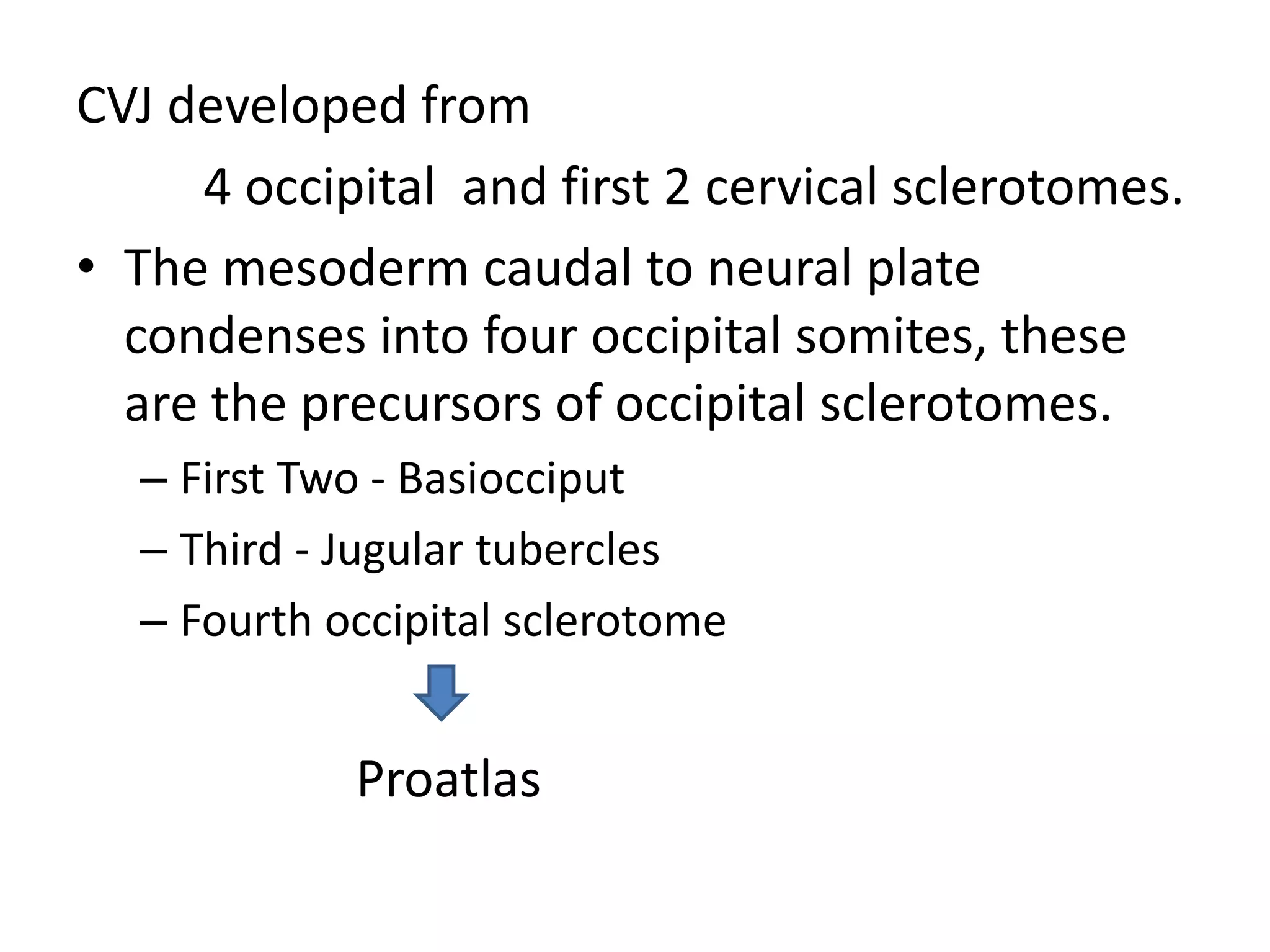
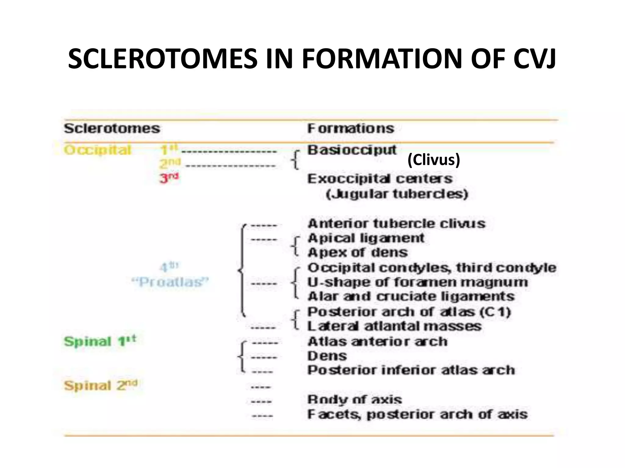
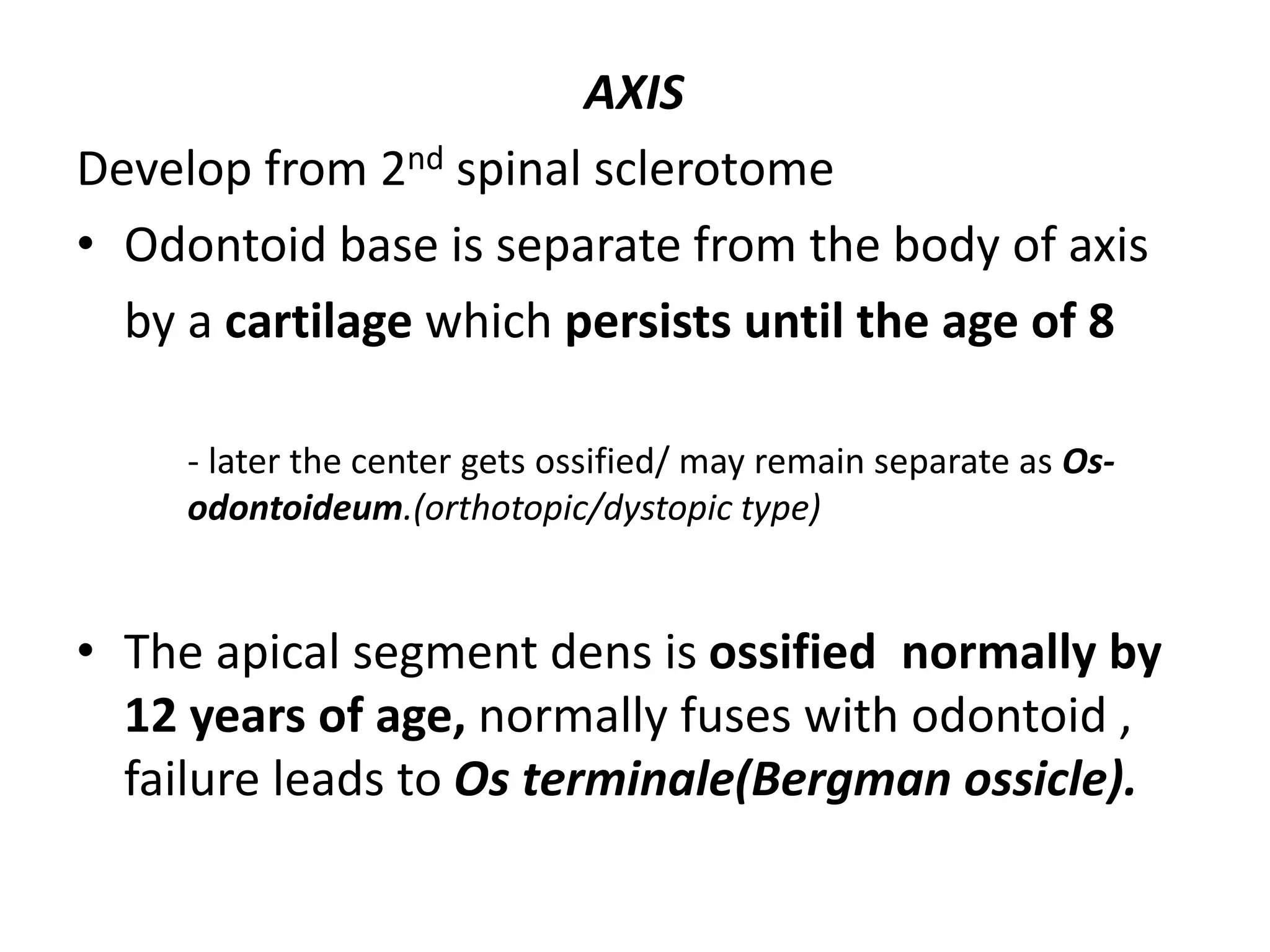
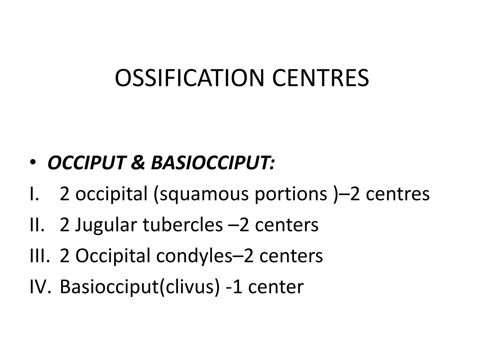
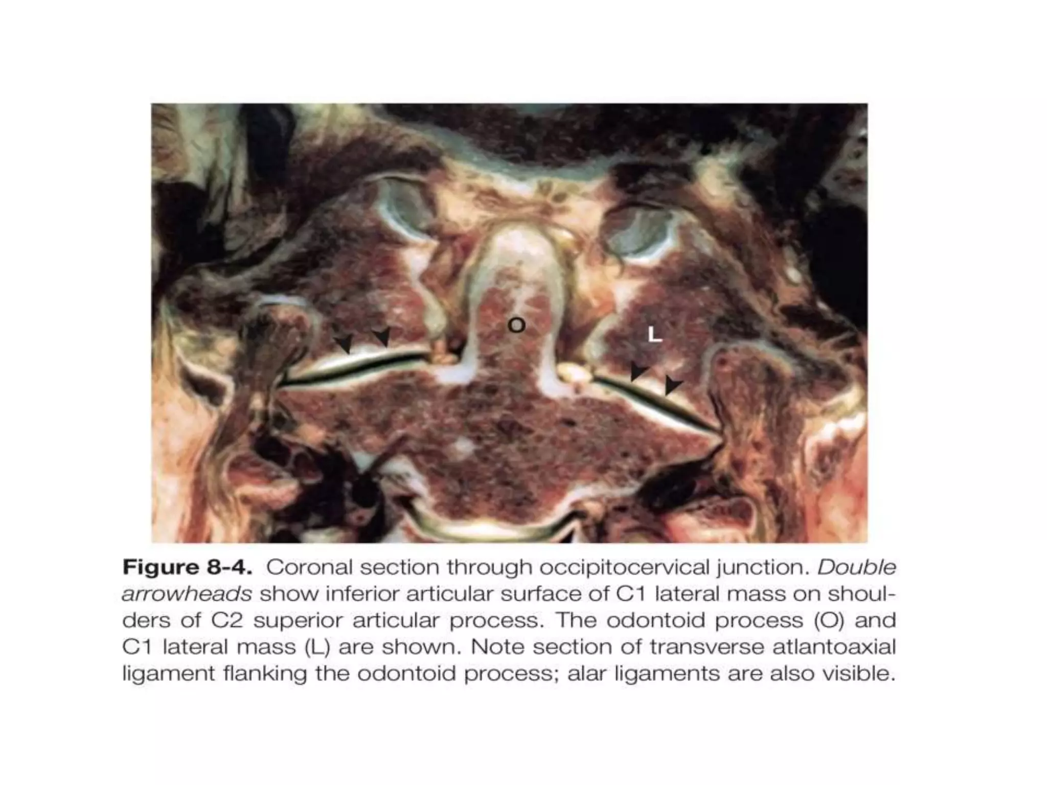
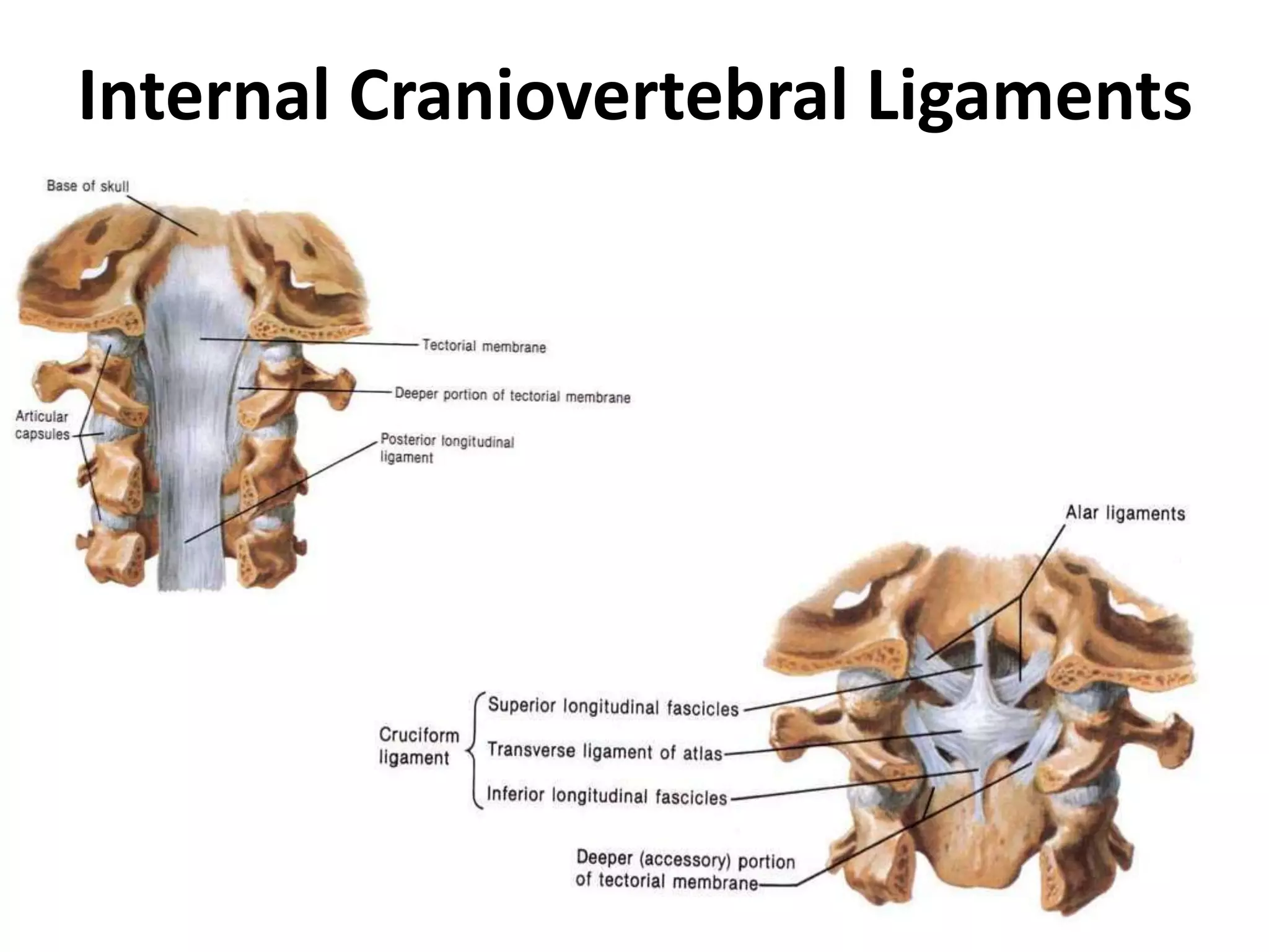
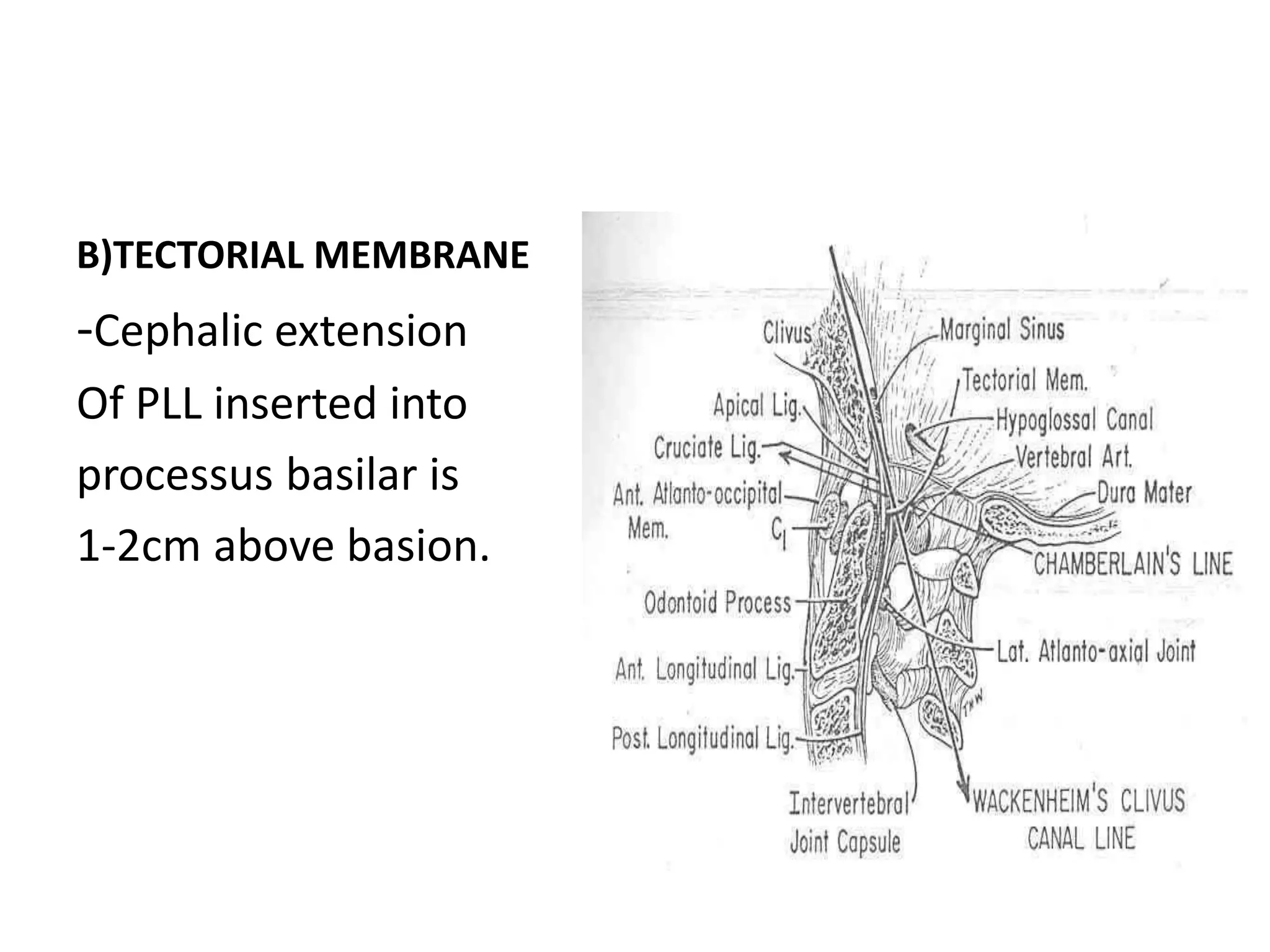
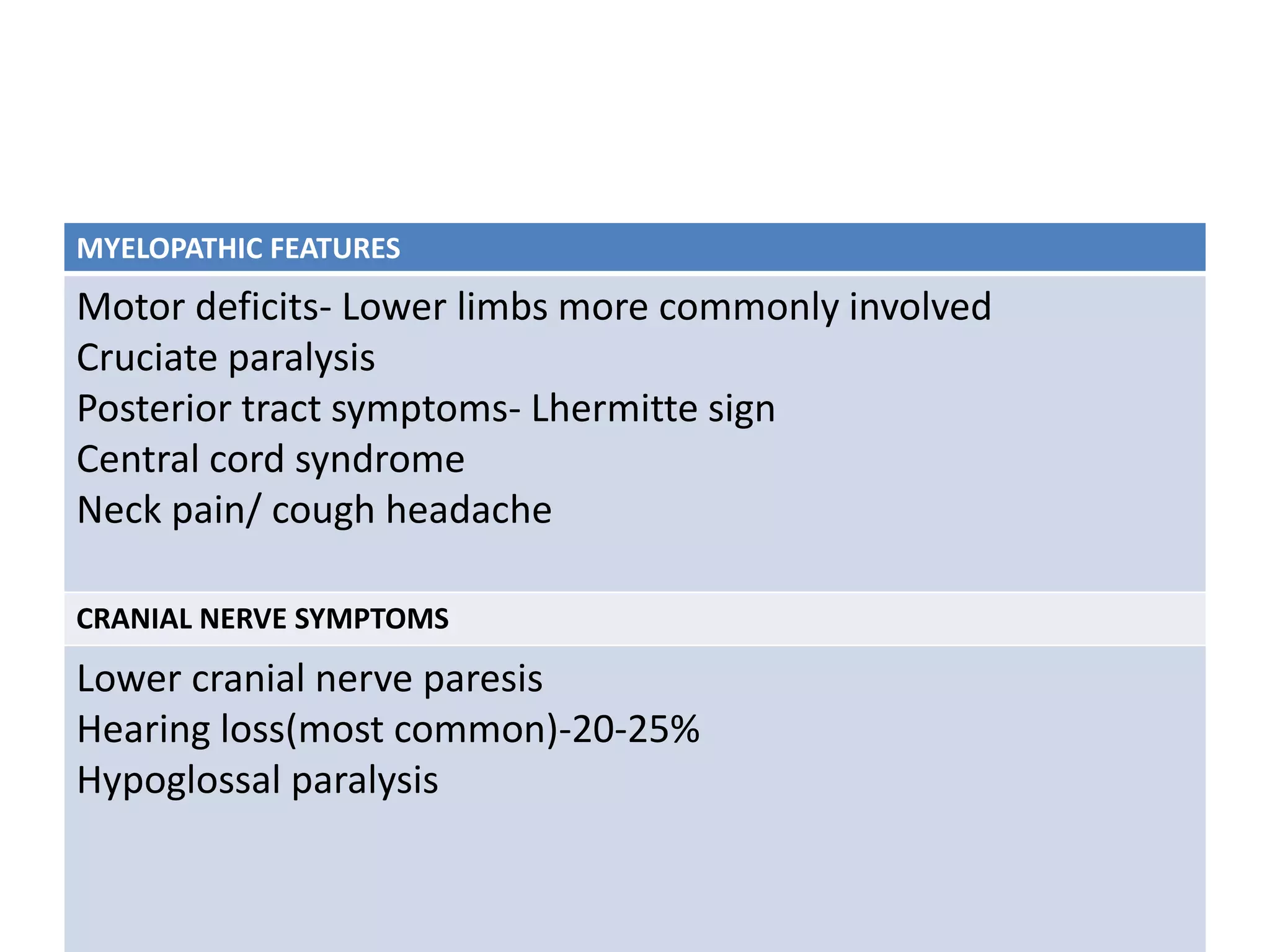
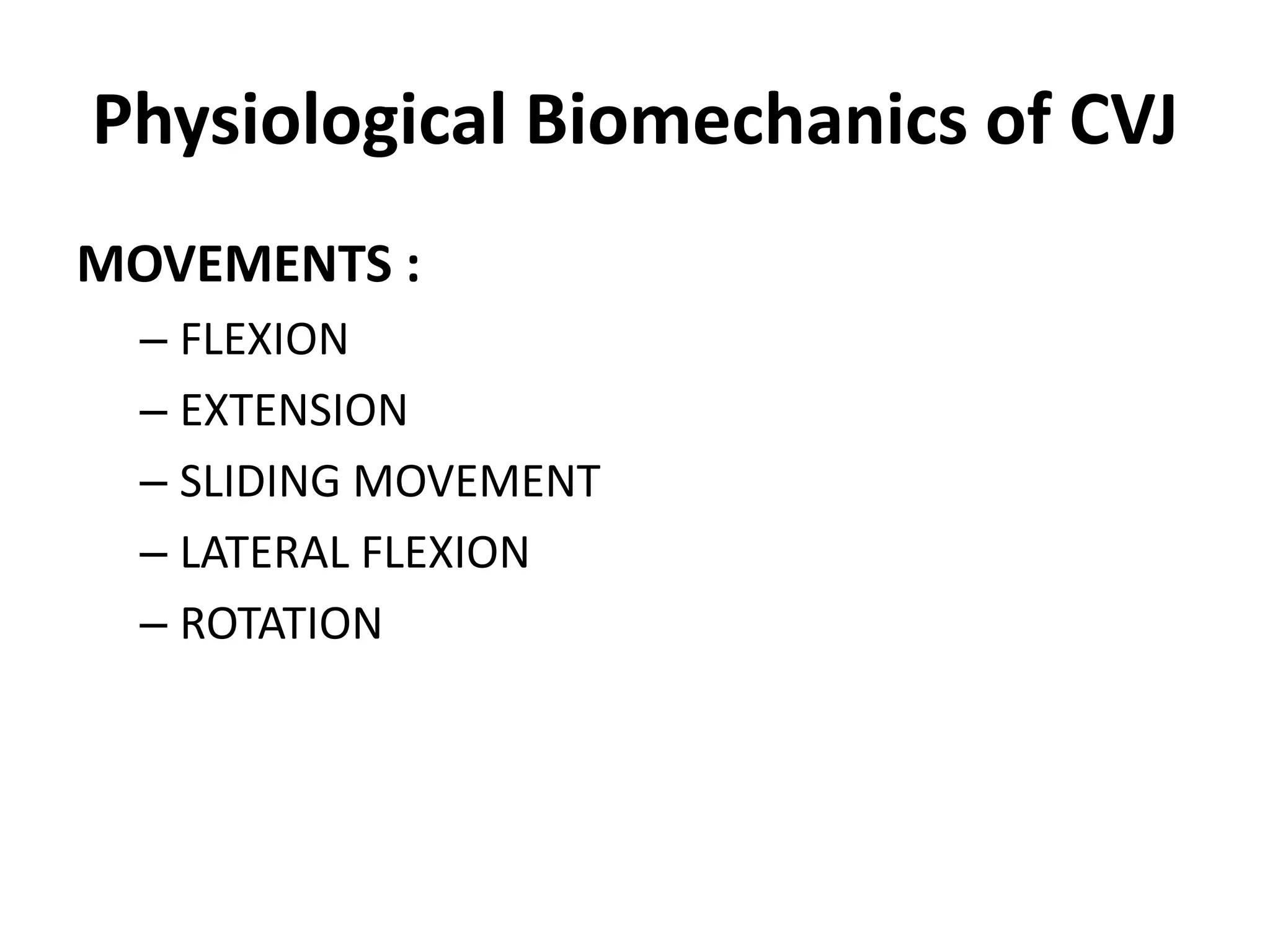
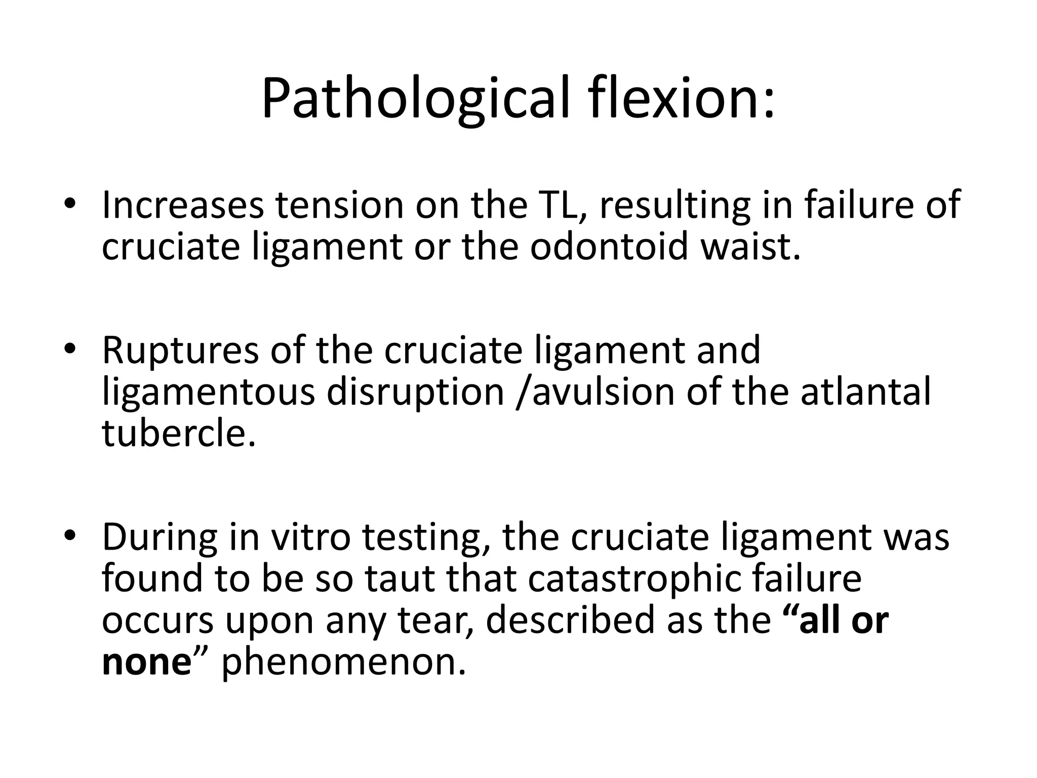
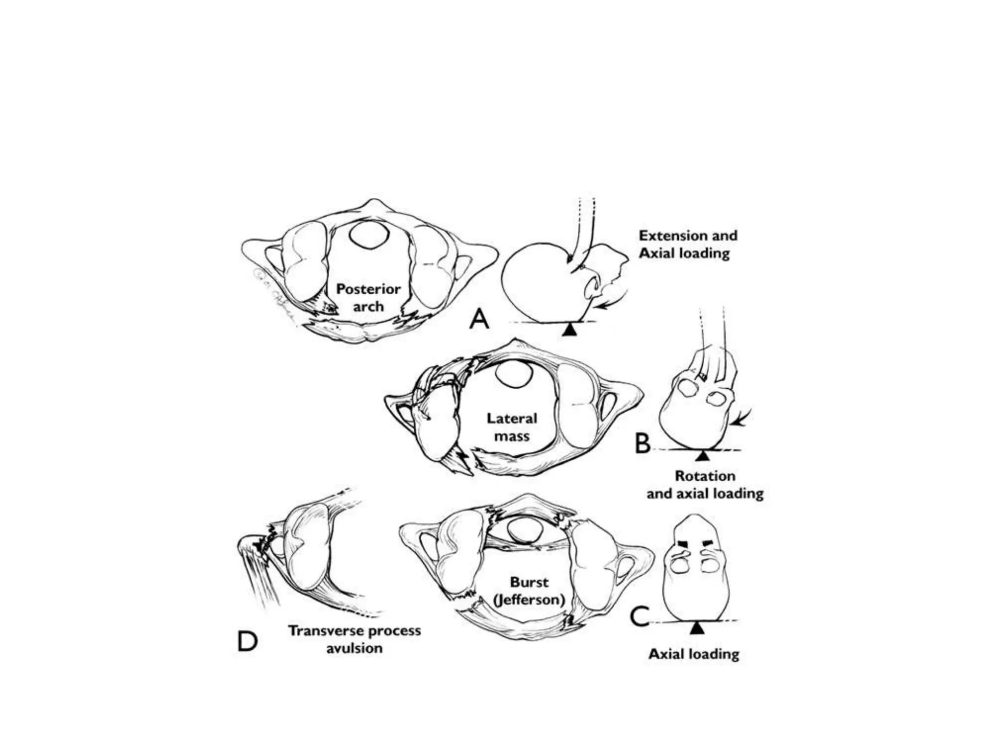
EMBRYOLOGY,ANATOMY,
BIOMECHANICS OF CVJ
Presenter: Dr. Tripurari Pandey
Moderator: Dr. Ankur Bajaj (M.Ch)
HISTORY
• Meckel , 1815 : manifestation of occipital Vertebrae.
• Bell , 1830 : first described spontaneous AAD.
• 1886 – Giacomini described the first case of congenital
AAD.
• Chamberlain , 1937 : Basilar invagination.
• Carl List, in 1941, described the neurological
syndromes accompanying developmental anomalies of
the occipital bone, the atlas, and the axis vertebrae.
• Atul Goel 2014 . Goel's Classification of Atlantoaxial
‘facetal’ dislocation.
• The CVJ is a collective term that refers occipital
bone (surrounds the foramen magnum), atlas,
axis, and supporting ligaments.
• It’s a transition zone b/w a mobile cranium &
relatively less mobile spinal column.
• Accounts approximately 25% of the vertical
height of the entire cervical spine.
• Development of the cartilaginous cranium & the
adjacent structures begins during the early weeks.
• 2nd week: Mesoderm cells condense in midline
:notochordal process.
• 3rdGestational week:
-notochordal process invaginates in b/w ecto &
endoderm : notochord.
-dorsal ectoderm thickens to form neural groove
which folds, fuses, : neural tube.
• B/W 3rd& 5thweek: Paraxial mesoderm gives rise
to somites(Segmentation).
• Total 42 somites(4 occipital, 8 cervical, 12
thoracic, 5lumbar, 5 sacral, 8 coccygeal) form at
4thweek.
-Ventromedial portion of somite is k/a
sclerotome which forms vertebral bodies around
notocord.
• Mesenchymal cells around the notochord to form the
intervertebral disc.
• Notochord disappears at the vertebral bodies, but
persist as nucleus pulposus at disc.
• The first 4 sclerotomes (normally forms VB) do not
follow this course & fuse to form the occipital bone &
Post. portion of FM.
• This membranous stage is f/b stages of chondrification
& ossification.
CVJ developed from
4 occipital and first 2 cervical sclerotomes.
• The mesoderm caudal to neural plate
condenses into four occipital somites, these
are the precursors of occipital sclerotomes.
– First Two - Basiocciput
– Third - Jugular tubercles
– Fourth occipital sclerotome
Proatlas
SCLEROTOMES IN FORMATION OF CVJ
(Clivus)
Proatlas
• Hypocentrum: anterior tubercle of the clivus.
• Centrum : apex of the dens and apical
ligament
• Neural Arch: two parts
-ventrorostral part: ant margin of FM, 2 occipital
condyles and alar/cruciate ligaments.
-dorsocaudal part : paired superior articular facets/
lateral masses of C1 / superior part of the post. arch of
the C1.
ATLAS
• major portion : by first spinal sclerotome.
• Centrum of sclerotome is separated to fuse with
the axis body forming the odontoid process.
• Hypocentrum of 1st spinal sclerotome: the
anterior arch of the atlas.
• Neural arch of the first spinal sclerotome forms
the inferior portion of the posterior arch of atlas.
First spinal sclerotome
Atlas vertebra is primarily formed from this
sclerotome.
Sclerotome division
Hypocentrum Centrum Neural Arch
Anterior arch C1 Dens Inferior
portion of
posterior
arch.
Second spinal sclerotome
Develops into axis vertebra
Sclerotome division
Hypocentrum Centrum Neural Arch
Disappears Body of axis Facets & Posterior
arch of axis
Body of Axis
Mid part of Dens
Apical part of Dens
AXIS
Develop from 2nd spinal sclerotome
• Odontoid base is separate from the body of axis
by a cartilage which persists until the age of 8
- later the center gets ossified/ may remain separate as Os-
odontoideum.(orthotopic/dystopic type)
• The apical segment dens is ossified normally by
12 years of age, normally fuses with odontoid ,
failure leads to Os terminale(Bergman ossicle).
• OCCIPUT & BASIOCCIPUT:
I. 2 occipital (squamous portions )–2 centres
II. 2 Jugular tubercles –2 centers
III. 2 Occipital condyles–2 centers
IV. Basiocciput(clivus) -1 center
OSSIFICATION CENTRES
ATLAS: ossifies from 3 centres
• Each half of post. arch along with lateral mass
– at 7 to 9 weeks centre appears,
– unites at 3 –4 years.
• Anterior arch :
– at 1 to 2 years centre appears,
– unites with lateral mass at 6 –8 years
• AXIS: having 5 primary & 2 secondary centres for
ossification.
– 2 Neural arches –2 centers(appear at 7 –8 wk)
– Body of axis –1 centre (appear at 4 –5 months)
– Body of dens –2 centers (appear at 6 –7 months)
Above 4 parts (at birth) unite at age of 3 –6 years
– Tip of odontoid appears at 3 –6 years and unites
with the body of odontoid at 12 years.
Applied:
• Dysplasia of the occipital segments may
flatten the clivus-platybasia.
• >1420 basal angle.(normal range 1240-1420)
-When the basiocciput and rim of foramen magnum are
underdeveloped, the odontoid and arch of atlas may
invaginate-Basilar invagination.
-If it is due to softening of bone or fibrous band k/a Basilar
impression.
• Bicornuate dens : dens body may fail to fuse in utero
resulting in a V-shaped cleft found radiographically at
birth , rare in adults.
• Failure of segmentation b/w the axis & the 3rd cervical
vertebra involves both the ant. & the post. Vertebral
segments, associated with other anomalies like
Klippel–Feil syndrome.
ANATOMY OF CVJ (ARTICULAR)
• Upper surfaces of C1 lateral masses are cup-like or
concave which fit into the ball & socket configuration,
united by articular capsules
• 3 synovial joints b/w atlas & axis –
• 1 median –between dens and atlas (Pivot variety)
• 2 lateral –b/w opposing articular facets (Plane variety)
Each joint has its own capsule & synovial cavity.
00-450 lateral ro
ANATOMY OF CVJ(LIGAMENTOUS)
• Principal stabilizing ligaments :
-Transverse Atlantal ligament
-Alar ligaments
• Secondary stabilizing ligaments(more elastic and weaker
than primary)
-Apical ligament
-Anterior & posterior A-O membranes
-Tectorial membrane
-Ligamentum flavum
-Capsular Ligament
External craniovertebral ligaments
Internal Craniovertebral Ligaments
ATLANTO-OCCIPITAL LIGAMENTS:
• A) Anterior Atlanto-occipital Membrane-Extends from
anterior edge of Foramen Magnum to anterior arch of
C1.
About 2 cm wide
Central fibres are thicker than the lateral portion.
• Ligament is continuous caudally with ALL of the spinal
column.
• acts as a tension band that stretches during extension,
serving as a secondary stabilizer against this motion.
B)TECTORIAL MEMBRANE
-Cephalic extension
Of PLL inserted into
processus basilar is
1-2cm above basion.
C)POSTERIOR ATLANTO-OCCIPITAL MEMBRANE:
• extends from occipital bone to posterior arch of atlas.
• A less strong ligament containing no significant elastic
tissue.
• Ligament is loose b/w the bones.
• Does not limit their motion.
• Firmly attached anteriorly to the duramater.
• Ligament invests itself :a canal through which the vertebral
artery, veins & 12th cranial nerve pass.
Transverse ligament
• Maintains the position of dens in sagittal
& craniocaudal direction.
• Inserted laterally in Bony prominence
in inner aspect of condyles.
• It is 8mm in height and 2-3 mm thick in
midline.
• Helps in free gliding motion to occur
over the posterior facet of the dens.
• posterior fibres of this ligament are
arcuate, & the more ventral fibres
are circular in configuration.
ALAR LIGAMENTS
• 2cords that extends from
dens tip to lateral part of
rim of FM.
• About 8mm wide.
• They are ventral & cranial to
the transverse ligament.
• Alar lig allow an anterior
shift of C1 from 3 to 5 mm.
• They limit the head –atlas
rotatory movement on the
odontoid-axis
APICAL LIGAMENT
• slender band of
fibres.
• 2-5mm wide & 2-
8mm.
• No mechanical
significance
CRANIOMETRY
• Uses series of lines, planes and angles to
define the normal anatomic relationships of
the CVJ.
• Plain X-rays,3DCT or on MRI
• Disadvantages: anatomic structures and
planes vary within normal range.
• Craniocervical junction malformations (CCJM) have been
described more frequently as Arnold Chiari Malformation
(CM) and BI.
1-Chiari malformation –herniation of the cerebellar tonsils in
the FM with compression of the neural structures and / or
of CSF flow.
2-BI -developmental anomaly of the CVJ in which odontoid
abnormally prolapses into the FM.
• In angular Craniometric Point (mainly uses four
points i.e. Nasion, Top of dorsum sellae,Basion and
Opistion) have been measured in sagittal view of MRI
to findout Cranio-cervical junction angulations .
primary cranial angles
Basal angle-Platybasia
Boogard´s angle-clival horizontalization
secondary craniocervical angles
Clivus canal angle-for kyphosis of craniocervical
junction
Cervical lordosis angle-for cervical spine lordosis
CRANIOMETRIC -LINE
CHAMBERLAIN’S LINE
• Posterior margin of hard
palate to opisthion
• Normal- tip of dens less
than 5mm below this line
• Abnormal- in basilar
invagination
MCRAE’S LINE
• Line from basion to
opisthion
• Normal – tip of dens below
this line
• Abnormal-in basilar
invagination
CRANIOMETRIC -LINE
MCGREGOR’S LINE
• Posterior margin of hard
palate to lowest part of
occipital bone.
• Normal- tip of dens less
than 7mm below this line
• Abnormal- in BI
WACKENHEIM’S LINE
• Line extrapolated along
dorsal surface of clivus
• Normal – dens should be
tangential or anterior to this
line.
• Abnormal-in BI
CRANIOMETRIC -LINE
DIGASTRIC LINE
• Line between incisurae
mastoidae
• Normal- 10mm above
atlanto-occipital joint
BIMASTOID LINE
• Line between tips of
mastoid processes
• Normal – intersects atlanto-
occipital joint
CRANIOMETRIC -LINE
WELCHER BASAL ANGLE
• Angle at junction of nasion-
tuberculum and tuberculum-
basion lines.
• Normal- 132-140 degree
• Abnormal->143 degree in
platybasia
CLIVUS CANAL ANGLE
• Angle at junction of
Wackenheim’s line and posterior
vertebral body line
• Normal – 150-180degree
• Abnormal-<150 degree in
platybasia
PLATYBASIA
• Skull base flattening
• Primary and secondary
• Bow string deformity
• Increased basal angle
• Decreased clivus canal
angle
• Association – basilar
invagination
ATLANTOAXIAL DISLOCATION
Congenital
Acquired
Traumatic
Atlantodens interval
3mm - adults
5mm – children
20 year old man with type
2 dens fracture(irregular
margins and atlantoaxial
dislocation(TRAUMA)
47 year old lady with
rheumatoid arthritis with
basilar impression, sclerosis of
atlantoaxial joint and
atlantoaxial dislocation
18 year old lady with TB,
retropharyngeal collection,
lytic area in dens and
atlantoaxial
dislocation(INFECTIVE)
Lines and angles used in radiologic diagnosis of C.V
anomalies
Parameter Normal range limits
A. PLATYBASIA
 Basal angle
 Boogard’s angle (Angle b/w clivus line and McRae's line)
 Bull’s angle (Angle b/w Line joining the mid point of
posterior and anterior arch of C1 and prolongation of
hard palate) normal<100
B. BASILAR INVAGINATION
• Chamberlain’s line
• Mcgregor’s line
• Mcrae line
C. ATLANTO-AXIAL DISLOCATION *
•Atlanto-odontoid space
< 142 degree
< 136 degree
< 13 degree
>3 mm above this line
>5 mm above this line
Tip above this line
upto 3 mm in adults
upto 5 mm in children
CLINICAL PRESENTATION OF CVJ ANOMALIES
• The most interesting feature variability in
clinical presentation due to compression of
the lower brainstem, cervical spinal cord,
cranial nerves, cervical nerve roots & vascular
supply.
• The most common symptom is neck pain -
85%.
• False localising signs: Usually motor
monoparesis, paraparesis, & quadripresis
MYELOPATHIC FEATURES
Motor deficits- Lower limbs more commonly involved
Cruciate paralysis
Posterior tract symptoms- Lhermitte sign
Central cord syndrome
Neck pain/ cough headache
CRANIAL NERVE SYMPTOMS
Lower cranial nerve paresis
Hearing loss(most common)-20-25%
Hypoglossal paralysis
BRAIN STEM/CEREBELLAR SIGNS
Sleep apnea and dysphagia
Nystagmus
Gait ataxia
VASCULAR SYMPTOMS
Syncope
Vertigo
Episodic paresis
Transient visual loss-10-25% of cases
(Due to vertebro basilar insufficiency)
Physiological Biomechanics of CVJ
MOVEMENTS :
– FLEXION
– EXTENSION
– SLIDING MOVEMENT
– LATERAL FLEXION
– ROTATION
FLEXION & EXTENSION :
– joints involved : occipitoatlantal & atlantoaxial
– average range at occipitoatlantal jt. :13 – 15 degrees
– atlantoaxial jt. : 10 degrees
• FLEXION IS LIMITED BY : tectorial membrane and
dens basion contact
• EXTENSION LIMITED BY :
– stretching of tectorial membrane
– opisthion
– post. arch of atlas contact.
• TL prevents pathological flexion of the C1-C2
• extension is inhibited by the bony elements of
the C1-C2 joint facets
• ANTERO-POSTERIOR TRANSLOCATION
BETWEEN DENS & ANT. RING OF ATLAS :
– adults : 3mm
– young children : 5mm
• In adults if
– upto 5 mm : rupture of cruciate lig.
– > 5 mm : rupture of both cruciate & alar lig.
ROTATION
JOINT INVOLVED : ATLANTOAXIAL JOINT
–maximum range : 37 – 420
–> rotation leads facet jt. interlocking
–rotation > 32 – 350 : angulation of contr.
vert. artery
–> 450 : ipsilateral Vertebral artery occlusion
• JOINT INVOLVED LATERAL ROTATION :
ATLANTOAXIAL JOINT + LOWER C-SPINE
– Max. Rotation :900
• LATERAL FLEXION : small amplitude 5 – 100
• SLIDING MOVEMENT :forward or backward movement of
head without flexion or extension of neck
• Forward slide :
– axis inclines forward
– post. displacement of axis
– ant. arch of atlas slides up
– atlantoodontoid space gap
– occipitoodontoid space gap
– n : 3 – 6 mm
– double in forward slide
• Backward slide :ant. arch of atlas slides down post.
atlantooccipital space reduces
Biomechanics of CVJ Pathology
• In trauma, the CVJ exhibits predictable
patterns of failure based on the mechanism of
injury.
Pathological flexion:
• Increases tension on the TL, resulting in failure of
cruciate ligament or the odontoid waist.
• Ruptures of the cruciate ligament and
ligamentous disruption /avulsion of the atlantal
tubercle.
• During in vitro testing, the cruciate ligament was
found to be so taut that catastrophic failure
occurs upon any tear, described as the “all or
none” phenomenon.
• Failure of the tectorial membrane has also
been seen.
• May lead to Dural tears, as the superior-most
part is continuous with the dura.
• Isolated tectorial membrane failure can occur
with minor instability in flexion and
extension.
Pathological extension:
• Hyperextension : Fracture of the atlas at the
posterior ring / fracture of the axis at the pars
interarticularis / the odontoid.
• Ligaments injury of the anterior CVJ, alar
ligaments, cruciform ligament, and tectorial
membranes
Supraphysiological rotation
• Isolated rupture of the alar ligament is rare:
hyperflexion paired with rotation
• Supraphysiological rotation at the atlantoaxial
junction can predict/ even diagnose: alar
ligament disruption.
• introduces instability in rotation and increase in
flexion/extension/lateral bending.
THANK YOU
MCQ`S
• Somoites/sclerotomes are derived from?
1. Endoderm
2. Ectoderm
3. Mesoderm
4. none
• Mesoderm
(Schimidik and sweet /6`th edition vol 2/pg
2055)
• Occipital bone is formed by fusion of ?
1. 2 sclerotomes
2. 3 sclerotomes
3. 4 sclerotomes
4. 5 sclerotomes
• 4 sclerotomes
(Schimidik and sweet /6`th edition vol 2/pg
2055)
• Apex of dens is derived from??
1. 1st occipital sclerotome
2. 2nd occipital sclerotome
3. 3d occipital sclerotome
4. Proatlas
• Proatlas
(Schimidik and sweet /6`th edition vol 2/pg
2055)
• Atlas is derived from??
1. Proatlas
2. 1st cervical sclerotome
3. Both
4. none
• Both
(Schimidik and sweet /6`th edition vol 2/pg
2055)
• How many ossifications centres does atlas
have?
1. 1
2. 2
3. 3
4. 4
• 3
(Schimidik and sweet /6`th edition vol 2/pg
2055)
• Apex of Dens gets Ossified by
1. 1yr
2. 1.5 yr
3. 2 yr
4. 3 yr
• 3 yr
(Schimidik and sweet /6`th edition vol 2/pg
2056)
• Odontoid gets fully ossified by??
1. 8 yrs
2. 10 yrs
3. 5 yrs
4. 12 yrs
• 12 yrs
• Schimidik and sweet /6`th edition vol 2/pg
2056
• Tectorial membrane is continuation of ??
1. ALL
2. PLL
3. TL
4. None
• PLL
(Schimidik and sweet /6`th edition vol 2/pg
2057)
• The toughest ligament of cvj complex?
1. Apical
2. Transverse ligament
3. Tectorial ligament
4. C1 C2 capsular ligament
• Transverse ligament
(Schimidik and sweet /6`th edition vol 2/pg
2057)
• About Ossification of the spine
A. At birth, most vertebrae have 3 primary and 5
secondary ossification centers(OC) connected by
hyaline
B. Exception to typical ossification occur at C1,C2,C7,
lumbar , sacrum and coccyx
C. C1 vertebrae has no secondary OC
D. Lumbar vertebrae has 3 primary OC pervertebrae and
7 secondary OC pervertebrae
E. Sacrum has 5 primary OC per vertebrae and 4
secondary OC.
Ans. All options are true.
Centrum ossification starts at lower thoracic/upper lumbar spine of
foetus. Moves in both direction while
Neural arch ossification begins at cervico-thoracic level and then upper
cervical region and lastly thoracolumbar region.
Atlas has two to five (three most common) primary ossification centers
and no secondary ossification centers.
Axis has five primary ossification centers and two secondary
ossification centers.
C3-6 has 3 primary ossification centers and 5 secondary ossification
centers per each vertebrae .
Co1 has 3 primary ossification centers. Co2–Co4 have one primary
ossification center each and no secondary ossification center.
Next Seminar – Dr Ravi Prakash
Topic – GCS ( Glasgow Coma Scale ).
Moderator- Dr. Awdesh Yadav (M.Ch)

CRANIOVERTEBRAL JUNCTION

  • 1.
    EMBRYOLOGY,ANATOMY, BIOMECHANICS OF CVJ Presenter:Dr. Tripurari Pandey Moderator: Dr. Ankur Bajaj (M.Ch)
  • 2.
    HISTORY • Meckel ,1815 : manifestation of occipital Vertebrae. • Bell , 1830 : first described spontaneous AAD. • 1886 – Giacomini described the first case of congenital AAD. • Chamberlain , 1937 : Basilar invagination. • Carl List, in 1941, described the neurological syndromes accompanying developmental anomalies of the occipital bone, the atlas, and the axis vertebrae. • Atul Goel 2014 . Goel's Classification of Atlantoaxial ‘facetal’ dislocation.
  • 3.
    • The CVJis a collective term that refers occipital bone (surrounds the foramen magnum), atlas, axis, and supporting ligaments. • It’s a transition zone b/w a mobile cranium & relatively less mobile spinal column. • Accounts approximately 25% of the vertical height of the entire cervical spine.
  • 5.
    • Development ofthe cartilaginous cranium & the adjacent structures begins during the early weeks. • 2nd week: Mesoderm cells condense in midline :notochordal process. • 3rdGestational week: -notochordal process invaginates in b/w ecto & endoderm : notochord. -dorsal ectoderm thickens to form neural groove which folds, fuses, : neural tube.
  • 6.
    • B/W 3rd&5thweek: Paraxial mesoderm gives rise to somites(Segmentation). • Total 42 somites(4 occipital, 8 cervical, 12 thoracic, 5lumbar, 5 sacral, 8 coccygeal) form at 4thweek. -Ventromedial portion of somite is k/a sclerotome which forms vertebral bodies around notocord.
  • 7.
    • Mesenchymal cellsaround the notochord to form the intervertebral disc. • Notochord disappears at the vertebral bodies, but persist as nucleus pulposus at disc. • The first 4 sclerotomes (normally forms VB) do not follow this course & fuse to form the occipital bone & Post. portion of FM. • This membranous stage is f/b stages of chondrification & ossification.
  • 8.
    CVJ developed from 4occipital and first 2 cervical sclerotomes. • The mesoderm caudal to neural plate condenses into four occipital somites, these are the precursors of occipital sclerotomes. – First Two - Basiocciput – Third - Jugular tubercles – Fourth occipital sclerotome Proatlas
  • 9.
    SCLEROTOMES IN FORMATIONOF CVJ (Clivus)
  • 10.
    Proatlas • Hypocentrum: anteriortubercle of the clivus. • Centrum : apex of the dens and apical ligament • Neural Arch: two parts -ventrorostral part: ant margin of FM, 2 occipital condyles and alar/cruciate ligaments. -dorsocaudal part : paired superior articular facets/ lateral masses of C1 / superior part of the post. arch of the C1.
  • 11.
    ATLAS • major portion: by first spinal sclerotome. • Centrum of sclerotome is separated to fuse with the axis body forming the odontoid process. • Hypocentrum of 1st spinal sclerotome: the anterior arch of the atlas. • Neural arch of the first spinal sclerotome forms the inferior portion of the posterior arch of atlas.
  • 12.
    First spinal sclerotome Atlasvertebra is primarily formed from this sclerotome. Sclerotome division Hypocentrum Centrum Neural Arch Anterior arch C1 Dens Inferior portion of posterior arch.
  • 13.
    Second spinal sclerotome Developsinto axis vertebra Sclerotome division Hypocentrum Centrum Neural Arch Disappears Body of axis Facets & Posterior arch of axis Body of Axis Mid part of Dens Apical part of Dens
  • 14.
    AXIS Develop from 2ndspinal sclerotome • Odontoid base is separate from the body of axis by a cartilage which persists until the age of 8 - later the center gets ossified/ may remain separate as Os- odontoideum.(orthotopic/dystopic type) • The apical segment dens is ossified normally by 12 years of age, normally fuses with odontoid , failure leads to Os terminale(Bergman ossicle).
  • 16.
    • OCCIPUT &BASIOCCIPUT: I. 2 occipital (squamous portions )–2 centres II. 2 Jugular tubercles –2 centers III. 2 Occipital condyles–2 centers IV. Basiocciput(clivus) -1 center OSSIFICATION CENTRES
  • 17.
    ATLAS: ossifies from3 centres • Each half of post. arch along with lateral mass – at 7 to 9 weeks centre appears, – unites at 3 –4 years. • Anterior arch : – at 1 to 2 years centre appears, – unites with lateral mass at 6 –8 years
  • 19.
    • AXIS: having5 primary & 2 secondary centres for ossification. – 2 Neural arches –2 centers(appear at 7 –8 wk) – Body of axis –1 centre (appear at 4 –5 months) – Body of dens –2 centers (appear at 6 –7 months) Above 4 parts (at birth) unite at age of 3 –6 years – Tip of odontoid appears at 3 –6 years and unites with the body of odontoid at 12 years.
  • 20.
    Applied: • Dysplasia ofthe occipital segments may flatten the clivus-platybasia. • >1420 basal angle.(normal range 1240-1420)
  • 21.
    -When the basiocciputand rim of foramen magnum are underdeveloped, the odontoid and arch of atlas may invaginate-Basilar invagination. -If it is due to softening of bone or fibrous band k/a Basilar impression.
  • 22.
    • Bicornuate dens: dens body may fail to fuse in utero resulting in a V-shaped cleft found radiographically at birth , rare in adults. • Failure of segmentation b/w the axis & the 3rd cervical vertebra involves both the ant. & the post. Vertebral segments, associated with other anomalies like Klippel–Feil syndrome.
  • 23.
    ANATOMY OF CVJ(ARTICULAR) • Upper surfaces of C1 lateral masses are cup-like or concave which fit into the ball & socket configuration, united by articular capsules • 3 synovial joints b/w atlas & axis – • 1 median –between dens and atlas (Pivot variety) • 2 lateral –b/w opposing articular facets (Plane variety) Each joint has its own capsule & synovial cavity.
  • 26.
  • 27.
    ANATOMY OF CVJ(LIGAMENTOUS) •Principal stabilizing ligaments : -Transverse Atlantal ligament -Alar ligaments • Secondary stabilizing ligaments(more elastic and weaker than primary) -Apical ligament -Anterior & posterior A-O membranes -Tectorial membrane -Ligamentum flavum -Capsular Ligament
  • 29.
  • 30.
  • 32.
    ATLANTO-OCCIPITAL LIGAMENTS: • A)Anterior Atlanto-occipital Membrane-Extends from anterior edge of Foramen Magnum to anterior arch of C1. About 2 cm wide Central fibres are thicker than the lateral portion. • Ligament is continuous caudally with ALL of the spinal column. • acts as a tension band that stretches during extension, serving as a secondary stabilizer against this motion.
  • 33.
    B)TECTORIAL MEMBRANE -Cephalic extension OfPLL inserted into processus basilar is 1-2cm above basion.
  • 34.
    C)POSTERIOR ATLANTO-OCCIPITAL MEMBRANE: •extends from occipital bone to posterior arch of atlas. • A less strong ligament containing no significant elastic tissue. • Ligament is loose b/w the bones. • Does not limit their motion. • Firmly attached anteriorly to the duramater. • Ligament invests itself :a canal through which the vertebral artery, veins & 12th cranial nerve pass.
  • 35.
    Transverse ligament • Maintainsthe position of dens in sagittal & craniocaudal direction. • Inserted laterally in Bony prominence in inner aspect of condyles. • It is 8mm in height and 2-3 mm thick in midline. • Helps in free gliding motion to occur over the posterior facet of the dens. • posterior fibres of this ligament are arcuate, & the more ventral fibres are circular in configuration.
  • 36.
    ALAR LIGAMENTS • 2cordsthat extends from dens tip to lateral part of rim of FM. • About 8mm wide. • They are ventral & cranial to the transverse ligament. • Alar lig allow an anterior shift of C1 from 3 to 5 mm. • They limit the head –atlas rotatory movement on the odontoid-axis
  • 37.
    APICAL LIGAMENT • slenderband of fibres. • 2-5mm wide & 2- 8mm. • No mechanical significance
  • 38.
    CRANIOMETRY • Uses seriesof lines, planes and angles to define the normal anatomic relationships of the CVJ. • Plain X-rays,3DCT or on MRI • Disadvantages: anatomic structures and planes vary within normal range.
  • 39.
    • Craniocervical junctionmalformations (CCJM) have been described more frequently as Arnold Chiari Malformation (CM) and BI. 1-Chiari malformation –herniation of the cerebellar tonsils in the FM with compression of the neural structures and / or of CSF flow. 2-BI -developmental anomaly of the CVJ in which odontoid abnormally prolapses into the FM.
  • 40.
    • In angularCraniometric Point (mainly uses four points i.e. Nasion, Top of dorsum sellae,Basion and Opistion) have been measured in sagittal view of MRI to findout Cranio-cervical junction angulations .
  • 41.
    primary cranial angles Basalangle-Platybasia Boogard´s angle-clival horizontalization secondary craniocervical angles Clivus canal angle-for kyphosis of craniocervical junction Cervical lordosis angle-for cervical spine lordosis
  • 42.
    CRANIOMETRIC -LINE CHAMBERLAIN’S LINE •Posterior margin of hard palate to opisthion • Normal- tip of dens less than 5mm below this line • Abnormal- in basilar invagination MCRAE’S LINE • Line from basion to opisthion • Normal – tip of dens below this line • Abnormal-in basilar invagination
  • 43.
    CRANIOMETRIC -LINE MCGREGOR’S LINE •Posterior margin of hard palate to lowest part of occipital bone. • Normal- tip of dens less than 7mm below this line • Abnormal- in BI WACKENHEIM’S LINE • Line extrapolated along dorsal surface of clivus • Normal – dens should be tangential or anterior to this line. • Abnormal-in BI
  • 44.
    CRANIOMETRIC -LINE DIGASTRIC LINE •Line between incisurae mastoidae • Normal- 10mm above atlanto-occipital joint BIMASTOID LINE • Line between tips of mastoid processes • Normal – intersects atlanto- occipital joint
  • 45.
    CRANIOMETRIC -LINE WELCHER BASALANGLE • Angle at junction of nasion- tuberculum and tuberculum- basion lines. • Normal- 132-140 degree • Abnormal->143 degree in platybasia CLIVUS CANAL ANGLE • Angle at junction of Wackenheim’s line and posterior vertebral body line • Normal – 150-180degree • Abnormal-<150 degree in platybasia
  • 46.
    PLATYBASIA • Skull baseflattening • Primary and secondary • Bow string deformity • Increased basal angle • Decreased clivus canal angle • Association – basilar invagination
  • 47.
    ATLANTOAXIAL DISLOCATION Congenital Acquired Traumatic Atlantodens interval 3mm- adults 5mm – children 20 year old man with type 2 dens fracture(irregular margins and atlantoaxial dislocation(TRAUMA) 47 year old lady with rheumatoid arthritis with basilar impression, sclerosis of atlantoaxial joint and atlantoaxial dislocation 18 year old lady with TB, retropharyngeal collection, lytic area in dens and atlantoaxial dislocation(INFECTIVE)
  • 48.
    Lines and anglesused in radiologic diagnosis of C.V anomalies Parameter Normal range limits A. PLATYBASIA  Basal angle  Boogard’s angle (Angle b/w clivus line and McRae's line)  Bull’s angle (Angle b/w Line joining the mid point of posterior and anterior arch of C1 and prolongation of hard palate) normal<100 B. BASILAR INVAGINATION • Chamberlain’s line • Mcgregor’s line • Mcrae line C. ATLANTO-AXIAL DISLOCATION * •Atlanto-odontoid space < 142 degree < 136 degree < 13 degree >3 mm above this line >5 mm above this line Tip above this line upto 3 mm in adults upto 5 mm in children
  • 49.
    CLINICAL PRESENTATION OFCVJ ANOMALIES • The most interesting feature variability in clinical presentation due to compression of the lower brainstem, cervical spinal cord, cranial nerves, cervical nerve roots & vascular supply. • The most common symptom is neck pain - 85%. • False localising signs: Usually motor monoparesis, paraparesis, & quadripresis
  • 50.
    MYELOPATHIC FEATURES Motor deficits-Lower limbs more commonly involved Cruciate paralysis Posterior tract symptoms- Lhermitte sign Central cord syndrome Neck pain/ cough headache CRANIAL NERVE SYMPTOMS Lower cranial nerve paresis Hearing loss(most common)-20-25% Hypoglossal paralysis
  • 51.
    BRAIN STEM/CEREBELLAR SIGNS Sleepapnea and dysphagia Nystagmus Gait ataxia VASCULAR SYMPTOMS Syncope Vertigo Episodic paresis Transient visual loss-10-25% of cases (Due to vertebro basilar insufficiency)
  • 52.
    Physiological Biomechanics ofCVJ MOVEMENTS : – FLEXION – EXTENSION – SLIDING MOVEMENT – LATERAL FLEXION – ROTATION
  • 53.
    FLEXION & EXTENSION: – joints involved : occipitoatlantal & atlantoaxial – average range at occipitoatlantal jt. :13 – 15 degrees – atlantoaxial jt. : 10 degrees • FLEXION IS LIMITED BY : tectorial membrane and dens basion contact • EXTENSION LIMITED BY : – stretching of tectorial membrane – opisthion – post. arch of atlas contact.
  • 54.
    • TL preventspathological flexion of the C1-C2 • extension is inhibited by the bony elements of the C1-C2 joint facets
  • 55.
    • ANTERO-POSTERIOR TRANSLOCATION BETWEENDENS & ANT. RING OF ATLAS : – adults : 3mm – young children : 5mm • In adults if – upto 5 mm : rupture of cruciate lig. – > 5 mm : rupture of both cruciate & alar lig.
  • 56.
    ROTATION JOINT INVOLVED :ATLANTOAXIAL JOINT –maximum range : 37 – 420 –> rotation leads facet jt. interlocking –rotation > 32 – 350 : angulation of contr. vert. artery –> 450 : ipsilateral Vertebral artery occlusion
  • 57.
    • JOINT INVOLVEDLATERAL ROTATION : ATLANTOAXIAL JOINT + LOWER C-SPINE – Max. Rotation :900 • LATERAL FLEXION : small amplitude 5 – 100
  • 58.
    • SLIDING MOVEMENT:forward or backward movement of head without flexion or extension of neck • Forward slide : – axis inclines forward – post. displacement of axis – ant. arch of atlas slides up – atlantoodontoid space gap – occipitoodontoid space gap – n : 3 – 6 mm – double in forward slide • Backward slide :ant. arch of atlas slides down post. atlantooccipital space reduces
  • 59.
    Biomechanics of CVJPathology • In trauma, the CVJ exhibits predictable patterns of failure based on the mechanism of injury.
  • 60.
    Pathological flexion: • Increasestension on the TL, resulting in failure of cruciate ligament or the odontoid waist. • Ruptures of the cruciate ligament and ligamentous disruption /avulsion of the atlantal tubercle. • During in vitro testing, the cruciate ligament was found to be so taut that catastrophic failure occurs upon any tear, described as the “all or none” phenomenon.
  • 61.
    • Failure ofthe tectorial membrane has also been seen. • May lead to Dural tears, as the superior-most part is continuous with the dura. • Isolated tectorial membrane failure can occur with minor instability in flexion and extension.
  • 62.
    Pathological extension: • Hyperextension: Fracture of the atlas at the posterior ring / fracture of the axis at the pars interarticularis / the odontoid. • Ligaments injury of the anterior CVJ, alar ligaments, cruciform ligament, and tectorial membranes
  • 65.
    Supraphysiological rotation • Isolatedrupture of the alar ligament is rare: hyperflexion paired with rotation • Supraphysiological rotation at the atlantoaxial junction can predict/ even diagnose: alar ligament disruption. • introduces instability in rotation and increase in flexion/extension/lateral bending.
  • 66.
  • 67.
  • 68.
    • Somoites/sclerotomes arederived from? 1. Endoderm 2. Ectoderm 3. Mesoderm 4. none
  • 69.
    • Mesoderm (Schimidik andsweet /6`th edition vol 2/pg 2055)
  • 70.
    • Occipital boneis formed by fusion of ? 1. 2 sclerotomes 2. 3 sclerotomes 3. 4 sclerotomes 4. 5 sclerotomes
  • 71.
    • 4 sclerotomes (Schimidikand sweet /6`th edition vol 2/pg 2055)
  • 72.
    • Apex ofdens is derived from?? 1. 1st occipital sclerotome 2. 2nd occipital sclerotome 3. 3d occipital sclerotome 4. Proatlas
  • 73.
    • Proatlas (Schimidik andsweet /6`th edition vol 2/pg 2055)
  • 74.
    • Atlas isderived from?? 1. Proatlas 2. 1st cervical sclerotome 3. Both 4. none
  • 75.
    • Both (Schimidik andsweet /6`th edition vol 2/pg 2055)
  • 76.
    • How manyossifications centres does atlas have? 1. 1 2. 2 3. 3 4. 4
  • 77.
    • 3 (Schimidik andsweet /6`th edition vol 2/pg 2055)
  • 78.
    • Apex ofDens gets Ossified by 1. 1yr 2. 1.5 yr 3. 2 yr 4. 3 yr
  • 79.
    • 3 yr (Schimidikand sweet /6`th edition vol 2/pg 2056)
  • 80.
    • Odontoid getsfully ossified by?? 1. 8 yrs 2. 10 yrs 3. 5 yrs 4. 12 yrs
  • 81.
    • 12 yrs •Schimidik and sweet /6`th edition vol 2/pg 2056
  • 82.
    • Tectorial membraneis continuation of ?? 1. ALL 2. PLL 3. TL 4. None
  • 83.
    • PLL (Schimidik andsweet /6`th edition vol 2/pg 2057)
  • 84.
    • The toughestligament of cvj complex? 1. Apical 2. Transverse ligament 3. Tectorial ligament 4. C1 C2 capsular ligament
  • 85.
    • Transverse ligament (Schimidikand sweet /6`th edition vol 2/pg 2057)
  • 86.
    • About Ossificationof the spine A. At birth, most vertebrae have 3 primary and 5 secondary ossification centers(OC) connected by hyaline B. Exception to typical ossification occur at C1,C2,C7, lumbar , sacrum and coccyx C. C1 vertebrae has no secondary OC D. Lumbar vertebrae has 3 primary OC pervertebrae and 7 secondary OC pervertebrae E. Sacrum has 5 primary OC per vertebrae and 4 secondary OC.
  • 87.
    Ans. All optionsare true. Centrum ossification starts at lower thoracic/upper lumbar spine of foetus. Moves in both direction while Neural arch ossification begins at cervico-thoracic level and then upper cervical region and lastly thoracolumbar region. Atlas has two to five (three most common) primary ossification centers and no secondary ossification centers. Axis has five primary ossification centers and two secondary ossification centers. C3-6 has 3 primary ossification centers and 5 secondary ossification centers per each vertebrae . Co1 has 3 primary ossification centers. Co2–Co4 have one primary ossification center each and no secondary ossification center.
  • 88.
    Next Seminar –Dr Ravi Prakash Topic – GCS ( Glasgow Coma Scale ). Moderator- Dr. Awdesh Yadav (M.Ch)