Downloaded 149 times


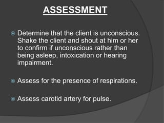


The document discusses cardio pulmonary resuscitation (CPR), which is a technique used to artificially support breathing and heart function when they have ceased. It involves clearing the airway, providing rescue breathing through mouth-to-mouth or with a bag and mask, and performing external chest compressions to manually pump the heart. The key steps of CPR include assessing for responsiveness, breathing, and pulse; opening the airway; giving breaths; and administering compressions at a rate of 100 per minute with a depth of 1.5-2 inches until emergency services arrive or the person starts breathing on their own.


