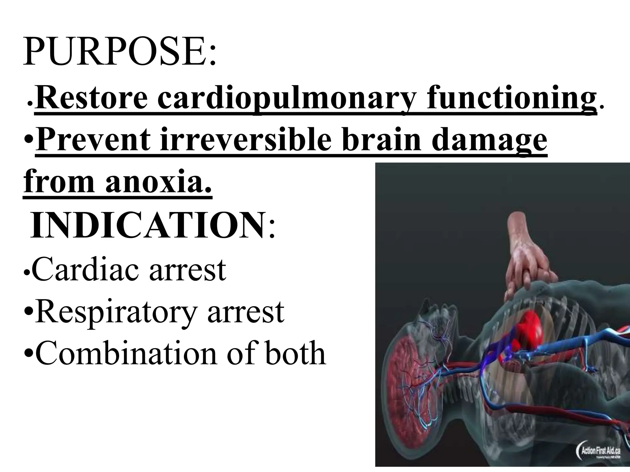
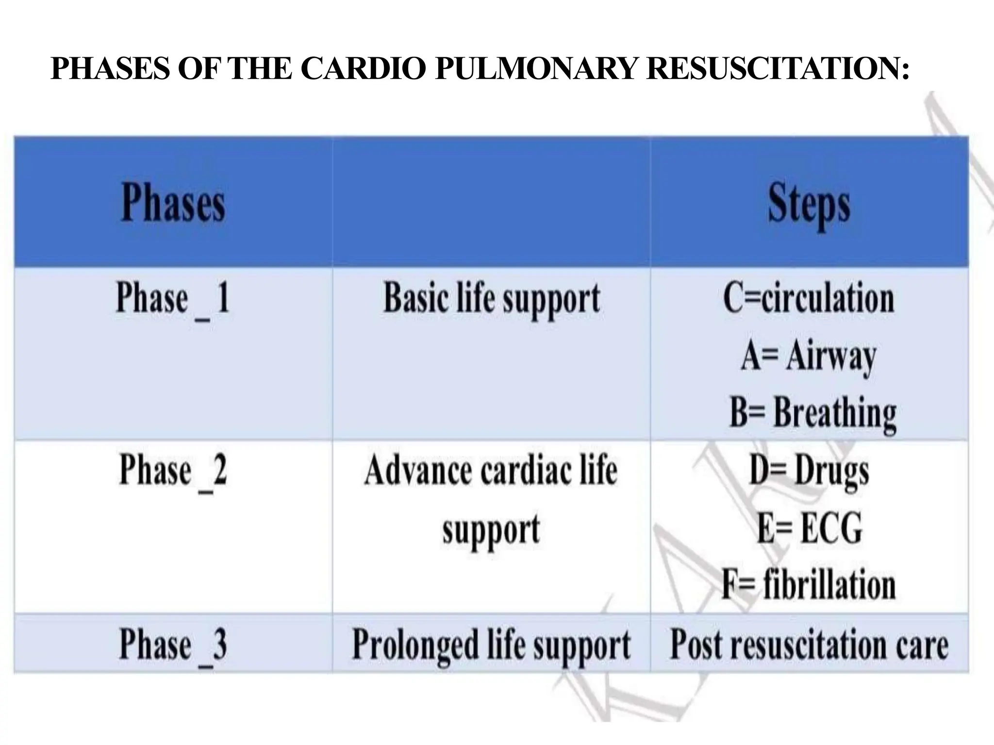
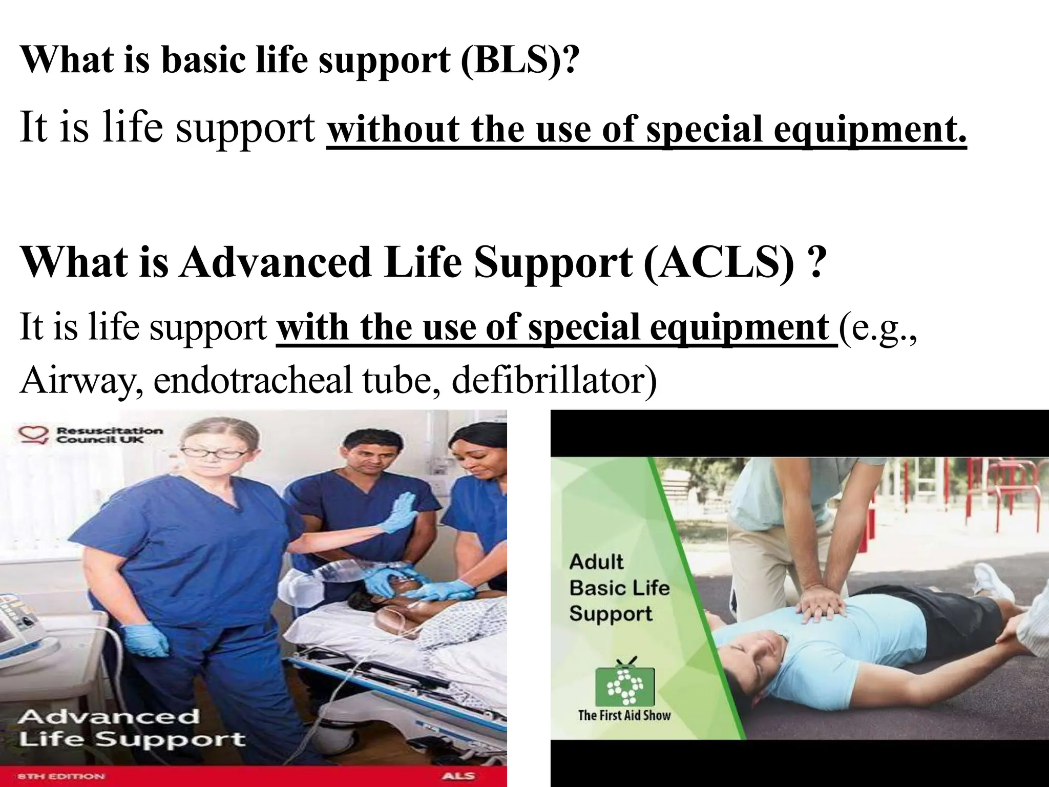
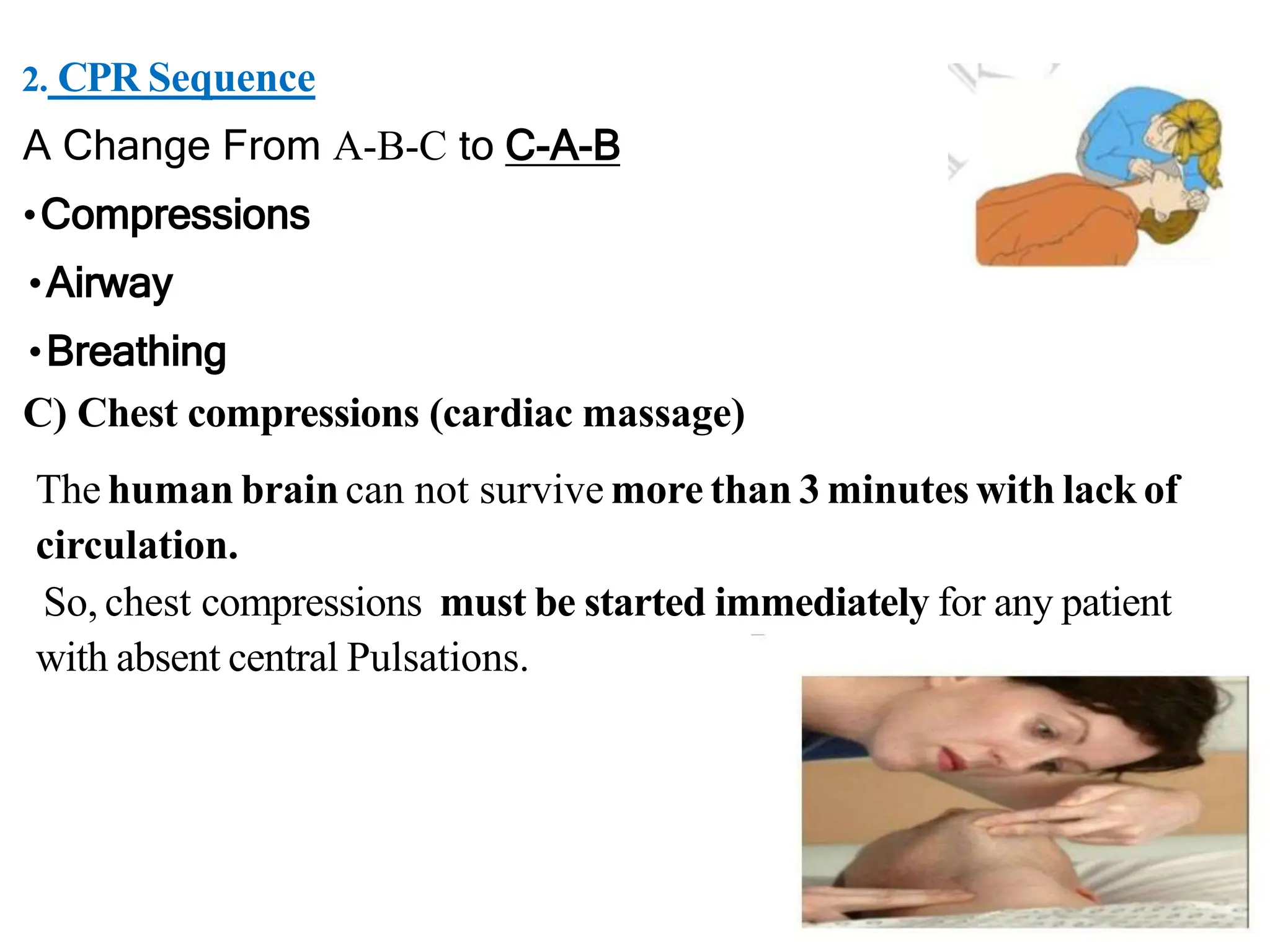
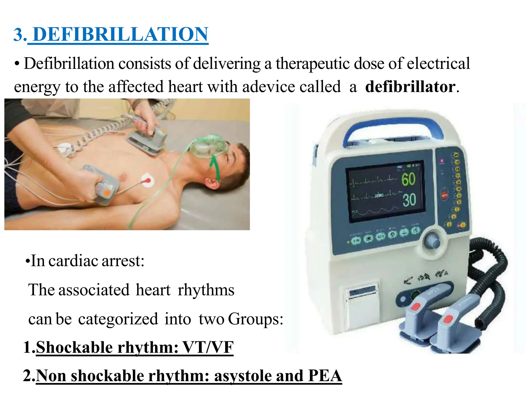
Cardio pulmonary resuscitation (CPR)
DEFINITION:
Cardio pulmonary resuscitation (CPR)
Is a technique of basic life support for the purpose of
oxygenation to the heart, lungs and brain until
and unless the appropriate medical treatment can
come and restore the normal cardiopulmonary
function .
Cardio pulmonary resuscitation is a series of steps
used to establish artificial ventilation and
circulation in the patient who is not breathing and
has no pulse
PURPOSE:
•Restore cardiopulmonary functioning.
•Prevent irreversible brain damage
from anoxia.
INDICATION:
•Cardiac arrest
•Respiratory arrest
•Combination of both
Causes of cardiac arrest
(6H & 4T)
1. Hypoxia
2. Hypotension .
3. Hypothermia
4. Hypoglycemia
5. Acidosis (H)
6. Hypokalemia (electrolyte disturbance )
1. Cardiac tamponed
2. Tension pneumothorax
3. Thromboembolism
4. Toxicity (digoxin, local anesthetic, Insecticides)
Diagnosis of cardiac arrest
 Blood pressure measurement.
 Taking the pulse on peripheral arteries
 Auscultation of cardiac tones
Loss of time!!!
Diagnosis of cardiac arrest (TRIAD)
1. Loss of consciousness.
2. Loss of apical & central pulsations (carotid, femoral).
3. Apnea.
HOW CPR WORKS:
•Air we breathe in, travels to our lungs were oxygen is pickedup byour
bloodandthen pumped by the heart to our tissue and organs.
•When a person experiences cardiac arrest-whether due to heart failure
in adults or the elderly or an injury such as
•neardrowning,
•Severe trauma in achild
-The heart goes from anormal arrhythmic Pattern called ventricular
fibrillation and eventually ceases to beat altogether.
•This prevents oxygen from circulating throughout the body,
rapidly killing cells and tissue.
•Inessence, cardio (heart) pulmonary (lung) resuscitation
(revive, revitalize) serves as an artificial heart beat and an
artificial respirator.
•CPR may not save the victim even when Performed
properly, but if started within 4 minute of cardiac arrest
And defibrillation is provided within 10minutes ,aperson
has a 40% chance of survival
EQUIPMENTS
A hard flat surface
No additional equipment is necessary but in hospital
setting, an emergency crash cart with defibrillator and
cardiac monitor should be brought to the bed side.
A crash cart contains:
1.Airway equipment.
2.Suction equipment
3.Intravenous equipment.
4.Laboratory tubes and syringes. Pre packed medication for
advanced life support.
Emergency crash cart
PHASES OFTHE CARDIO PULMONARY RESUSCITATION:
What is basic life support (BLS)?
It is life support without the use of special equipment.
What is Advanced Life Support (ACLS) ?
It is life support with the use of special equipment (e.g.,
Airway, endotracheal tube, defibrillator)
1. EARLY RECOGNITION
Assessment is of crucial importance. It Includes:
1.Unresponsiveness
•Check the victim for a response.
•Shake shoulders gently
•Ask"Are you allright"?
2.No breathing or no normal breathing (i.e, only gasping)
3. No pulse felt within 10 seconds.
2. CPR Sequence
A Change From A-B-C to C-A-B
•Compressions
•Airway
•Breathing
C) Chest compressions (cardiac massage)
The human brain can not survive more than 3 minutes with lack of
circulation.
So, chest compressions must be started immediately for any patient
with absent central Pulsations.
TECHNIQUE OF CHEST COMPRESSION
-Patient must be placed on a hard surface (wooden Board)
-The palmof one hand is placedin the Concavity of the lower half of the
sternum2 fingers above the xiphoid process .(AVOID Xiphisterna
junction fracture & injury)
-The other hand is placed over the hand on the sternum.
-Shoulders should be positioned directly over the hands With the
elbows locked straight and arms extended.
Use your upper body weight to compress.
-Sternum must be depressed at least 5 cm in adults ,
and 2-4 cm in children, 1-2 cm in infants
Must be performed at a rate of 100-120/min
During CPR the ratio of chest
compressions to ventilation
should as follows:
-Single rescuer = 30:2
-In the presence of 2 rescuers
chest Compressions must not be
interrupted for ventilation
Chest compressions in infant (0-12 Months)
Chest compressions must be continued for 2
minutes before reassessment of cardiac
rhythm.
(2minutes = equivalent to 5 cycles 30:2)
Golden rules:
❖Ensure high quality chest compressions:
rate, depth recoil
❖Plan actions before interrupting CPR.
❖MINIMIZE interruption of chest
compressions.
❖Early defibrillation of shockable rhythm.
Assessment of the adequacy of chest compressions:
Systolic BP: 60-80 mmHg.
Diastolic BP: > 40mmHg
COP 30% of normal
Problems and complications of chest compressions
1.Rib fractures
2.Fracture sternum
3.Rib separation
4.Pneumothorax
5.Hemothorax
6.Lung contusions.
7.Liver lacerations
8.Fat emboli
(A)Airway
Loss of consciousness often results in airway obstruction due to loss of tone in the muscles
of the airway and falling back of the tongue.
CLEAR THE AIRWAY
( A) Basic techniques for airway patency:
1.Headtilt,chinlift:onehandisplacedontheforeheadandtheotheronthechinthe
head is tilted upwards to cause anterior displacement of the tongue.
3. Finger sweep: Sweep out foreign body in the mouth
by index finger (in unconscious pt only). This is NOT
advised in a conscious or convulsing patient
4. Heimlich maneuvers:
if the patient is conscious and the foreign body can not be removed by
a finger sweep.It is done while the patient is standing up or lying
down.
This is asubdiaphragmatic abdominal thrust that elevates the
diaphragm expelling a blast of air from the lungs that displaces the
foreign body. In infants his can be done by a series of blows on the
back and chest thrusts .
Heimlich maneuvers :
(B)Breathing
Breathing: Breathe for the person
Rescue breathing can be mouth-to-mouth breathing or mouth-to-nose
breathing if the mouth is seriously injured or can't be opened.
With the airway open (using the head-tilt,chin-lift maneuver)
Pinch the nostrils shut for mouth-to-mouth breathing and cover the
person's mouth with yours, making aseal.
Basic techniques include:
1.Mouth to mouth breathing: with the airway held open, pinch the nostrils
closed, take a deep breath and seal your lips over the patient's mouth.
Blow steadilyinto the patient's mouth watching the chest rise as if the
patient was taking a deep breath.
2.Mouth to nose breathing: seal the mouth shut and breathe steadily
through the nose.
3.Mouth to mouth and nose: is used in infants and small children.
Assessment of restoration of breathing and circulation
•Contraction of pupil
•Improved color of the skin
•Free movement of the chest wall
•Swallowing attempts Struggling movements
Signs of restored ventilation and circulation Include:
•Struggling movements
•Improved color
•Return of or strong pulse
•Return of systemic blood pressure
When to terminate BLS?
•Pulse and respiration returns
•Emergency medical helparrives
•Physician declared patient is deceased
• In a non-health setting.
• another indication to stop BLS would be that the Rescuer was
Exhausted and physically unable to Continue to perform BLS.
ADVANCED LIFE SUPPORT
ALS includes:
❖Circulation by cardiac massage
❖Airway management by equipment's
❖Breathing by advanced techniques
❖Defibrillation by manual defibrillator
❖Drugs.
(B)Advanced techniques for airway patency
1) Face Mask
2)Oropharyngeal airway
3)Nasopharyngeal airway
4)Laryngeal mask (LMA)
5)Endotracheal tube
6)Cricothyrotomy (SurgicalAirway)
7)Tracheostomy (SurgicalAirway)
BREATHING
•Expiredair contains 16% O2, so supplemental100% O2should be
used as soonas possible.
• Successful breathing is achieved by delivery of a tidal volume of 800-
1200 ml in adults at a rate of 10-12 Breaths/min in adults
(B)Advanced techniques include:
1)Self-inflating resuscitation bag (Ambu Bag)
2)Mechanical ventilator in OR or in ICU
Expired air =16%O2.
Ambu Bag (room air) = 21% O2.
Ambu bag + O2 (10-15L) = 45%O2
3. DEFIBRILLATION
• Defibrillation consists of delivering a therapeutic dose of electrical
energy to the affected heart with adevice called a defibrillator.
•In cardiac arrest:
The associated heart rhythms
can be categorized into two Groups:
1.Shockable rhythm: VT/VF
2.Non shockable rhythm: asystole and PEA
•The basic difference in the treatment of these two groups of
arrythmia is the need for defibrillation in patients with VT/VF
C) Defibrillation
Position of Paddles:
• One paddle is placed in the right infraclavicular
region, while the other is placed in the
left 5th -6th intercostal space anterior axillary line.
•Alternatively antero- posterior position may be used:
one paddle is placed in the left intrascapular region
while the other is placed in the left 5th-6th
intercostal space anteriorAxillary line
•ActionCompletely depolarizeall myocardial cells so SA node
can re-Establish as pacemaker
•Voltage of electricity discharge High from. 150 J to 360 J
(biphasic) 360 J (monophasic)
Drugs used in CPR
Adrenaline:
❖Given as a vasopressor a-1 effect (not as an Inotrope)
❖Dose: 1 mg (0.01 mg/kg) I V every 4 minutes (alternatingcycles)
while continuing CPR.
❖Given:
 Adrenaline 1mg IV (10ml of 1:10,000)
 When to give?
 Shockable rhythm : give after 3rd shock (during CPR).
Flush with 20ml saline.
 Non-shockable rhythm : give as soon as IV access is
established. Flush with 20ml saline.
 Repeat adrenaline dose during every other CPR cycle t hereafter
(i.e., repeat every 3-5 minutes once given, regardless of rhythm)
Amiodarone:
•Dose: 300 mg IV bolus (5 mg/kg)
•Given: in shockable rhythm After the 3rd shock.
•If unavailable give lidocaine 100 mg IV (1-1.5 mg/kg)
Vasopressin (ADH): 40 IU single dose once.
Magnesium:
•Dose: 2 g IV.
1.Given: VF/VT with hypomagnesemia.
2.Torsade de pointes.
3.Digoxin toxicity
Calcium:
•Dose: 10 ml of 10% Calcium chloride IV.
•Indications: PEAcaused by: hyperkalemia,Hypocalcemia,
hypermagnesemia, and over dose of calcium channel
blockers .
•Do NOT give calcium solutions and NaHCO3 simultaneously by
the same route
IV Fluids:
• Infuse fluids rapidly if hypovolemia is suspected.
• Use normal saline (0.9% NaCl) or Ringer's solution.
• Avoid dextrose which is redistributed away from the intravascular space rapidly
and causes hyperglycemia which may worsen neurological Outcome aftercardiac
arrest.
• Dextrose is indicated only if there is documented Hypoglycemia
Thrombolytics:
• Fibrinolytic therapy isconsideredwhencardiacArrestiscausedbyprovenor suspected
acute pulmonary embolism.
• If a fibrinolytic drug is usedin thesecircumstances ConsiderperformingCPRfor at least
60-90 Minutes before termination of resuscitationAttempts.
• Eg:Alteplase, tenecteplase (old generation: Streptokinase)
Sodium bicarbonate:
•Used in:
1.Severe metabolic acidosis (pH < 7.1)
2.Life-threatening hyperkalemia.
3.Tricyclic antidepressant over dose.
•Dose: (half correction) 1/2 Base Deficit x 1/3 Body weight
Avoid its routine use due to Its complications:
1.Increases CO2 load:
2.Inhibits release of O2 to tissues.
3.Impairs myocardial contractility.
4.Causes hypernatremia.
5.Adrenaline works better in acidic medium
Atropine:
•Its routine use in PEAand asystole is not beneficial and
has become obsolete.
Indicated in:
• sinus bradycardia
•AV block Causing hemodynamic instability.
•Dose: 0.5 mg IV. Repeated up to a maximum of 3 mg (full
atropinization)
Return of circulation
 Full ABCDE assessment
 Controlled oxygenation (aim92-96%)
 Consider therapeutic hypothermia 32-36˚ for
24 hours (avoid hyperthermia)
 Post-arrest investigations
 Treat cause
 Consider transfer to intensive care if still
requiring ventilation or high-
dependency care
No return of circulation
 In general, CPR should be continued as longas there is a
shockable rhythm (mechanical compression device may be
used)
 Only stop if a registrar or above makes the decision with the team
 Extracorporeal CPR using extracorporeal membrane oxygenation
(ECMO) device may be considered where available for select
patients to facilitate other definitive treatments, e.g., PCI,
pulmonary thrombectomy for massive PE, rewarming for
hypothermia
 Afterwards → Retrospectively document everything that happened
Algorithm differences in children
 Pulse check
 Infant (<1 year): feel brachial pulse
 Children (>1 year): feel carotid pulse
 Compression: ventilation ratio
 At birth: 3:1 ratio
 Infants/children: start with 5 rescue breaths, then
15:2 ratio
 Compressions
 Compress to at least one-third of the AP chest diameter
 Infant (<1 year):
 Encircling technique (preferred):
performed by placing both thumbs flat on the lower
sternum pointing towards the infant’s head and the
fingers around the rib cage
 Two-finger technique (may be easier if only one
rescuer):
compress the sternum with the tips of two fingers
 Children (>1 year):
as for an adult but only use one hand (unless need 2nd to
achieve target depth)
 Defibrillation
 Energy:
 Manual defibrillator: 4 J/kg
 If using automated defibrillat or for child <8 years:
use pediatric-attenuated adult shock energy
 If using automated defibrillator for child > 8 years:
use adult shock energy
 Infants: 4.5 cm pads
 Children: 8-12 cm pads
 If pediatric electrodes are unavailable, it is acceptable to
use the adult defibrillator and settings – ensure the pads are
not touching each other
 Drug doses
Adrenaline 10 mcg/kg (0.1ml/kg of 1: 10,000
solution)
 Amiodarone 5mg/kg – repeat same dose after 5th
shock if still in shockable rhythm

CARDIOPULMONARY RESUSCITATION (CPR).pptx

  • 1.
  • 2.
    DEFINITION: Cardio pulmonary resuscitation(CPR) Is a technique of basic life support for the purpose of oxygenation to the heart, lungs and brain until and unless the appropriate medical treatment can come and restore the normal cardiopulmonary function . Cardio pulmonary resuscitation is a series of steps used to establish artificial ventilation and circulation in the patient who is not breathing and has no pulse
  • 3.
    PURPOSE: •Restore cardiopulmonary functioning. •Preventirreversible brain damage from anoxia. INDICATION: •Cardiac arrest •Respiratory arrest •Combination of both
  • 4.
    Causes of cardiacarrest (6H & 4T) 1. Hypoxia 2. Hypotension . 3. Hypothermia 4. Hypoglycemia 5. Acidosis (H) 6. Hypokalemia (electrolyte disturbance ) 1. Cardiac tamponed 2. Tension pneumothorax 3. Thromboembolism 4. Toxicity (digoxin, local anesthetic, Insecticides)
  • 6.
    Diagnosis of cardiacarrest  Blood pressure measurement.  Taking the pulse on peripheral arteries  Auscultation of cardiac tones Loss of time!!! Diagnosis of cardiac arrest (TRIAD) 1. Loss of consciousness. 2. Loss of apical & central pulsations (carotid, femoral). 3. Apnea.
  • 7.
    HOW CPR WORKS: •Airwe breathe in, travels to our lungs were oxygen is pickedup byour bloodandthen pumped by the heart to our tissue and organs. •When a person experiences cardiac arrest-whether due to heart failure in adults or the elderly or an injury such as •neardrowning, •Severe trauma in achild -The heart goes from anormal arrhythmic Pattern called ventricular fibrillation and eventually ceases to beat altogether.
  • 8.
    •This prevents oxygenfrom circulating throughout the body, rapidly killing cells and tissue. •Inessence, cardio (heart) pulmonary (lung) resuscitation (revive, revitalize) serves as an artificial heart beat and an artificial respirator. •CPR may not save the victim even when Performed properly, but if started within 4 minute of cardiac arrest And defibrillation is provided within 10minutes ,aperson has a 40% chance of survival
  • 9.
    EQUIPMENTS A hard flatsurface No additional equipment is necessary but in hospital setting, an emergency crash cart with defibrillator and cardiac monitor should be brought to the bed side. A crash cart contains: 1.Airway equipment. 2.Suction equipment 3.Intravenous equipment. 4.Laboratory tubes and syringes. Pre packed medication for advanced life support.
  • 10.
  • 11.
    PHASES OFTHE CARDIOPULMONARY RESUSCITATION:
  • 12.
    What is basiclife support (BLS)? It is life support without the use of special equipment. What is Advanced Life Support (ACLS) ? It is life support with the use of special equipment (e.g., Airway, endotracheal tube, defibrillator)
  • 13.
  • 14.
    Assessment is ofcrucial importance. It Includes: 1.Unresponsiveness •Check the victim for a response. •Shake shoulders gently •Ask"Are you allright"? 2.No breathing or no normal breathing (i.e, only gasping) 3. No pulse felt within 10 seconds.
  • 15.
    2. CPR Sequence AChange From A-B-C to C-A-B •Compressions •Airway •Breathing C) Chest compressions (cardiac massage) The human brain can not survive more than 3 minutes with lack of circulation. So, chest compressions must be started immediately for any patient with absent central Pulsations.
  • 16.
    TECHNIQUE OF CHESTCOMPRESSION -Patient must be placed on a hard surface (wooden Board) -The palmof one hand is placedin the Concavity of the lower half of the sternum2 fingers above the xiphoid process .(AVOID Xiphisterna junction fracture & injury) -The other hand is placed over the hand on the sternum. -Shoulders should be positioned directly over the hands With the elbows locked straight and arms extended. Use your upper body weight to compress. -Sternum must be depressed at least 5 cm in adults , and 2-4 cm in children, 1-2 cm in infants
  • 17.
    Must be performedat a rate of 100-120/min During CPR the ratio of chest compressions to ventilation should as follows: -Single rescuer = 30:2 -In the presence of 2 rescuers chest Compressions must not be interrupted for ventilation
  • 18.
    Chest compressions ininfant (0-12 Months) Chest compressions must be continued for 2 minutes before reassessment of cardiac rhythm. (2minutes = equivalent to 5 cycles 30:2)
  • 19.
    Golden rules: ❖Ensure highquality chest compressions: rate, depth recoil ❖Plan actions before interrupting CPR. ❖MINIMIZE interruption of chest compressions. ❖Early defibrillation of shockable rhythm. Assessment of the adequacy of chest compressions: Systolic BP: 60-80 mmHg. Diastolic BP: > 40mmHg COP 30% of normal
  • 20.
    Problems and complicationsof chest compressions 1.Rib fractures 2.Fracture sternum 3.Rib separation 4.Pneumothorax 5.Hemothorax 6.Lung contusions. 7.Liver lacerations 8.Fat emboli
  • 21.
    (A)Airway Loss of consciousnessoften results in airway obstruction due to loss of tone in the muscles of the airway and falling back of the tongue. CLEAR THE AIRWAY ( A) Basic techniques for airway patency: 1.Headtilt,chinlift:onehandisplacedontheforeheadandtheotheronthechinthe head is tilted upwards to cause anterior displacement of the tongue.
  • 22.
    3. Finger sweep:Sweep out foreign body in the mouth by index finger (in unconscious pt only). This is NOT advised in a conscious or convulsing patient 4. Heimlich maneuvers: if the patient is conscious and the foreign body can not be removed by a finger sweep.It is done while the patient is standing up or lying down. This is asubdiaphragmatic abdominal thrust that elevates the diaphragm expelling a blast of air from the lungs that displaces the foreign body. In infants his can be done by a series of blows on the back and chest thrusts .
  • 23.
  • 24.
    (B)Breathing Breathing: Breathe forthe person Rescue breathing can be mouth-to-mouth breathing or mouth-to-nose breathing if the mouth is seriously injured or can't be opened. With the airway open (using the head-tilt,chin-lift maneuver) Pinch the nostrils shut for mouth-to-mouth breathing and cover the person's mouth with yours, making aseal. Basic techniques include: 1.Mouth to mouth breathing: with the airway held open, pinch the nostrils closed, take a deep breath and seal your lips over the patient's mouth. Blow steadilyinto the patient's mouth watching the chest rise as if the patient was taking a deep breath.
  • 25.
    2.Mouth to nosebreathing: seal the mouth shut and breathe steadily through the nose. 3.Mouth to mouth and nose: is used in infants and small children. Assessment of restoration of breathing and circulation •Contraction of pupil •Improved color of the skin •Free movement of the chest wall •Swallowing attempts Struggling movements
  • 26.
    Signs of restoredventilation and circulation Include: •Struggling movements •Improved color •Return of or strong pulse •Return of systemic blood pressure When to terminate BLS? •Pulse and respiration returns •Emergency medical helparrives •Physician declared patient is deceased • In a non-health setting. • another indication to stop BLS would be that the Rescuer was Exhausted and physically unable to Continue to perform BLS.
  • 27.
    ADVANCED LIFE SUPPORT ALSincludes: ❖Circulation by cardiac massage ❖Airway management by equipment's ❖Breathing by advanced techniques ❖Defibrillation by manual defibrillator ❖Drugs.
  • 28.
    (B)Advanced techniques forairway patency 1) Face Mask 2)Oropharyngeal airway 3)Nasopharyngeal airway 4)Laryngeal mask (LMA) 5)Endotracheal tube 6)Cricothyrotomy (SurgicalAirway) 7)Tracheostomy (SurgicalAirway)
  • 29.
    BREATHING •Expiredair contains 16%O2, so supplemental100% O2should be used as soonas possible. • Successful breathing is achieved by delivery of a tidal volume of 800- 1200 ml in adults at a rate of 10-12 Breaths/min in adults
  • 30.
    (B)Advanced techniques include: 1)Self-inflatingresuscitation bag (Ambu Bag) 2)Mechanical ventilator in OR or in ICU Expired air =16%O2. Ambu Bag (room air) = 21% O2. Ambu bag + O2 (10-15L) = 45%O2
  • 31.
    3. DEFIBRILLATION • Defibrillationconsists of delivering a therapeutic dose of electrical energy to the affected heart with adevice called a defibrillator. •In cardiac arrest: The associated heart rhythms can be categorized into two Groups: 1.Shockable rhythm: VT/VF 2.Non shockable rhythm: asystole and PEA
  • 32.
    •The basic differencein the treatment of these two groups of arrythmia is the need for defibrillation in patients with VT/VF
  • 34.
    C) Defibrillation Position ofPaddles: • One paddle is placed in the right infraclavicular region, while the other is placed in the left 5th -6th intercostal space anterior axillary line. •Alternatively antero- posterior position may be used: one paddle is placed in the left intrascapular region while the other is placed in the left 5th-6th intercostal space anteriorAxillary line
  • 35.
    •ActionCompletely depolarizeall myocardialcells so SA node can re-Establish as pacemaker •Voltage of electricity discharge High from. 150 J to 360 J (biphasic) 360 J (monophasic)
  • 36.
    Drugs used inCPR Adrenaline: ❖Given as a vasopressor a-1 effect (not as an Inotrope) ❖Dose: 1 mg (0.01 mg/kg) I V every 4 minutes (alternatingcycles) while continuing CPR. ❖Given:  Adrenaline 1mg IV (10ml of 1:10,000)  When to give?  Shockable rhythm : give after 3rd shock (during CPR). Flush with 20ml saline.  Non-shockable rhythm : give as soon as IV access is established. Flush with 20ml saline.  Repeat adrenaline dose during every other CPR cycle t hereafter (i.e., repeat every 3-5 minutes once given, regardless of rhythm)
  • 39.
    Amiodarone: •Dose: 300 mgIV bolus (5 mg/kg) •Given: in shockable rhythm After the 3rd shock. •If unavailable give lidocaine 100 mg IV (1-1.5 mg/kg) Vasopressin (ADH): 40 IU single dose once. Magnesium: •Dose: 2 g IV. 1.Given: VF/VT with hypomagnesemia. 2.Torsade de pointes. 3.Digoxin toxicity
  • 40.
    Calcium: •Dose: 10 mlof 10% Calcium chloride IV. •Indications: PEAcaused by: hyperkalemia,Hypocalcemia, hypermagnesemia, and over dose of calcium channel blockers . •Do NOT give calcium solutions and NaHCO3 simultaneously by the same route IV Fluids: • Infuse fluids rapidly if hypovolemia is suspected. • Use normal saline (0.9% NaCl) or Ringer's solution. • Avoid dextrose which is redistributed away from the intravascular space rapidly and causes hyperglycemia which may worsen neurological Outcome aftercardiac arrest. • Dextrose is indicated only if there is documented Hypoglycemia
  • 41.
    Thrombolytics: • Fibrinolytic therapyisconsideredwhencardiacArrestiscausedbyprovenor suspected acute pulmonary embolism. • If a fibrinolytic drug is usedin thesecircumstances ConsiderperformingCPRfor at least 60-90 Minutes before termination of resuscitationAttempts. • Eg:Alteplase, tenecteplase (old generation: Streptokinase) Sodium bicarbonate: •Used in: 1.Severe metabolic acidosis (pH < 7.1) 2.Life-threatening hyperkalemia. 3.Tricyclic antidepressant over dose. •Dose: (half correction) 1/2 Base Deficit x 1/3 Body weight
  • 42.
    Avoid its routineuse due to Its complications: 1.Increases CO2 load: 2.Inhibits release of O2 to tissues. 3.Impairs myocardial contractility. 4.Causes hypernatremia. 5.Adrenaline works better in acidic medium
  • 43.
    Atropine: •Its routine usein PEAand asystole is not beneficial and has become obsolete. Indicated in: • sinus bradycardia •AV block Causing hemodynamic instability. •Dose: 0.5 mg IV. Repeated up to a maximum of 3 mg (full atropinization)
  • 44.
    Return of circulation Full ABCDE assessment  Controlled oxygenation (aim92-96%)  Consider therapeutic hypothermia 32-36˚ for 24 hours (avoid hyperthermia)  Post-arrest investigations  Treat cause  Consider transfer to intensive care if still requiring ventilation or high- dependency care
  • 45.
    No return ofcirculation  In general, CPR should be continued as longas there is a shockable rhythm (mechanical compression device may be used)  Only stop if a registrar or above makes the decision with the team  Extracorporeal CPR using extracorporeal membrane oxygenation (ECMO) device may be considered where available for select patients to facilitate other definitive treatments, e.g., PCI, pulmonary thrombectomy for massive PE, rewarming for hypothermia  Afterwards → Retrospectively document everything that happened
  • 46.
    Algorithm differences inchildren  Pulse check  Infant (<1 year): feel brachial pulse  Children (>1 year): feel carotid pulse  Compression: ventilation ratio  At birth: 3:1 ratio  Infants/children: start with 5 rescue breaths, then 15:2 ratio
  • 47.
     Compressions  Compressto at least one-third of the AP chest diameter  Infant (<1 year):  Encircling technique (preferred): performed by placing both thumbs flat on the lower sternum pointing towards the infant’s head and the fingers around the rib cage  Two-finger technique (may be easier if only one rescuer): compress the sternum with the tips of two fingers  Children (>1 year): as for an adult but only use one hand (unless need 2nd to achieve target depth)
  • 48.
     Defibrillation  Energy: Manual defibrillator: 4 J/kg  If using automated defibrillat or for child <8 years: use pediatric-attenuated adult shock energy  If using automated defibrillator for child > 8 years: use adult shock energy  Infants: 4.5 cm pads  Children: 8-12 cm pads  If pediatric electrodes are unavailable, it is acceptable to use the adult defibrillator and settings – ensure the pads are not touching each other
  • 49.
     Drug doses Adrenaline10 mcg/kg (0.1ml/kg of 1: 10,000 solution)  Amiodarone 5mg/kg – repeat same dose after 5th shock if still in shockable rhythm