Table of Contents
Tableof Contents................................................................................................................................................1
A 24-year-old contact lens wearer presents with a 2-day history of eye pain......................................................2
A 20-year-old man, a contact lens wearer, comes in with a 5-day history of eye redness and pain......................3
A 45-year-old, diabetic patient, presents with a 1-week history of RE redness and blurring of vision...................4
A 40-year-old lady presents with a 3-day history of eye discomfort.....................................................................5
A 50-year-old man presents with a 3-day history of skin rash over the face........................................................6
A 40-year-old man presents with sudden onset eye discomfort...........................................................................7
A 45-year-old man presents with sudden onset eye discomfort...........................................................................8
A 45-year-old lady presents with blurring of vision that is worse in the morning.................................................9
A 20-year-old man presents with a 2-month history of painless progressive blurring of vision..........................10
A 50-year-old lady complains about an increasing whitish lesion in the eye. She gives a history of chronic
glaucoma.........................................................................................................................................................11
A 50-year-old man presents with a fleshy growth that has been on the eye for the past few years...................12
A 50-year-old man presents with a 3-day history of pain, blurring of vision and redness of the eye. He
underwent a corneal graft surgery 3 months ago.............................................................................................13
A 35-year-old man complains of double vision in the left eye............................................................................14
A 50-year-old presents with eye pain and redness. He underwent an uncomplicated cataract surgery 4 days ago
.........................................................................................................................................................................15
A 25-year-old man presents with discomfort in both eyes.................................................................................16
2.
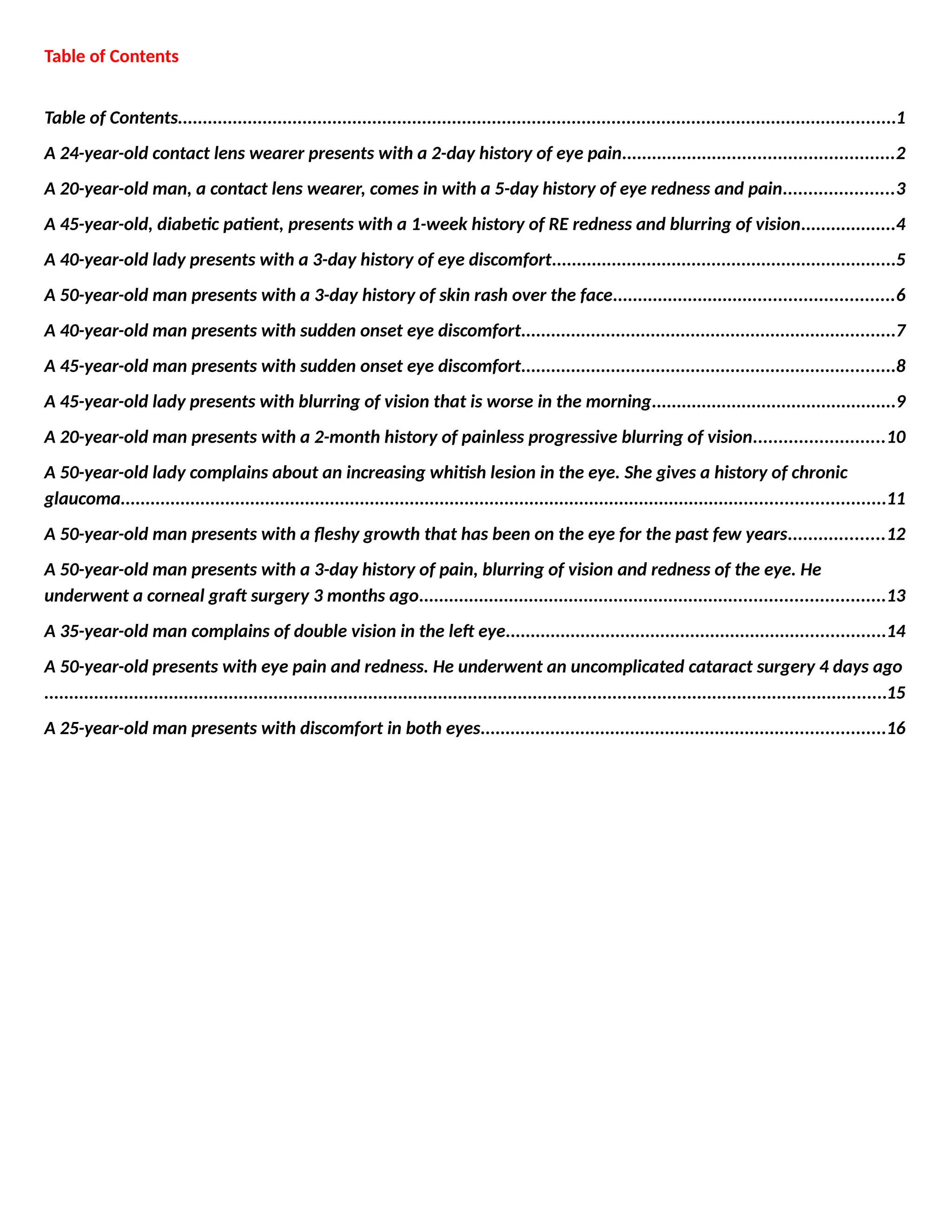
A 24-year-old contactlens wearer presents with a 2-day history of eye pain
What else do you want
to know about in the
history?
-Blurring of vision
-Contact lens history
-Contact with soil or contaminated water
-Immunosuppression: diabetes, human immunodeficiency virus, steroids,
chemotherapy onset
-Progression
-Previous treatment
-Pain
-Trauma
What are the signs? -Conjunctiva: injected
-Corneal ulcer/infiltrate involving the visual axis
-Central epithelial defect
-Hypopyon
What are the
differential diagnoses?
-Contact lens-related infective keratitis
-Exposure infective keratitis/neurotrophic infective keratitis
How do you manage
this patient?
-Admit the patient
-Perform a corneal scrape and send for microscopy and cultures
-I -intensive topical antibiotic treatment: gentamicin 14 mg/ml hourly,
cephazolin 50 mg/ml hourly through the night
- -Systemic antibiotic treatment if the infiltrate is near the limbus (oral
ciprofloxacin 500 mg twice a day for a week)
What do you send the
corneal scrapings for?
-Gram stain
-Blood agar
-Chocolate agar
-Thioglycate
-Brain heart infusion broth (BHIB)
-Sabouraud dextrose
-Others: Suspicious for fungal infection: giemsa stain, methenamine silver
stain
What are the
complications of a
corneal ulcer?
-Acute: thinning of the cornea resulting in corneal perforation leading to
endophthalmitis
-Long-term: scar, astigmatism, blindness
3.
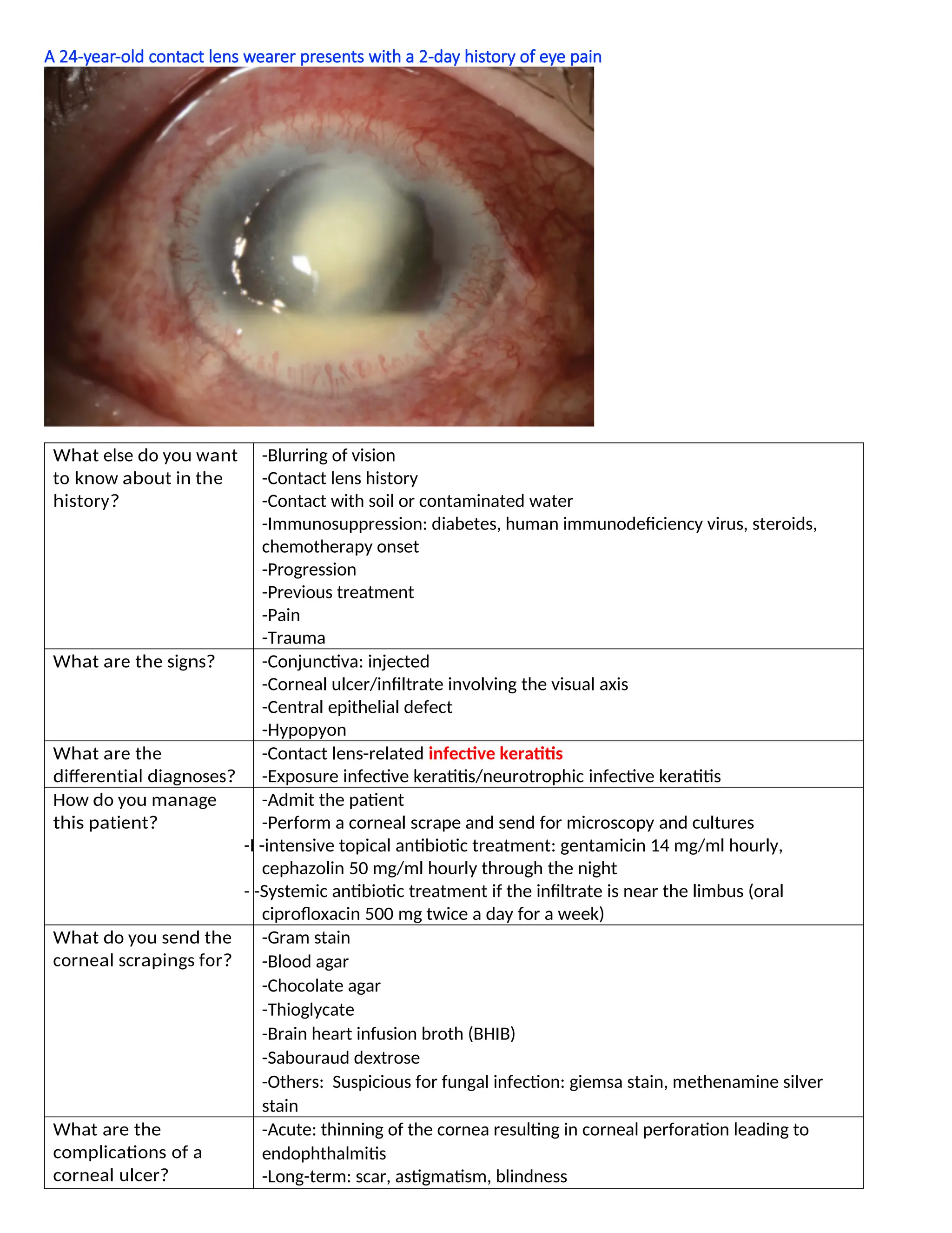
A 20-year-old man,a contact lens wearer, comes in with a 5-day history of eye redness and pain
What are the signs? - Conjunctiva: injected
- Cornea: surrounding stromal haze, radial keratoneuritis (yellow arrow)
What is the diagnoses? Acanthamoeba keratitis
What do you send the
corneal scrapings for?
Microscopy: calcofluor white/acridine orange stain (double-walled cysts)
Culture: non-nutrient agar with Escherichia coli overlay
How would you treat
this patient?
Biguanides: polyhexamethylene biguanide (PHMB), chlorhexidine
Diamidines: propamidine, hexamidine
Treatment is prolonged and requires a combination of biguanides and diamidines
4.
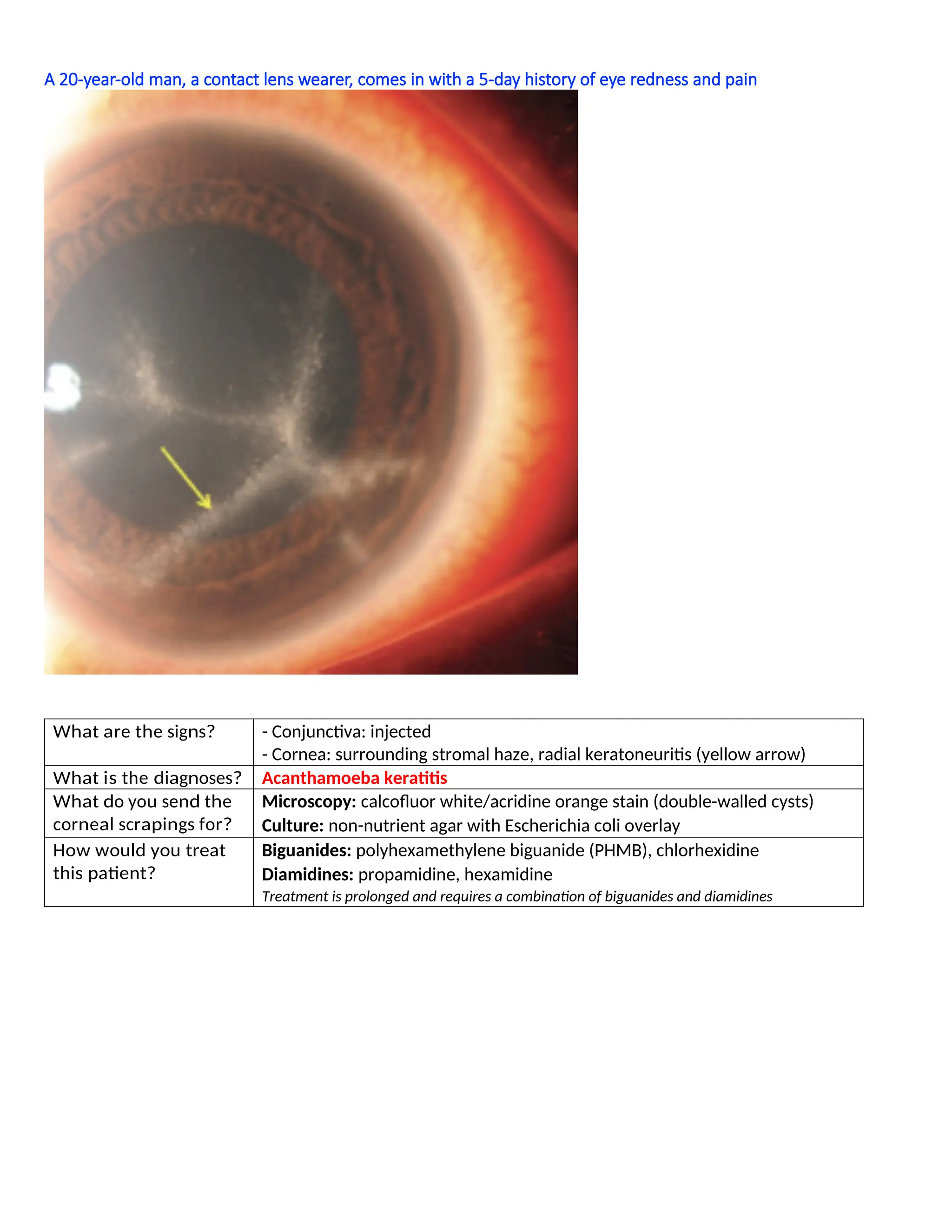
A 45-year-old, diabeticpatient, presents with a 1-week history of RE redness and blurring of vision
What are the signs? Conjunctiva: injected
Cornea
- Large corneal infiltrate inferior to the visual axis, involving the limbus
- Edges of the infiltrates are feathery with presence of satellite lesions (yellow
arrow)
What is the diagnosis? Fungal keratitis
What further test will
you do?
Scrape and send for fungal microscopy and culture
How do you treat it? - Topical: amphotericin B (yeast), natamycin (filamentous fungi)
- Consider intrastromal and/or systemic anti-fungals due to poor ocular
penetration of topical anti-fungal eyedrops
5.
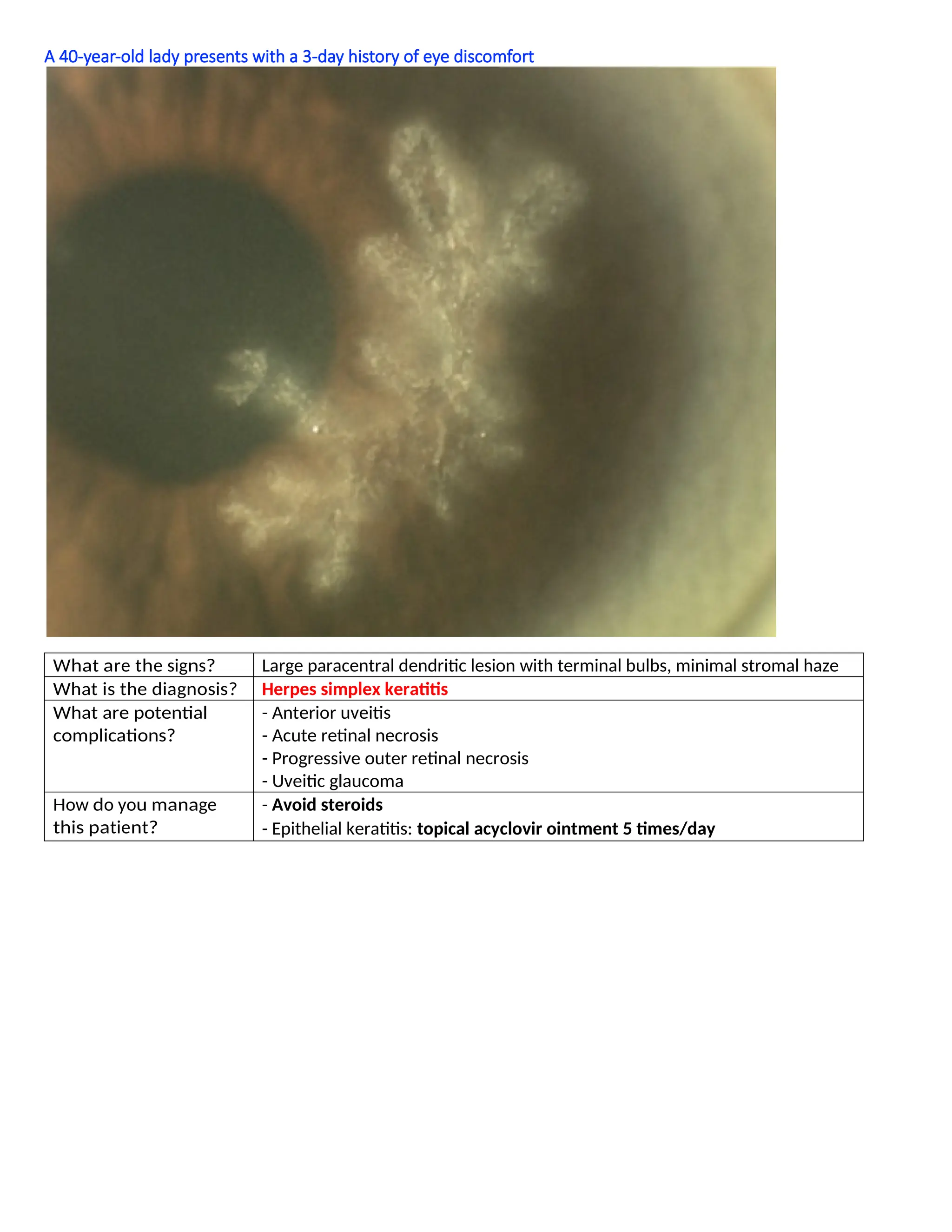
A 40-year-old ladypresents with a 3-day history of eye discomfort
What are the signs? Large paracentral dendritic lesion with terminal bulbs, minimal stromal haze
What is the diagnosis? Herpes simplex keratitis
What are potential
complications?
- Anterior uveitis
- Acute retinal necrosis
- Progressive outer retinal necrosis
- Uveitic glaucoma
How do you manage
this patient?
- Avoid steroids
- Epithelial keratitis: topical acyclovir ointment 5 times/day
6.
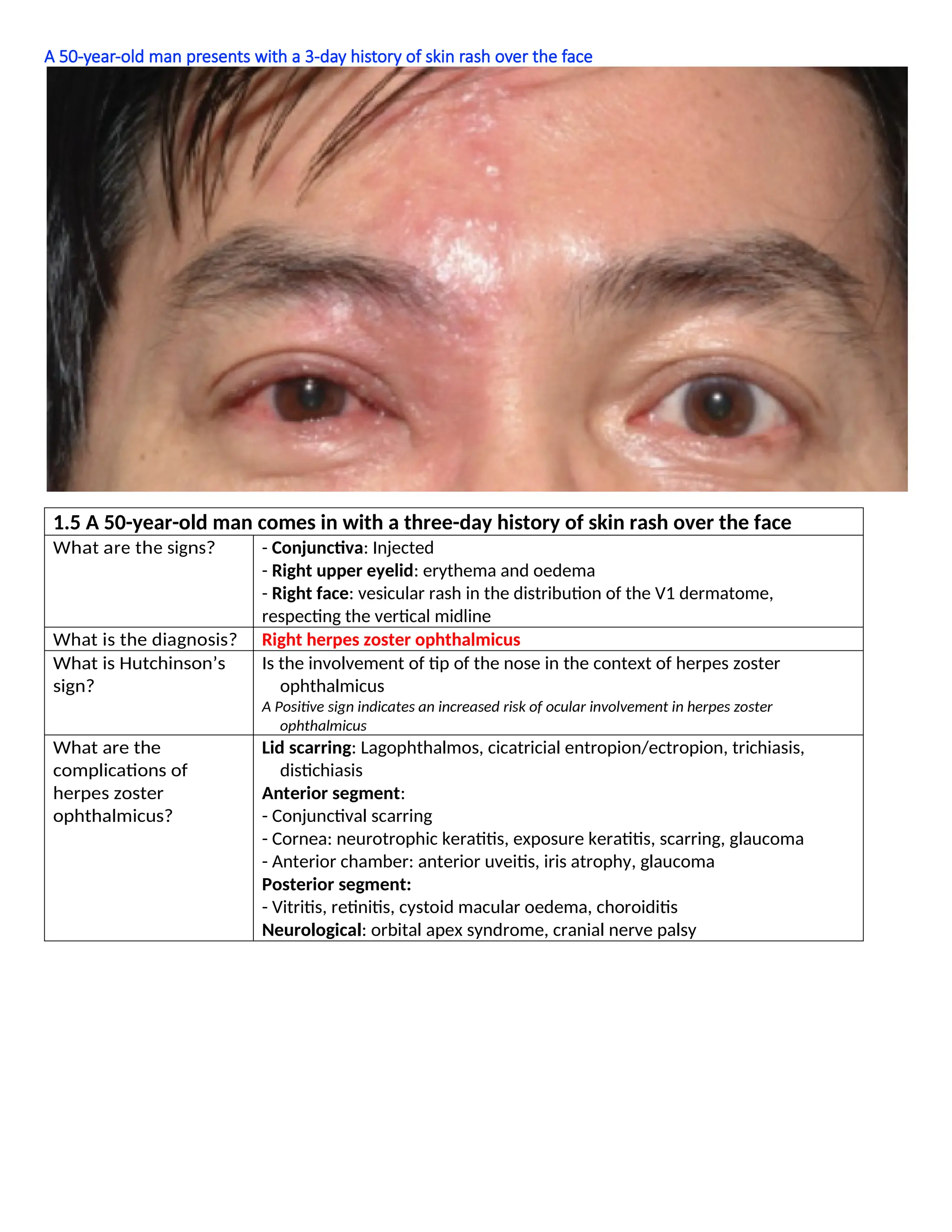
A 50-year-old manpresents with a 3-day history of skin rash over the face
1.5 A 50-year-old man comes in with a three-day history of skin rash over the face
What are the signs? - Conjunctiva: Injected
- Right upper eyelid: erythema and oedema
- Right face: vesicular rash in the distribution of the V1 dermatome,
respecting the vertical midline
What is the diagnosis? Right herpes zoster ophthalmicus
What is Hutchinson’s
sign?
Is the involvement of tip of the nose in the context of herpes zoster
ophthalmicus
A Positive sign indicates an increased risk of ocular involvement in herpes zoster
ophthalmicus
What are the
complications of
herpes zoster
ophthalmicus?
Lid scarring: Lagophthalmos, cicatricial entropion/ectropion, trichiasis,
distichiasis
Anterior segment:
- Conjunctival scarring
- Cornea: neurotrophic keratitis, exposure keratitis, scarring, glaucoma
- Anterior chamber: anterior uveitis, iris atrophy, glaucoma
Posterior segment:
- Vitritis, retinitis, cystoid macular oedema, choroiditis
Neurological: orbital apex syndrome, cranial nerve palsy
7.
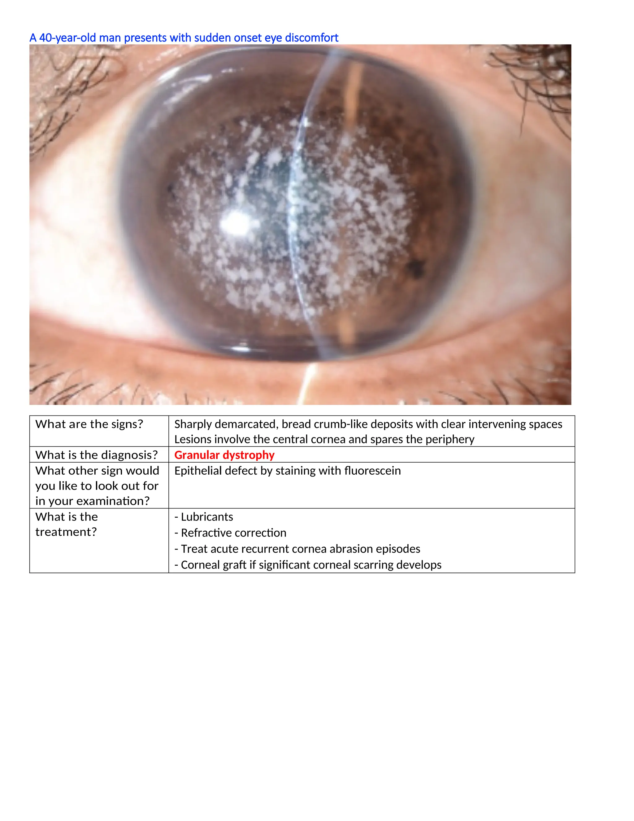
A 40-year-old manpresents with sudden onset eye discomfort
What are the signs? Sharply demarcated, bread crumb-like deposits with clear intervening spaces
Lesions involve the central cornea and spares the periphery
What is the diagnosis? Granular dystrophy
What other sign would
you like to look out for
in your examination?
Epithelial defect by staining with fluorescein
What is the
treatment?
- Lubricants
- Refractive correction
- Treat acute recurrent cornea abrasion episodes
- Corneal graft if significant corneal scarring develops
8.
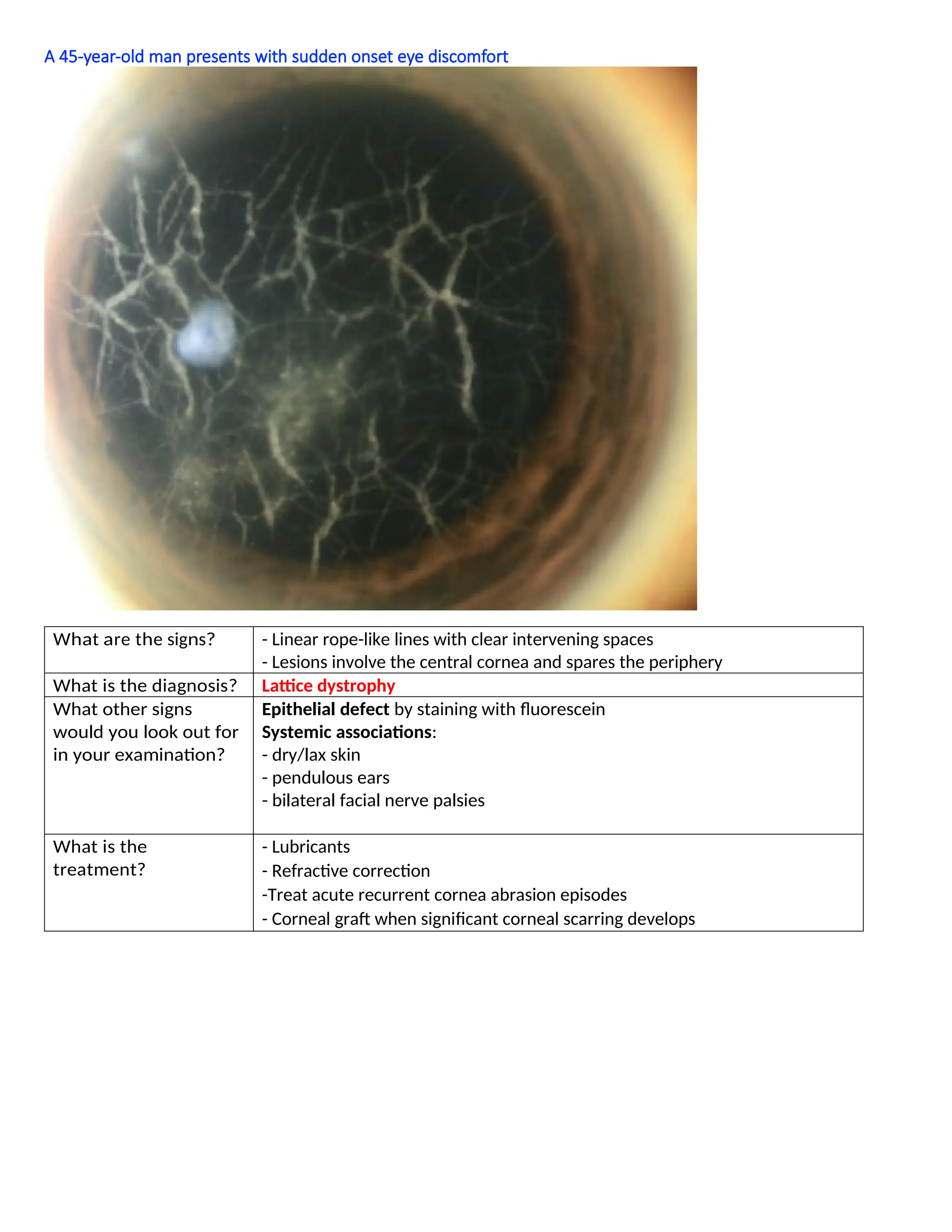
A 45-year-old manpresents with sudden onset eye discomfort
What are the signs? - Linear rope-like lines with clear intervening spaces
- Lesions involve the central cornea and spares the periphery
What is the diagnosis? Lattice dystrophy
What other signs
would you look out for
in your examination?
Epithelial defect by staining with fluorescein
Systemic associations:
- dry/lax skin
- pendulous ears
- bilateral facial nerve palsies
What is the
treatment?
- Lubricants
- Refractive correction
-Treat acute recurrent cornea abrasion episodes
- Corneal graft when significant corneal scarring develops
9.
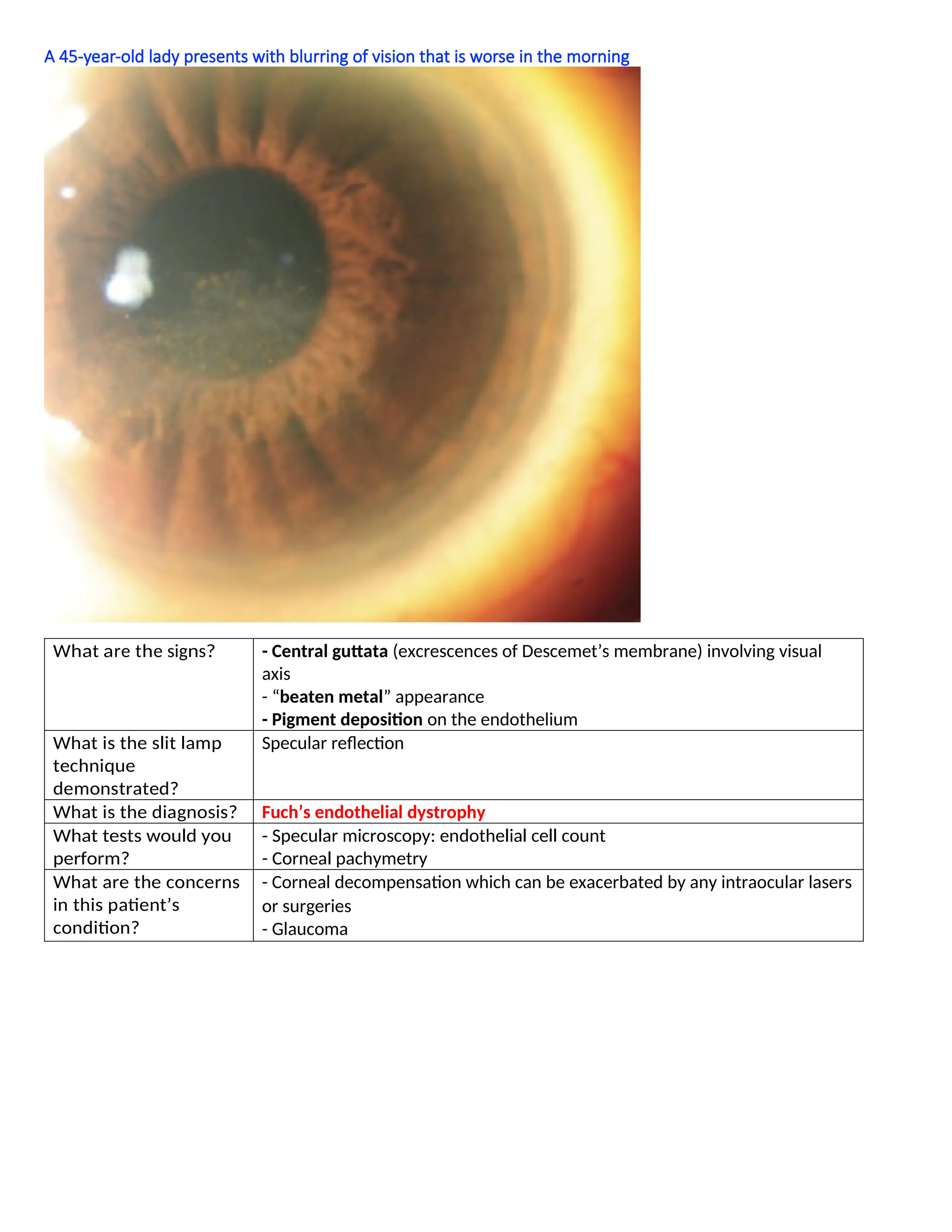
A 45-year-old ladypresents with blurring of vision that is worse in the morning
What are the signs? - Central guttata (excrescences of Descemet’s membrane) involving visual
axis
- “beaten metal” appearance
- Pigment deposition on the endothelium
What is the slit lamp
technique
demonstrated?
Specular reflection
What is the diagnosis? Fuch’s endothelial dystrophy
What tests would you
perform?
- Specular microscopy: endothelial cell count
- Corneal pachymetry
What are the concerns
in this patient’s
condition?
- Corneal decompensation which can be exacerbated by any intraocular lasers
or surgeries
- Glaucoma
10.
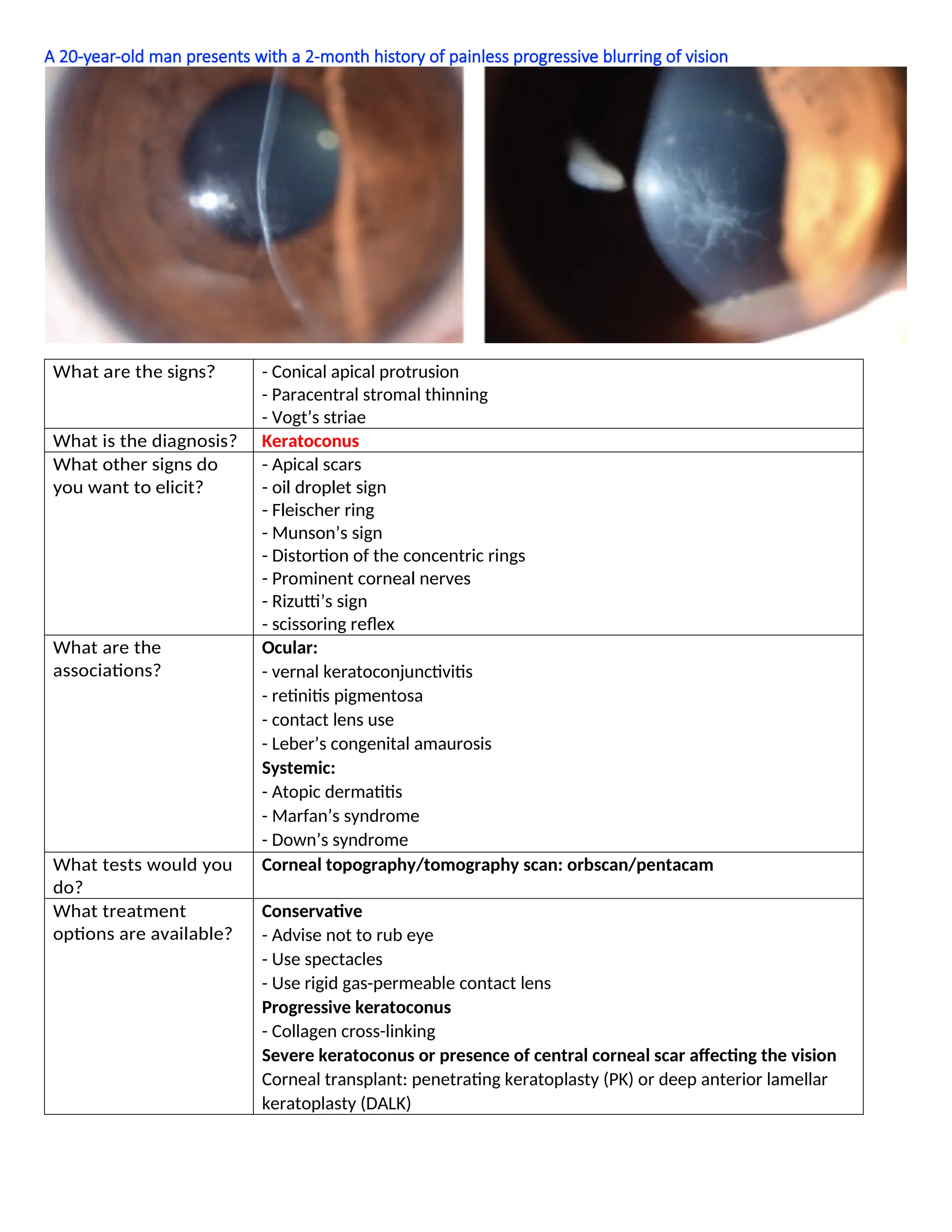
A 20-year-old manpresents with a 2-month history of painless progressive blurring of vision
What are the signs? - Conical apical protrusion
- Paracentral stromal thinning
- Vogt’s striae
What is the diagnosis? Keratoconus
What other signs do
you want to elicit?
- Apical scars
- oil droplet sign
- Fleischer ring
- Munson’s sign
- Distortion of the concentric rings
- Prominent corneal nerves
- Rizutti’s sign
- scissoring reflex
What are the
associations?
Ocular:
- vernal keratoconjunctivitis
- retinitis pigmentosa
- contact lens use
- Leber’s congenital amaurosis
Systemic:
- Atopic dermatitis
- Marfan’s syndrome
- Down’s syndrome
What tests would you
do?
Corneal topography/tomography scan: orbscan/pentacam
What treatment
options are available?
Conservative
- Advise not to rub eye
- Use spectacles
- Use rigid gas-permeable contact lens
Progressive keratoconus
- Collagen cross-linking
Severe keratoconus or presence of central corneal scar affecting the vision
Corneal transplant: penetrating keratoplasty (PK) or deep anterior lamellar
keratoplasty (DALK)
11.
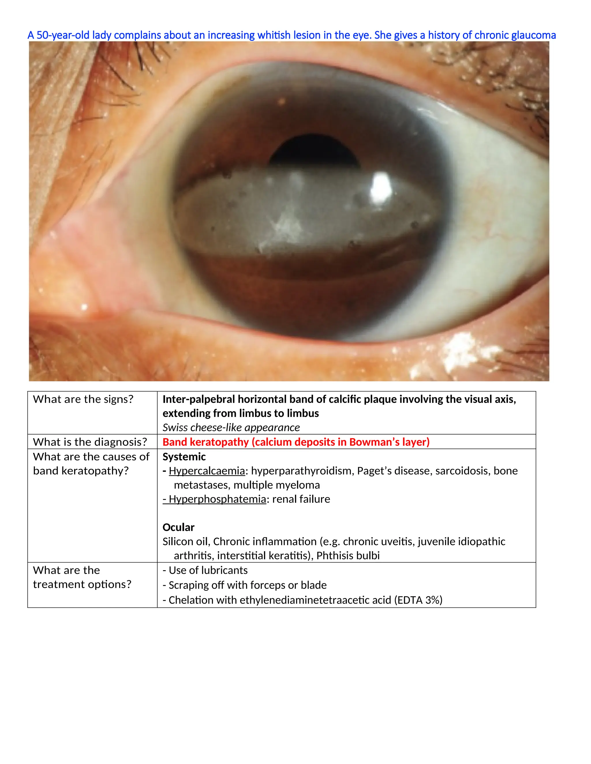
A 50-year-old ladycomplains about an increasing whitish lesion in the eye. She gives a history of chronic glaucoma
What are the signs? Inter-palpebral horizontal band of calcific plaque involving the visual axis,
extending from limbus to limbus
Swiss cheese-like appearance
What is the diagnosis? Band keratopathy (calcium deposits in Bowman’s layer)
What are the causes of
band keratopathy?
Systemic
- Hypercalcaemia: hyperparathyroidism, Paget’s disease, sarcoidosis, bone
metastases, multiple myeloma
- Hyperphosphatemia: renal failure
Ocular
Silicon oil, Chronic inflammation (e.g. chronic uveitis, juvenile idiopathic
arthritis, interstitial keratitis), Phthisis bulbi
What are the
treatment options?
- Use of lubricants
- Scraping off with forceps or blade
- Chelation with ethylenediaminetetraacetic acid (EDTA 3%)
12.
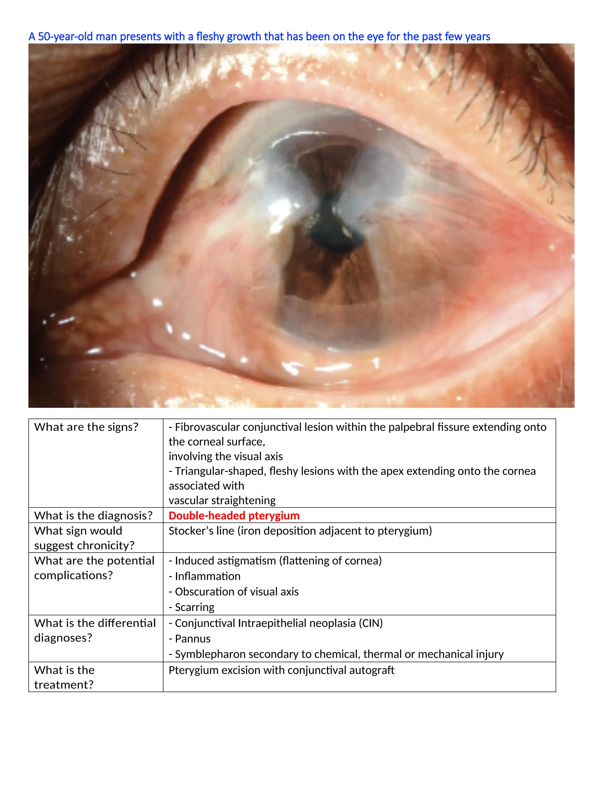
A 50-year-old manpresents with a fleshy growth that has been on the eye for the past few years
What are the signs? - Fibrovascular conjunctival lesion within the palpebral fissure extending onto
the corneal surface,
involving the visual axis
- Triangular-shaped, fleshy lesions with the apex extending onto the cornea
associated with
vascular straightening
What is the diagnosis? Double-headed pterygium
What sign would
suggest chronicity?
Stocker’s line (iron deposition adjacent to pterygium)
What are the potential
complications?
- Induced astigmatism (flattening of cornea)
- Inflammation
- Obscuration of visual axis
- Scarring
What is the differential
diagnoses?
- Conjunctival Intraepithelial neoplasia (CIN)
- Pannus
- Symblepharon secondary to chemical, thermal or mechanical injury
What is the
treatment?
Pterygium excision with conjunctival autograft
13.
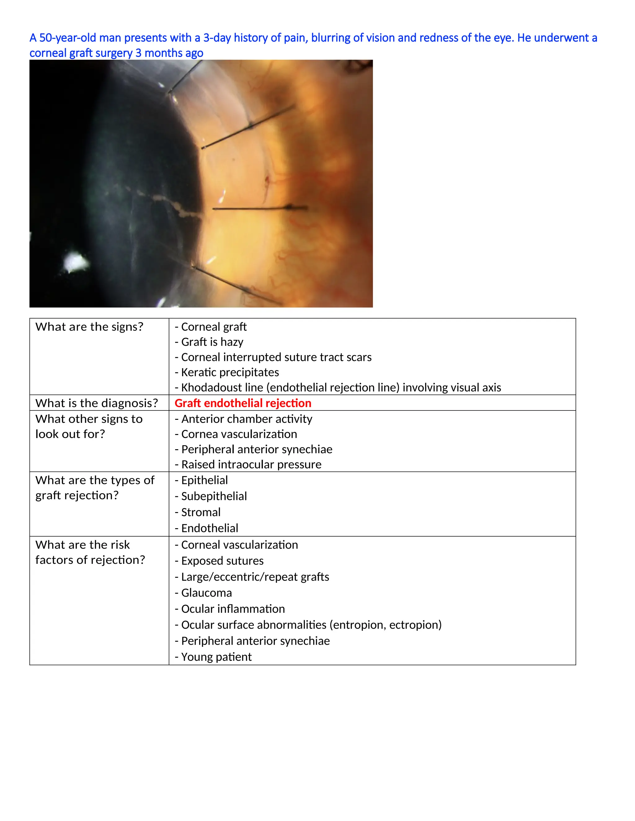
A 50-year-old manpresents with a 3-day history of pain, blurring of vision and redness of the eye. He underwent a
corneal graft surgery 3 months ago
What are the signs? - Corneal graft
- Graft is hazy
- Corneal interrupted suture tract scars
- Keratic precipitates
- Khodadoust line (endothelial rejection line) involving visual axis
What is the diagnosis? Graft endothelial rejection
What other signs to
look out for?
- Anterior chamber activity
- Cornea vascularization
- Peripheral anterior synechiae
- Raised intraocular pressure
What are the types of
graft rejection?
- Epithelial
- Subepithelial
- Stromal
- Endothelial
What are the risk
factors of rejection?
- Corneal vascularization
- Exposed sutures
- Large/eccentric/repeat grafts
- Glaucoma
- Ocular inflammation
- Ocular surface abnormalities (entropion, ectropion)
- Peripheral anterior synechiae
- Young patient
14.
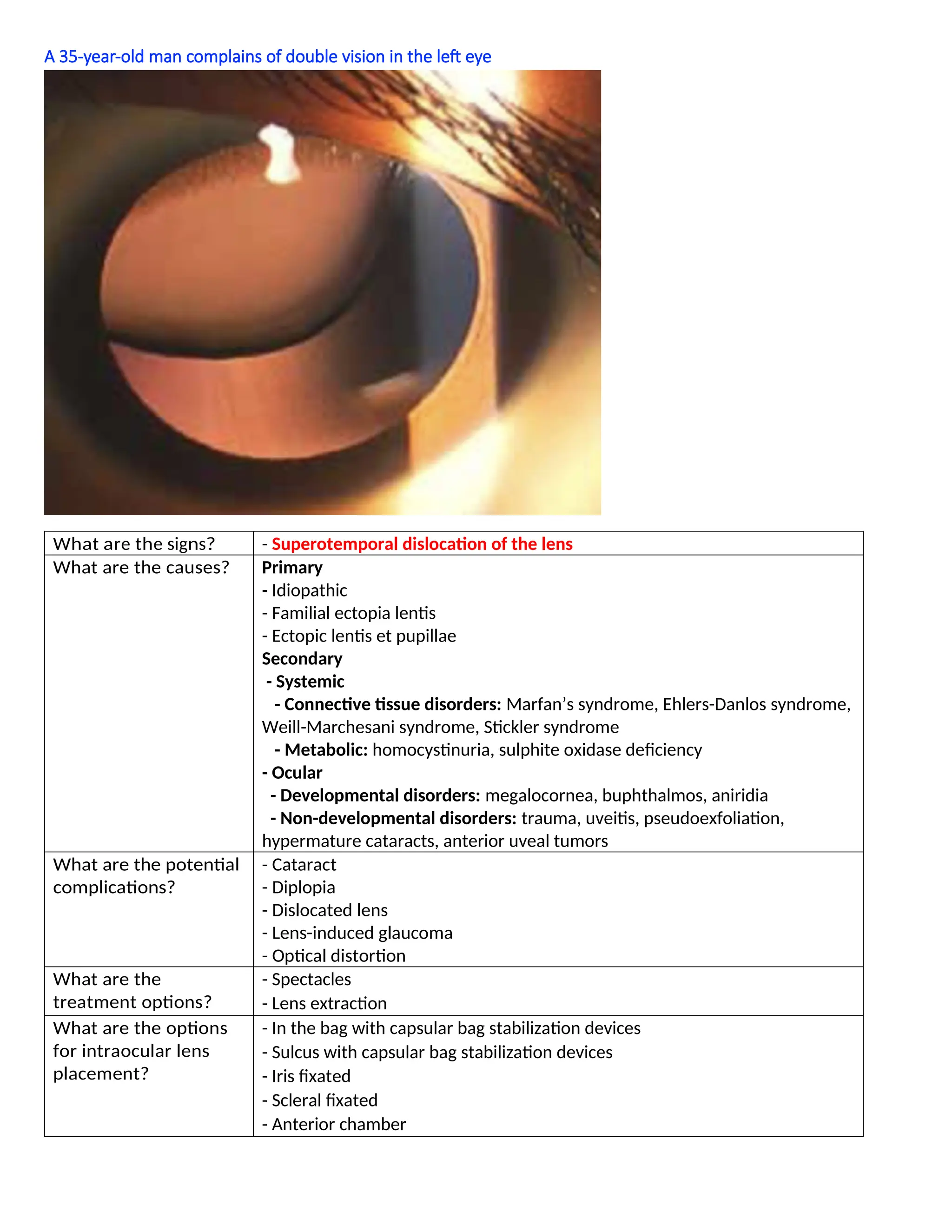
A 35-year-old mancomplains of double vision in the left eye
What are the signs? - Superotemporal dislocation of the lens
What are the causes? Primary
- Idiopathic
- Familial ectopia lentis
- Ectopic lentis et pupillae
Secondary
- Systemic
- Connective tissue disorders: Marfan’s syndrome, Ehlers-Danlos syndrome,
Weill-Marchesani syndrome, Stickler syndrome
- Metabolic: homocystinuria, sulphite oxidase deficiency
- Ocular
- Developmental disorders: megalocornea, buphthalmos, aniridia
- Non-developmental disorders: trauma, uveitis, pseudoexfoliation,
hypermature cataracts, anterior uveal tumors
What are the potential
complications?
- Cataract
- Diplopia
- Dislocated lens
- Lens-induced glaucoma
- Optical distortion
What are the
treatment options?
- Spectacles
- Lens extraction
What are the options
for intraocular lens
placement?
- In the bag with capsular bag stabilization devices
- Sulcus with capsular bag stabilization devices
- Iris fixated
- Scleral fixated
- Anterior chamber
15.
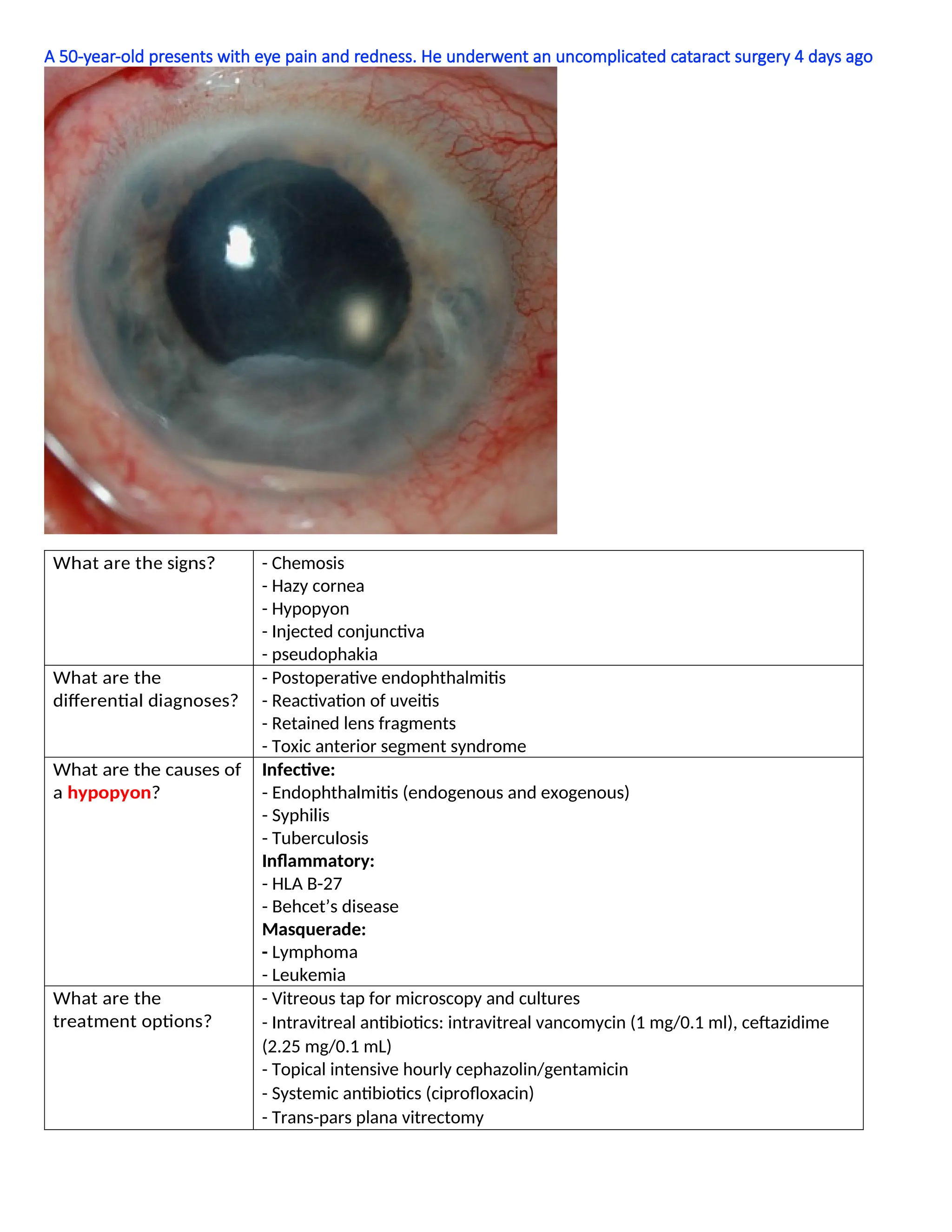
A 50-year-old presentswith eye pain and redness. He underwent an uncomplicated cataract surgery 4 days ago
What are the signs? - Chemosis
- Hazy cornea
- Hypopyon
- Injected conjunctiva
- pseudophakia
What are the
differential diagnoses?
- Postoperative endophthalmitis
- Reactivation of uveitis
- Retained lens fragments
- Toxic anterior segment syndrome
What are the causes of
a hypopyon?
Infective:
- Endophthalmitis (endogenous and exogenous)
- Syphilis
- Tuberculosis
Inflammatory:
- HLA B-27
- Behcet’s disease
Masquerade:
- Lymphoma
- Leukemia
What are the
treatment options?
- Vitreous tap for microscopy and cultures
- Intravitreal antibiotics: intravitreal vancomycin (1 mg/0.1 ml), ceftazidime
(2.25 mg/0.1 mL)
- Topical intensive hourly cephazolin/gentamicin
- Systemic antibiotics (ciprofloxacin)
- Trans-pars plana vitrectomy
16.
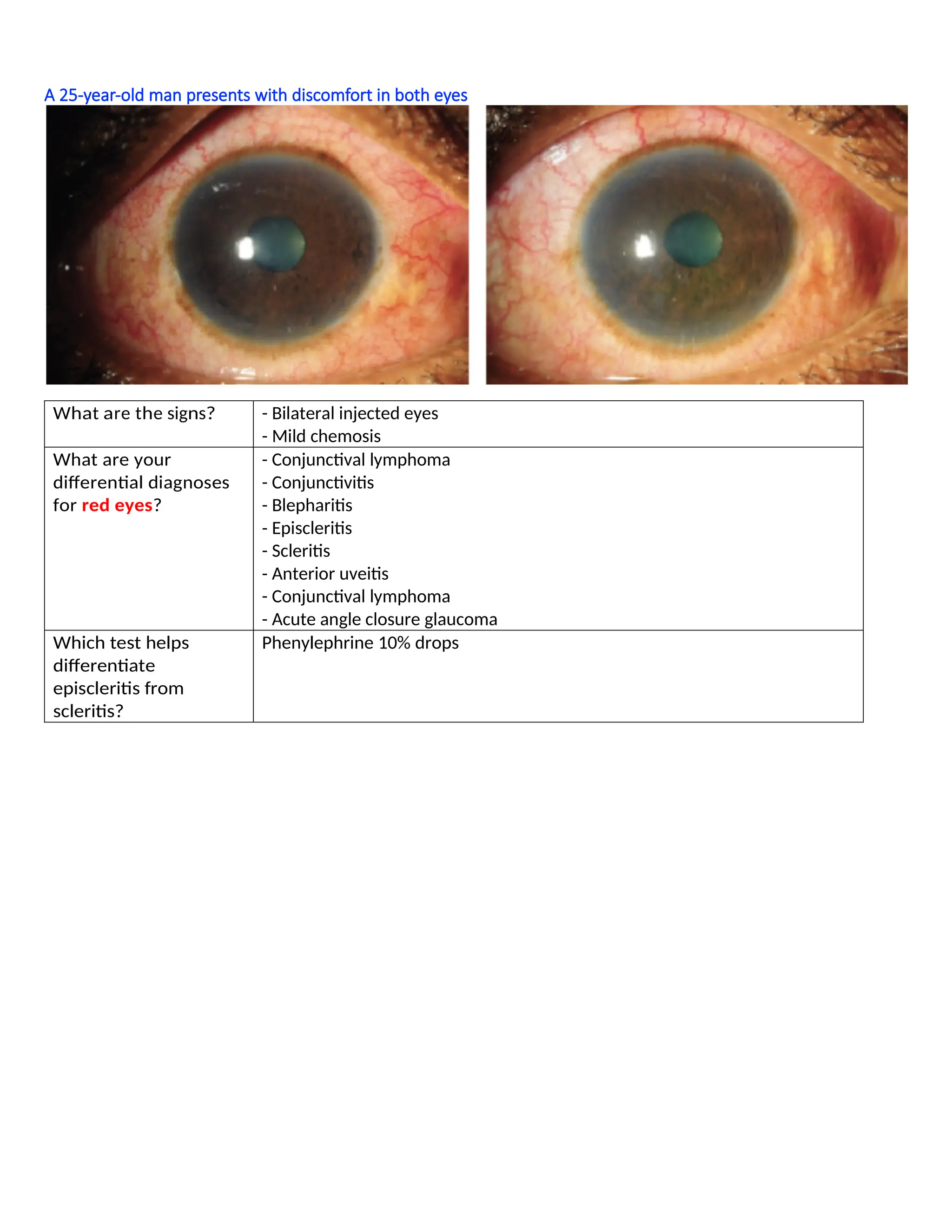
A 25-year-old manpresents with discomfort in both eyes
What are the signs? - Bilateral injected eyes
- Mild chemosis
What are your
differential diagnoses
for red eyes?
- Conjunctival lymphoma
- Conjunctivitis
- Blepharitis
- Episcleritis
- Scleritis
- Anterior uveitis
- Conjunctival lymphoma
- Acute angle closure glaucoma
Which test helps
differentiate
episcleritis from
scleritis?
Phenylephrine 10% drops


![keratitis [Autosaved].pptx](https://cdn.slidesharecdn.com/ss_thumbnails/keratitisautosaved-220807175619-8bc9d2cd-thumbnail.jpg?width=640&height=640&fit=bounds)


