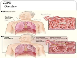
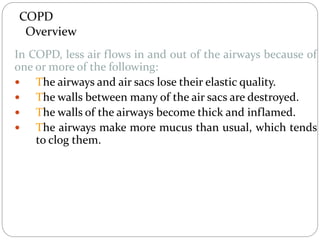
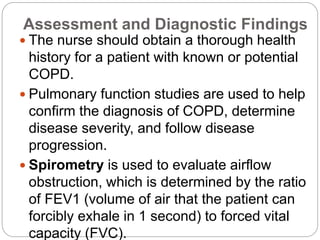
Chronic obstructive
pulmonary disease
(COPD)
COPD
Out line
 What is COPD?
 Overview
 Causes of COPD
 Symptoms of COPD
 What's the difference between COPD and asthma?
 Diagnostic tests needed for COPD
 Medical management of COPD
 Preventive measures
 Nursing intervention
 Outlook and Prognosis
COPD
Definition
 COPD, or chronic obstructive pulmonary disease, is a
progressive disease that makes it hard to breathe.
"Progressive" means the disease gets worse over time.
is a type of obstructive lung disease characterized by
chronically poor airflow. It typically worsens over time.
COPD
Overview
COPD
Overview
In COPD, less air flows in and out of the airways because of
one or more of the following:
 The airways and air sacs lose their elastic quality.
 The walls between many of the air sacs are destroyed.
 The walls of the airways become thick and inflamed.
 The airways make more mucus than usual, which tends
to clog them.
COPD
Causes
 Smoking
 Air pollution
 genetic (hereditary) risk Currently, the only clearly
inherited risk factor is alpha 1-antitrypsin deficiency
(AAT).
COPD
Symptoms
 Productive cough
 Breathlessness
 Chest infection
 Other symptoms of COPD can be more vague, weight
loss, tiredness and ankle swelling.
 It is unclear if different types of COPD exist. While
previously divided into emphysema & chronic
bronchitis, emphysema is only a description of lung
changes rather than a disease itself, & chronic
bronchitis is simply a descriptor of symptoms that may
or may not occur with COPD
Difference between COPD and Asthma
 In COPD there is permanent damage to the airways. The
narrowed airways are fixed, & so symptoms are chronic
(persistent). Rx to open up the airways, is therefore
limited.
 In asthma there is inflammation in the airways which
makes the muscles in the airways constrict. This causes
the airways to narrow. The symptoms tend to come & go,
& vary in severity from time to time. Rx to reduce
inflammation & to open up the airways usually works
well.
 COPD is more likely than asthma to cause a chronic
(ongoing) cough with sputum.
 Night time waking with breathlessness or wheeze is
common in asthma and uncommon in COPD.
 COPD is rare before the age of 35 whilst asthma is
common in under-35.
Difference between COPD and Asthma
 COPD is a type of obstructive lung disease in which
chronic incompletely reversible poor airflow (airflow
limitation) & inability to breathe out fully (air trapping)
exist.
 The poor airflow is the result of breakdown of lung
tissue (known as emphysema) & small airways
disease known as obstructive bronchiolitis.
 The relative contributions of these 2 factors vary
between people. Severe destruction of small airways
can lead to the formation of large air pockets—
known as bullae—that replace lung tissue. This form
Emphysema
 In emphysema, impaired gas exchange
(oxygen, carbon dioxide) results from
destruction of the walls of overdistended
alveoli.
 “Emphysema” is a pathological term that
describes an abnormal distention of the air
spaces beyond the terminal bronchioles,
with destruction of the walls of the alveoli.
 It is the end stage of a process that has
progressed slowly for many years.
Emphysema
 As the walls of the alveoli are destroyed (a process
accelerated by recurrent infections), the alveolar
surface area in direct contact with the pulmonary
capillaries continually decreases, causing an increase in
dead space (lung area where no gas exchange can
occur) and impaired oxygen diffusion, which leads to
hypoxemia.
 In the later stages of the disease, carbon
dioxide elimination is impaired, resulting in
increased carbon dioxide tension in arterial
blood (hypercapnia) and causing respiratory
acidosis.
Emphysema
 As the alveolar walls continue to break down, the
pulmonary capillary bed is reduced.
 Consequently, pulmonary blood flow is increased,
forcing the right ventricle to maintain a higher blood
pressure in the pulmonary artery.
 Hypoxemia may further increase pulmonary artery
pressure.
 Thus, right-sided heart failure (cor pulmonale) is one of
the complications of emphysema. Congestion,
dependent edema, distended neck veins, or pain in the
region of the liver suggests the development of cardiac
failure.
Risk Factors
COPD/Emphysema
 Exposure to tobacco smoke accounts for an
estimated 80% to 90% of COPD cases
 Passive smoking
 Occupational exposure
 Ambient air pollution
 Genetic abnormalities, including a
deficiency of alpha1-antitrypsin, an enzyme
inhibitor that normally counteracts the
destruction of lung tissue by certain other
enzymes
Clinical Manifestations
 COPD is characterized by three primary
symptoms: cough, sputum production, and
dyspnea on exertion.
 These symptoms often worsen over time.
 Chronic cough and sputum production often
precede the development of airflow limitation
by many years.
 However, not all individuals with cough and
sputum production will develop COPD.
Dyspnea may be severe and often interferes
with the patient’s activities.
Clinical Manifestations
 Weight loss is common because dyspnea
interferes with eating, and the work of
breathing is energy-depleting.
 Often the patient cannot participate in even
mild exercise because of dyspnea; as COPD
progresses, dyspnea occurs even at rest.
Clinical Manifestations
 As the work of breathing increases over
time, the accessory muscles are recruited
in an effort to breathe.
 The patient with COPD is at risk for
respiratory insufficiency and respiratory
infections, which in turn increase the risk
for acute and chronic respiratory failure.
Clinical Manifestations
 In COPD patients with a primary
emphysematous component, chronic
hyperinflation leads to the “barrel chest”
thorax configuration.
 This results from fixation of the ribs in the
inspiratory position (due to hyperinflation) and
from loss of lung elasticity (Fig. 24-3).
 Retraction of the supraclavicular fossae
occurs on inspiration, causing the shoulders
to heave upward (Fig. 24-4). In advanced
emphysema, the abdominal muscles also
Assessment and Diagnostic Findings
 The nurse should obtain a thorough health
history for a patient with known or potential
COPD.
 Pulmonary function studies are used to help
confirm the diagnosis of COPD, determine
disease severity, and follow disease
progression.
 Spirometry is used to evaluate airflow
obstruction, which is determined by the ratio
of FEV1 (volume of air that the patient can
forcibly exhale in 1 second) to forced vital
capacity (FVC).
Complications
Respiratory insufficiency and
Respiratory failure are major life-
threatening complications of COPD.
Medical Management
RISK REDUCTION
 Smoking cessation is the single most effective
intervention to prevent COPD or slow its progression.
 Recent surveys indicate that 25% of all American
adults smoke.
 Nurses play a key role in promoting smoking
cessation and educating patients about ways to do
so.
 Patients diagnosed with COPD who continue to
smoke must be encouraged and assisted to quit.
Medical Management
PHARMACOLOGIC THERAPY
 Bronchodilators: relieve bronchospasm
and reduce airway obstruction by allowing
increased oxygen distribution throughout the
lungs and improving alveolar ventilation.
 Corticosteroids: Inhaled and systemic
corticosteroids (oral or intravenous) may
also be used in COPD but are used more
frequently in asthma.
MANAGEMENT OF
EXACERBATION
 An exacerbation of COPD is difficult to
diagnose, but signs and symptoms may
include increased dyspnea, increased
sputum production and purulence,
respiratory failure, changes in mental
status, or worsening blood gas
abnormalities.
 Primary causes for an acute exacerbation
include tracheobronchial infection and air
pollution.
MANAGEMENT OF
EXACERBATION
 Secondary causes are pneumonia;
pulmonary embolism; pneumothorax; rib
fractures or chest trauma; inappropriate use
of sedative, opioid, or beta-blocking agents;
and right- or left-sided heart failure.
 First, the primary cause of the exacerbation
is identified, and then specific treatment is
administered.
MANAGEMENT OF
EXACERBATION
 Optimization of bronchodilator medications
is the first-line therapy and involves
identifying the best medication or
combinations of medications taken on a
regular schedule for that patient.
 Depending on the signs and symptoms,
corticosteroids, antibiotic agents, oxygen
therapy, and intensive respiratory
interventions may also be used.
OXYGEN THERAPY
 Oxygen therapy can be administered as long-term
continuous therapy, during exercise, or to prevent
acute dyspnea.
 Long-term oxygen therapy has been shown to
improve the patient’s quality of life and survival.
 Indications for oxygen supplementation include a
PaO2 of 55 mm Hg or less or evidence of tissue
hypoxia and organ damage such as cor pulmonale,
secondary polycythemia, edema from right heart
failure, or impaired mental status.
NURSING ALERT
 Because hypoxemia stimulates respiration in the
patient with severe COPD, increasing the oxygen flow
to a high rate may greatly raise the patient’s blood
oxygen level.
 At the same time, this will suppress the respiratory
drive, causing increased retention of carbon dioxide
and CO2 narcosis.
 The nurse should closely monitor the patient’s
respiratory response to oxygen administration via
physical assessment, pulse oximetry, and/or arterial
blood gases.
SURGICAL MANAGEMENT
 Bullectomy: is a surgical option for select patients
with bullous emphysema. Bullae are enlarged
airspaces that do not contribute to ventilation but
occupy space in the thorax; these areas may be
surgically excised.
 Lung Volume Reduction Surgery: Treatment options
for patients with end-stage COPD with a primary
emphysematous component are limited, although lung
volume reduction surgery is an option for a specific
subset of patients.
 Lung Transplantation: is a viable alternative for
definitive surgical treatment of end-stage emphysema.
COPD
Diagnostic tests
 Symptoms
 Physical examination
 Sample of sputum
 Chest x-ray
 High-resolution CT (HRCT scan)
 Pulmonary function test (spirometery)
 Arterial blood gases test
 Pulse oximeter
A lateral chest x-ray of a person
with emphysema. Note the barrel
chest and flat diaphragm
Chest X-ray demonstrating severe
COPD. Note the small heart size
in comparison to the lungs.
Axial CT image of the lung of a
person with end-stage bullous
emphysema.
A severe case of bullous
emphysema
Lung bulla as seen on CXR in
a person with severe COPD
COPD
Medical management
 Give antibiotics to treat infection
 Give bronchodilators to relieve bronchospasm, reduce
airway obstruction, mucosal edema and liquefy
secretions.
 Chest physiotherapy and postural drainage to improve
pulmonary ventilation.
 Proper hydration helps to cough up secretions or
tracheal suctioning when the patient is unable to cough.
 Steroid therapy if the patient fails to respond to more
conservative treatment.
COPD
Medical management (cont…)
 Stop smoking
 Oxygenation with low concentration during the acute
episodes
 In asthma adrenaline ( epinephrine) SC if the
bronchospasm not relieved.
 Aminophylins IV if the above treatment does not help.
 IV corticosteroids for patients with chronic asthma or
frequent attack.
 Sedative or tranquilizers to calm the patient.
 Increase fluids intake to correct loss of diaphoresis &
inaccessible loss of hyperventilation.
 Intubations & mechanical ventilation if there is
respiratory failure.
COPD
Preventive measures
To prevent irritation and infection of the airways,
instruct the patient to:
 Avoid exposure to cigarette, pipe, and cigar smoke as
well as to dusts and powders.
 Avoid use of aerosol sprays.
 Stay indoors when the pollen count is high.
 Stay indoors when temperature and humidity are
both high
COPD
Preventive measures (cont…)
 Use air conditioning to help decrease pollutants and
control temperature
 Avoid exposure to persons known to have colds or other
respiratory tract infection
 Avoid enclosed, crowded areas during cold and flu
season.
 Obtain immunization against influenza and
streptococcal pneumonia.
COPD
Preventive measures (cont…)
To ensure prompt, effective treatment of a developing
respiratory infection, instruct the patient to do the
following:-
 Report any change in sputum color character,
increased tightness of the chest, increased dyspnea,
or fatigue.
 Call the physician if ordered antibiotics do not
relieve symptoms within 24 hours.
COPD
Nursing intervention
 Assessment
 History
 Patient's environment
 Work history, exercise pattern, smoking habits
 The onset & development of symptoms
 Sleeping positions
COPD
Nursing intervention (cont…)
 Physical examination
Signs of heavy smokers
 Observe for clubbing
 Distended neck vein on expiration
 The presence of barrel chest
 Observe for abdominal breathing
 The use of pursed lips breathing and chest
movement
 Auscultate the chest& listen for musical wheezes
characteristics of chronic bronchitis
COPD
Nursing intervention (cont…)
review the results of diagnostic procedure:
 Arterial blood gases
 Pulmonary function tests
 X-ray films
 Nursing diagnosis
 Ineffective breathing pattern related to increase
need of O2
 Ineffective airway clearance related to excessive
accumulation of secretions
 Impaired gas exchange related to impaired
expiration &co2 retention
COPD
Nursing intervention (cont…)
 Activity intolerance related to inadequate oxygenation
 High risk for ineffective individual coping related to
chronic disease, its effects& its treatment
 High risk for altered health maintenance related to
insufficient knowledge of prevention, identification
and treatment of respiratory complication of COPD

2 COPD.ppt

  • 1.
  • 2.
    COPD Out line  Whatis COPD?  Overview  Causes of COPD  Symptoms of COPD  What's the difference between COPD and asthma?  Diagnostic tests needed for COPD  Medical management of COPD  Preventive measures  Nursing intervention  Outlook and Prognosis
  • 3.
    COPD Definition  COPD, orchronic obstructive pulmonary disease, is a progressive disease that makes it hard to breathe. "Progressive" means the disease gets worse over time. is a type of obstructive lung disease characterized by chronically poor airflow. It typically worsens over time.
  • 4.
  • 5.
    COPD Overview In COPD, lessair flows in and out of the airways because of one or more of the following:  The airways and air sacs lose their elastic quality.  The walls between many of the air sacs are destroyed.  The walls of the airways become thick and inflamed.  The airways make more mucus than usual, which tends to clog them.
  • 6.
    COPD Causes  Smoking  Airpollution  genetic (hereditary) risk Currently, the only clearly inherited risk factor is alpha 1-antitrypsin deficiency (AAT).
  • 7.
    COPD Symptoms  Productive cough Breathlessness  Chest infection  Other symptoms of COPD can be more vague, weight loss, tiredness and ankle swelling.
  • 8.
     It isunclear if different types of COPD exist. While previously divided into emphysema & chronic bronchitis, emphysema is only a description of lung changes rather than a disease itself, & chronic bronchitis is simply a descriptor of symptoms that may or may not occur with COPD
  • 9.
    Difference between COPDand Asthma  In COPD there is permanent damage to the airways. The narrowed airways are fixed, & so symptoms are chronic (persistent). Rx to open up the airways, is therefore limited.  In asthma there is inflammation in the airways which makes the muscles in the airways constrict. This causes the airways to narrow. The symptoms tend to come & go, & vary in severity from time to time. Rx to reduce inflammation & to open up the airways usually works well.
  • 10.
     COPD ismore likely than asthma to cause a chronic (ongoing) cough with sputum.  Night time waking with breathlessness or wheeze is common in asthma and uncommon in COPD.  COPD is rare before the age of 35 whilst asthma is common in under-35. Difference between COPD and Asthma
  • 11.
     COPD isa type of obstructive lung disease in which chronic incompletely reversible poor airflow (airflow limitation) & inability to breathe out fully (air trapping) exist.  The poor airflow is the result of breakdown of lung tissue (known as emphysema) & small airways disease known as obstructive bronchiolitis.  The relative contributions of these 2 factors vary between people. Severe destruction of small airways can lead to the formation of large air pockets— known as bullae—that replace lung tissue. This form
  • 12.
    Emphysema  In emphysema,impaired gas exchange (oxygen, carbon dioxide) results from destruction of the walls of overdistended alveoli.  “Emphysema” is a pathological term that describes an abnormal distention of the air spaces beyond the terminal bronchioles, with destruction of the walls of the alveoli.  It is the end stage of a process that has progressed slowly for many years.
  • 13.
    Emphysema  As thewalls of the alveoli are destroyed (a process accelerated by recurrent infections), the alveolar surface area in direct contact with the pulmonary capillaries continually decreases, causing an increase in dead space (lung area where no gas exchange can occur) and impaired oxygen diffusion, which leads to hypoxemia.  In the later stages of the disease, carbon dioxide elimination is impaired, resulting in increased carbon dioxide tension in arterial blood (hypercapnia) and causing respiratory acidosis.
  • 14.
    Emphysema  As thealveolar walls continue to break down, the pulmonary capillary bed is reduced.  Consequently, pulmonary blood flow is increased, forcing the right ventricle to maintain a higher blood pressure in the pulmonary artery.  Hypoxemia may further increase pulmonary artery pressure.  Thus, right-sided heart failure (cor pulmonale) is one of the complications of emphysema. Congestion, dependent edema, distended neck veins, or pain in the region of the liver suggests the development of cardiac failure.
  • 15.
    Risk Factors COPD/Emphysema  Exposureto tobacco smoke accounts for an estimated 80% to 90% of COPD cases  Passive smoking  Occupational exposure  Ambient air pollution  Genetic abnormalities, including a deficiency of alpha1-antitrypsin, an enzyme inhibitor that normally counteracts the destruction of lung tissue by certain other enzymes
  • 16.
    Clinical Manifestations  COPDis characterized by three primary symptoms: cough, sputum production, and dyspnea on exertion.  These symptoms often worsen over time.  Chronic cough and sputum production often precede the development of airflow limitation by many years.  However, not all individuals with cough and sputum production will develop COPD. Dyspnea may be severe and often interferes with the patient’s activities.
  • 17.
    Clinical Manifestations  Weightloss is common because dyspnea interferes with eating, and the work of breathing is energy-depleting.  Often the patient cannot participate in even mild exercise because of dyspnea; as COPD progresses, dyspnea occurs even at rest.
  • 18.
    Clinical Manifestations  Asthe work of breathing increases over time, the accessory muscles are recruited in an effort to breathe.  The patient with COPD is at risk for respiratory insufficiency and respiratory infections, which in turn increase the risk for acute and chronic respiratory failure.
  • 19.
    Clinical Manifestations  InCOPD patients with a primary emphysematous component, chronic hyperinflation leads to the “barrel chest” thorax configuration.  This results from fixation of the ribs in the inspiratory position (due to hyperinflation) and from loss of lung elasticity (Fig. 24-3).  Retraction of the supraclavicular fossae occurs on inspiration, causing the shoulders to heave upward (Fig. 24-4). In advanced emphysema, the abdominal muscles also
  • 21.
    Assessment and DiagnosticFindings  The nurse should obtain a thorough health history for a patient with known or potential COPD.  Pulmonary function studies are used to help confirm the diagnosis of COPD, determine disease severity, and follow disease progression.  Spirometry is used to evaluate airflow obstruction, which is determined by the ratio of FEV1 (volume of air that the patient can forcibly exhale in 1 second) to forced vital capacity (FVC).
  • 22.
    Complications Respiratory insufficiency and Respiratoryfailure are major life- threatening complications of COPD.
  • 23.
    Medical Management RISK REDUCTION Smoking cessation is the single most effective intervention to prevent COPD or slow its progression.  Recent surveys indicate that 25% of all American adults smoke.  Nurses play a key role in promoting smoking cessation and educating patients about ways to do so.  Patients diagnosed with COPD who continue to smoke must be encouraged and assisted to quit.
  • 24.
    Medical Management PHARMACOLOGIC THERAPY Bronchodilators: relieve bronchospasm and reduce airway obstruction by allowing increased oxygen distribution throughout the lungs and improving alveolar ventilation.  Corticosteroids: Inhaled and systemic corticosteroids (oral or intravenous) may also be used in COPD but are used more frequently in asthma.
  • 25.
    MANAGEMENT OF EXACERBATION  Anexacerbation of COPD is difficult to diagnose, but signs and symptoms may include increased dyspnea, increased sputum production and purulence, respiratory failure, changes in mental status, or worsening blood gas abnormalities.  Primary causes for an acute exacerbation include tracheobronchial infection and air pollution.
  • 26.
    MANAGEMENT OF EXACERBATION  Secondarycauses are pneumonia; pulmonary embolism; pneumothorax; rib fractures or chest trauma; inappropriate use of sedative, opioid, or beta-blocking agents; and right- or left-sided heart failure.  First, the primary cause of the exacerbation is identified, and then specific treatment is administered.
  • 27.
    MANAGEMENT OF EXACERBATION  Optimizationof bronchodilator medications is the first-line therapy and involves identifying the best medication or combinations of medications taken on a regular schedule for that patient.  Depending on the signs and symptoms, corticosteroids, antibiotic agents, oxygen therapy, and intensive respiratory interventions may also be used.
  • 28.
    OXYGEN THERAPY  Oxygentherapy can be administered as long-term continuous therapy, during exercise, or to prevent acute dyspnea.  Long-term oxygen therapy has been shown to improve the patient’s quality of life and survival.  Indications for oxygen supplementation include a PaO2 of 55 mm Hg or less or evidence of tissue hypoxia and organ damage such as cor pulmonale, secondary polycythemia, edema from right heart failure, or impaired mental status.
  • 29.
    NURSING ALERT  Becausehypoxemia stimulates respiration in the patient with severe COPD, increasing the oxygen flow to a high rate may greatly raise the patient’s blood oxygen level.  At the same time, this will suppress the respiratory drive, causing increased retention of carbon dioxide and CO2 narcosis.  The nurse should closely monitor the patient’s respiratory response to oxygen administration via physical assessment, pulse oximetry, and/or arterial blood gases.
  • 30.
    SURGICAL MANAGEMENT  Bullectomy:is a surgical option for select patients with bullous emphysema. Bullae are enlarged airspaces that do not contribute to ventilation but occupy space in the thorax; these areas may be surgically excised.  Lung Volume Reduction Surgery: Treatment options for patients with end-stage COPD with a primary emphysematous component are limited, although lung volume reduction surgery is an option for a specific subset of patients.  Lung Transplantation: is a viable alternative for definitive surgical treatment of end-stage emphysema.
  • 31.
    COPD Diagnostic tests  Symptoms Physical examination  Sample of sputum  Chest x-ray  High-resolution CT (HRCT scan)  Pulmonary function test (spirometery)  Arterial blood gases test  Pulse oximeter
  • 32.
    A lateral chestx-ray of a person with emphysema. Note the barrel chest and flat diaphragm Chest X-ray demonstrating severe COPD. Note the small heart size in comparison to the lungs.
  • 33.
    Axial CT imageof the lung of a person with end-stage bullous emphysema. A severe case of bullous emphysema
  • 34.
    Lung bulla asseen on CXR in a person with severe COPD
  • 35.
    COPD Medical management  Giveantibiotics to treat infection  Give bronchodilators to relieve bronchospasm, reduce airway obstruction, mucosal edema and liquefy secretions.  Chest physiotherapy and postural drainage to improve pulmonary ventilation.  Proper hydration helps to cough up secretions or tracheal suctioning when the patient is unable to cough.  Steroid therapy if the patient fails to respond to more conservative treatment.
  • 36.
    COPD Medical management (cont…) Stop smoking  Oxygenation with low concentration during the acute episodes  In asthma adrenaline ( epinephrine) SC if the bronchospasm not relieved.  Aminophylins IV if the above treatment does not help.  IV corticosteroids for patients with chronic asthma or frequent attack.  Sedative or tranquilizers to calm the patient.  Increase fluids intake to correct loss of diaphoresis & inaccessible loss of hyperventilation.  Intubations & mechanical ventilation if there is respiratory failure.
  • 37.
    COPD Preventive measures To preventirritation and infection of the airways, instruct the patient to:  Avoid exposure to cigarette, pipe, and cigar smoke as well as to dusts and powders.  Avoid use of aerosol sprays.  Stay indoors when the pollen count is high.  Stay indoors when temperature and humidity are both high
  • 38.
    COPD Preventive measures (cont…) Use air conditioning to help decrease pollutants and control temperature  Avoid exposure to persons known to have colds or other respiratory tract infection  Avoid enclosed, crowded areas during cold and flu season.  Obtain immunization against influenza and streptococcal pneumonia.
  • 39.
    COPD Preventive measures (cont…) Toensure prompt, effective treatment of a developing respiratory infection, instruct the patient to do the following:-  Report any change in sputum color character, increased tightness of the chest, increased dyspnea, or fatigue.  Call the physician if ordered antibiotics do not relieve symptoms within 24 hours.
  • 40.
    COPD Nursing intervention  Assessment History  Patient's environment  Work history, exercise pattern, smoking habits  The onset & development of symptoms  Sleeping positions
  • 41.
    COPD Nursing intervention (cont…) Physical examination Signs of heavy smokers  Observe for clubbing  Distended neck vein on expiration  The presence of barrel chest  Observe for abdominal breathing  The use of pursed lips breathing and chest movement  Auscultate the chest& listen for musical wheezes characteristics of chronic bronchitis
  • 43.
    COPD Nursing intervention (cont…) reviewthe results of diagnostic procedure:  Arterial blood gases  Pulmonary function tests  X-ray films  Nursing diagnosis  Ineffective breathing pattern related to increase need of O2  Ineffective airway clearance related to excessive accumulation of secretions  Impaired gas exchange related to impaired expiration &co2 retention
  • 44.
    COPD Nursing intervention (cont…) Activity intolerance related to inadequate oxygenation  High risk for ineffective individual coping related to chronic disease, its effects& its treatment  High risk for altered health maintenance related to insufficient knowledge of prevention, identification and treatment of respiratory complication of COPD