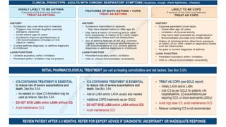
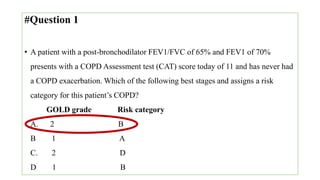
Chronic Obstructive Pulmonary
Disease (COPD)
By: Dr Ibrahim Ahmed Nur
Senior house officer (SHO)
KIU
Date: 09-09-2022
Outline
• Introduction
• Epidemiology
• Pathphysiology
• Clinical features
• Diagnosis
• Management
• Complication
• COPD exacerbation
• ACOS (Asthma-COPD Overlap Syndromes)
#Question 1
• A patient with a post-bronchodilator FEV1/FEVC of 65% and FEV1 of 70%
presents with a COPD Assessment test (CAT) score today of 11 and has never had
a COPD exacerbation. Which of the following best stages and assigns a risk
category for this patient’s COPD?
GOLD grade Risk category
A. 2 B
B 1 A
C. 2 D
D. 1 B
Introduction
• COPD is the 3rd leading cause of death in the world (GOLD Guidelines 2021).
• More than 3 million people died of COPD in 2012 accounting for 6% globally
(WHO)
• Prevalence of COPD in Uganda was 16% this was highest in people aged 30-39
years (Fresh air Uganda survey, Lancet Global Health 2015)
Burden of COPD
• COPD represents an important public health challenge that is both preventable
and treatable.
• The burden of COPD is projected to increase in coming decades due to
continued exposure to COPD risk factors and the aging of the world’s
population.
• COPD is associated with significant economic burden.
Definition of COPD
A common, preventable and treatable disease, characterized by persistent respiratory
symptoms and airflow limitation that are usually progressive and associated with an
enhanced chronic inflammatory response in the airways and/or alveoli due to
significant exposure to noxious particles or gases. (Vogelmeier et al., 2017).
COPD includes
1. Chronic Bronchitis
2. Emphysema
Chronic Bronchitis
Emphysema
Chronic bronchitis
Defined as a chronic productive cough for three months in each of the two
successive years in a patient in whom other causes of chronic cough have been
excluded.
Emphysema
Abnormal and permanent enlargement of the airspaces distal to the terminal
bronchioles that is accompanied by destruction of the airspaces walls , without
obvious fibrosis.
Emphysema has 4 morphologic patterns:
1. Centriacinar.
2. Distal acinar, or paraseptal
3. Panacinar.
4. irregular
Pathophysiology
• COPD is an inflammatory condition involving the airways, lung parenchyma, and
pulmonary vasculature
Emphysema: one of the structural changes seen in COPD where there is
destruction of the alveolar air sacs (gas-exchanging surfaces of the lungs) leading
to obstructive physiology.
• An irritant (e.g., smoking) causes an inflammatory response, Neutrophils and
macrophages are recruited and release multiple inflammatory mediators, Oxidants
and excess proteases leading to the destruction of the air sacs, The protease-
mediated destruction of elastin leads to a loss of elastic recoil and results in airway
collapse during exhalation Air trapping
Loss of elasticity Air trapping Increase in end expiratory volume Barrel chest
Genetics and COPD
Alpha 1-antitrypsin deficiency is a genetic condition (caused by misfolding of the
mutated protein which can accumulate in the liver) that is responsible for about 2%
of cases of COPD.
In this condition, the body does not make enough of a protein alpha 1-antitrypsin.
Alpha 1-antitrypsin protects the lungs from damage caused by protease enzymes,
such as elastase and trypsin, that can be released as a result of an inflammatory
response to tobacco smoke.
Chronic bronchitis
• Mucous gland hyperplasia is the histologic hallmark of chronic bronchitis.
• Airway structural changes include atrophy, focal squamous metaplasia, ciliary
abnormalities, variable amounts of airway smooth muscle hyperplasia, inflammation,
and bronchial wall thickening
• Damage to the endothelium impairs the mucociliary response that clears bacteria
and mucus.
• Inflammation and secretions provide the obstructive component of chronic
bronchitis.
• Neutrophilia develops in the airway lumen, and neutrophilic infiltrates accumulate
in the submucosa.
• The respiratory bronchioles display a mononuclear inflammatory process, lumen
occlusion by mucus plugging, goblet cell metaplasia, smooth muscle hyperplasia,
and distortion due to fibrosis.
• These changes, combined with loss of supporting alveolar attachments, cause
airflow limitation by allowing airway walls to deform and narrow the airway
lumen.
• The body responds by decreasing ventilation and increasing cardiac output.
• This V/Q mismatch results in rapid circulation in a poorly ventilated lung, leading
to hypoxemia and polycythemia.
• Eventually, hypercapnia and respiratory acidosis develop, leading to pulmonary
artery vasoconstriction and cor pulmonale.
Alveolar hypoxia pulmonary vasoconstriction Pulmonary HTN
Reduced blood to LV Reduce LV out put reduce in circulatory
volume Activation RAAS Fluid retention
• With the ensuing hypoxemia, polycythemia, and increased CO2 retention, these
patients have signs of right heart failure and are known as "blue bloaters."
Clinical features
Symptoms
The characteristic symptoms of COPD are chronic and progressive dyspnea, cough
and sputum production that can be variable from day to day.
Dyspnea: progressive persistent and worse with exercise.
Chronic cough: May be intermittent and may be unproductive.
Chronic sputum production: COPD patients commonly coughs up sputum.
Other symptoms
• Wheezing
• Chest tightness
• Wt. loss
• Respiratory infection
Examination
Bossily benign exam
• Barrel chest
• Prolonged expiratory phase
• Pursed lip breathing, accessory muscle use.
• Tachypnea.
• clubbing
• Hyper resonant
• Decreased breath sounds
• Wheezing
• Paradoxical retraction of the lower interspaces during inspiration (ie, hoover’sign)
• Tripod position- pt with end stage COPD adopt positions that relieve dyspnea,
such as leaning forward with arms outstretched and weight supported on the palms
or elbows.
Extreimities:
Clubbing
Lower extremity edema in RHF
Skin: central cyanosis
Diagnosis
• Full history
• Full examination
• CXR
• CBC
• ABGs
• PFT
• Alpha 1 AT level
• ECG and ECHO
CXR :-
 Hyperinflation, bullae, flat hemidiaphragm
 Also its important to exclude lung cancer
 interstitial markings & bullae
• Decreased FEV 1
• Decreased FEV1/FVC <0.7
• Minimal or no response to SABD (less than 12%)
Assessment of symptoms
Combined tool (ABCD assessment tool)
Example
Consider two patients:
 Both patients with FEV1 < 30% of predicted
 Both with CAT scores of 18 and mMRC dyspnoea scale of 2
 But, one with 0 exacerbations in the past year and the other with 3
exacerbations in the past year.
PATIENT 1:
 FEV1 < 30% of predicted: GOLD GRADE 4
 0 exacerbations in the past year: GOLD GROUP A or B
 CAT scores of 18 and mMRC dyspnoea scale of 2: GOLD GROUP B
PATIENT 1: GOLD GRADE 4 GROUP B
PATIENT2:
 FEV1 < 30% of predicted: GOLD GRADE 4
 3 exacerbations in the past year: GOLD GROUP C or D
 CAT scores of 18 and mMRC dyspnoea scale of 2: GOLD GROUP D
PATIENT 2: GOLD GARDE 4 GROUP D
BODe index
• BODe index is a simple grading system for COPD
• Using body mass index (BMI), airflow Obstruction, Dyspnoea, and Exercise
capacity as its scoring variables.
• It has been shown to be better than FeV1 at predicting risk of hospitalization and
death in patients with COPD, as it is multidimensional.
• Patients are scored as having a BODe index of 0–10
• Higher scores indicating a higher risk of death.
• It is being increasingly used, with recommendations to calculate it in the clinical
setting to give prognostic information (Celli Br et al. New Engl J Med 2004; 350:
1005–1012).
Differential diagnosis
•Management
Principles
• To relieve symptoms.
• To decrease the frequency and severity of acute attacks.
• To slow progression of disease
• Improve quality of life
• To prolong survival
management
General management
• Smoking cessation (critical in slowing lung function decline)
• Vaccinations for:
• Pneumococcal pneumonia
• Influenza
• Pulmonary rehabilitation:
• Improve symptoms and quality of life
• Reduces frequency of exacerbations
• Components: exercise training, nutritional counselling and psychosocial support
• O2 therapy:
• If O2 saturation is < 88% in a stable patient (PO₂ < 55 mm Hg)
• If concurrent pulmonary hypertension, right-sided heart failure, or polycythemia
Why low concentration O2 given in COPD? Or what happens
when high flow O2 given?
o In COPD, the patient is dependent on hypoxic drive for respiration.
o High flow oxygen blunts the chemo responsiveness of the respiratory center in the
medulla (part of the brainstem) and thus aggravates respiratory failure (Type 2
respiratory failure). To avoid this, low flow oxygen is given
Surgical intervention
• Surgery is reserved for severe cases not controlled with medical therapy to
improve quality of life.
• Bullectomy: removal of giant bullae to relieve local compression
• Lung volume reduction: resection of the most diseased parts of the lung to
decrease hyperinflation
• Lung transplant: indicated in end-stage lung disease
Oxford Handbook of
Respiratory Medicine
Page: 197
Exacerbation of COPD
• Canadian Thoracic Society defined: “a sustained worsening of dyspnea, cough
or sputum production leading to an increase in the use of maintenance medications
and/or supplementation with additional medications
• It is an acute event characterized by a worsening of the patient’s respiratory
symptoms that is beyond the normal day to day variations and leads to change of
medication.
Causes of exacerbation
• Infective
1. Viral e.g. RSV, Rhinoviruses, influenza, parainfluenza
2. Bacterial(50%) H.influenza, S.pneumoniae, moraxela cataharis,mycoplasma
pneumonia
• Non-infective –air pollution, pulmonary embolism, pneumothorax, sedatives, heart failure or
arrhythmia.
Assessment of severity of exacerbation
• Classified as:
• Mild: Treated with short acting bronchodilators(SABDs) only
• Moderate: Treated with SABDs plus antibiotics and/or oral corticosteroids)
• Severe: Patients require hospitalization or visit the emergency room, may be also
be associated with acute respiratory failure.
• Blood eosinophil count may also predict exacerbation rates (in patients treated
with LABA without ICS).
Oxford Handbook of
Respiratory Medicine
Page: 203
COPD AND COMMORBIDITIES
• The presence of comorbidities should not alter COPD treatment
• Comorbidities should be treated per usual standards regardless of the presence of
COPD
• Cardiovascular diseases are common and important comorbidities in COPD
• Lung cancer is frequently seen in patients with COPD and is a major cause of
death
Covid 19 and COPD
•Asthma-COPD Overlap Syndrome (ACOS)
ACOS
 characterized by persistent air flow limitation with:
 several features usually associated with asthma
 several features usually associated with COPD
 ACOS is therefore identified in clinical practice by the features that it shares with
both asthma and COPD.
#Question 1
• A patient with a post-bronchodilator FEV1/FVC of 65% and FEV1 of 70%
presents with a COPD Assessment test (CAT) score today of 11 and has never had
a COPD exacerbation. Which of the following best stages and assigns a risk
category for this patient’s COPD?
GOLD grade Risk category
A. 2 B
B 1 A
C. 2 D
D 1 B
REFERENCES
• GOLD Guidelines 2021
• Oxford Handbook of Respiratory Medicine (4th edition)
• Up-to-date 2021
• THANKS FOR LISTENING

Chronic Obstructive Pulmonary Disease (COPD).pptx

  • 1.
    Chronic Obstructive Pulmonary Disease(COPD) By: Dr Ibrahim Ahmed Nur Senior house officer (SHO) KIU Date: 09-09-2022
  • 2.
    Outline • Introduction • Epidemiology •Pathphysiology • Clinical features • Diagnosis • Management • Complication • COPD exacerbation • ACOS (Asthma-COPD Overlap Syndromes)
  • 3.
    #Question 1 • Apatient with a post-bronchodilator FEV1/FEVC of 65% and FEV1 of 70% presents with a COPD Assessment test (CAT) score today of 11 and has never had a COPD exacerbation. Which of the following best stages and assigns a risk category for this patient’s COPD? GOLD grade Risk category A. 2 B B 1 A C. 2 D D. 1 B
  • 4.
    Introduction • COPD isthe 3rd leading cause of death in the world (GOLD Guidelines 2021). • More than 3 million people died of COPD in 2012 accounting for 6% globally (WHO) • Prevalence of COPD in Uganda was 16% this was highest in people aged 30-39 years (Fresh air Uganda survey, Lancet Global Health 2015)
  • 6.
    Burden of COPD •COPD represents an important public health challenge that is both preventable and treatable. • The burden of COPD is projected to increase in coming decades due to continued exposure to COPD risk factors and the aging of the world’s population. • COPD is associated with significant economic burden.
  • 7.
    Definition of COPD Acommon, preventable and treatable disease, characterized by persistent respiratory symptoms and airflow limitation that are usually progressive and associated with an enhanced chronic inflammatory response in the airways and/or alveoli due to significant exposure to noxious particles or gases. (Vogelmeier et al., 2017).
  • 8.
    COPD includes 1. ChronicBronchitis 2. Emphysema Chronic Bronchitis Emphysema
  • 9.
    Chronic bronchitis Defined asa chronic productive cough for three months in each of the two successive years in a patient in whom other causes of chronic cough have been excluded.
  • 10.
    Emphysema Abnormal and permanentenlargement of the airspaces distal to the terminal bronchioles that is accompanied by destruction of the airspaces walls , without obvious fibrosis. Emphysema has 4 morphologic patterns: 1. Centriacinar. 2. Distal acinar, or paraseptal 3. Panacinar. 4. irregular
  • 14.
    Pathophysiology • COPD isan inflammatory condition involving the airways, lung parenchyma, and pulmonary vasculature Emphysema: one of the structural changes seen in COPD where there is destruction of the alveolar air sacs (gas-exchanging surfaces of the lungs) leading to obstructive physiology. • An irritant (e.g., smoking) causes an inflammatory response, Neutrophils and macrophages are recruited and release multiple inflammatory mediators, Oxidants and excess proteases leading to the destruction of the air sacs, The protease- mediated destruction of elastin leads to a loss of elastic recoil and results in airway collapse during exhalation Air trapping
  • 15.
    Loss of elasticityAir trapping Increase in end expiratory volume Barrel chest
  • 16.
    Genetics and COPD Alpha1-antitrypsin deficiency is a genetic condition (caused by misfolding of the mutated protein which can accumulate in the liver) that is responsible for about 2% of cases of COPD. In this condition, the body does not make enough of a protein alpha 1-antitrypsin. Alpha 1-antitrypsin protects the lungs from damage caused by protease enzymes, such as elastase and trypsin, that can be released as a result of an inflammatory response to tobacco smoke.
  • 17.
    Chronic bronchitis • Mucousgland hyperplasia is the histologic hallmark of chronic bronchitis. • Airway structural changes include atrophy, focal squamous metaplasia, ciliary abnormalities, variable amounts of airway smooth muscle hyperplasia, inflammation, and bronchial wall thickening
  • 18.
    • Damage tothe endothelium impairs the mucociliary response that clears bacteria and mucus. • Inflammation and secretions provide the obstructive component of chronic bronchitis. • Neutrophilia develops in the airway lumen, and neutrophilic infiltrates accumulate in the submucosa. • The respiratory bronchioles display a mononuclear inflammatory process, lumen occlusion by mucus plugging, goblet cell metaplasia, smooth muscle hyperplasia, and distortion due to fibrosis. • These changes, combined with loss of supporting alveolar attachments, cause airflow limitation by allowing airway walls to deform and narrow the airway lumen.
  • 19.
    • The bodyresponds by decreasing ventilation and increasing cardiac output. • This V/Q mismatch results in rapid circulation in a poorly ventilated lung, leading to hypoxemia and polycythemia. • Eventually, hypercapnia and respiratory acidosis develop, leading to pulmonary artery vasoconstriction and cor pulmonale. Alveolar hypoxia pulmonary vasoconstriction Pulmonary HTN Reduced blood to LV Reduce LV out put reduce in circulatory volume Activation RAAS Fluid retention • With the ensuing hypoxemia, polycythemia, and increased CO2 retention, these patients have signs of right heart failure and are known as "blue bloaters."
  • 21.
    Clinical features Symptoms The characteristicsymptoms of COPD are chronic and progressive dyspnea, cough and sputum production that can be variable from day to day. Dyspnea: progressive persistent and worse with exercise. Chronic cough: May be intermittent and may be unproductive. Chronic sputum production: COPD patients commonly coughs up sputum.
  • 22.
    Other symptoms • Wheezing •Chest tightness • Wt. loss • Respiratory infection
  • 23.
    Examination Bossily benign exam •Barrel chest • Prolonged expiratory phase • Pursed lip breathing, accessory muscle use. • Tachypnea. • clubbing
  • 24.
    • Hyper resonant •Decreased breath sounds • Wheezing
  • 25.
    • Paradoxical retractionof the lower interspaces during inspiration (ie, hoover’sign) • Tripod position- pt with end stage COPD adopt positions that relieve dyspnea, such as leaning forward with arms outstretched and weight supported on the palms or elbows.
  • 26.
    Extreimities: Clubbing Lower extremity edemain RHF Skin: central cyanosis
  • 27.
    Diagnosis • Full history •Full examination • CXR • CBC • ABGs • PFT • Alpha 1 AT level • ECG and ECHO
  • 28.
    CXR :-  Hyperinflation,bullae, flat hemidiaphragm  Also its important to exclude lung cancer  interstitial markings & bullae
  • 31.
    • Decreased FEV1 • Decreased FEV1/FVC <0.7 • Minimal or no response to SABD (less than 12%)
  • 34.
  • 36.
    Combined tool (ABCDassessment tool)
  • 38.
    Example Consider two patients: Both patients with FEV1 < 30% of predicted  Both with CAT scores of 18 and mMRC dyspnoea scale of 2  But, one with 0 exacerbations in the past year and the other with 3 exacerbations in the past year.
  • 39.
    PATIENT 1:  FEV1< 30% of predicted: GOLD GRADE 4  0 exacerbations in the past year: GOLD GROUP A or B  CAT scores of 18 and mMRC dyspnoea scale of 2: GOLD GROUP B PATIENT 1: GOLD GRADE 4 GROUP B PATIENT2:  FEV1 < 30% of predicted: GOLD GRADE 4  3 exacerbations in the past year: GOLD GROUP C or D  CAT scores of 18 and mMRC dyspnoea scale of 2: GOLD GROUP D PATIENT 2: GOLD GARDE 4 GROUP D
  • 40.
    BODe index • BODeindex is a simple grading system for COPD • Using body mass index (BMI), airflow Obstruction, Dyspnoea, and Exercise capacity as its scoring variables. • It has been shown to be better than FeV1 at predicting risk of hospitalization and death in patients with COPD, as it is multidimensional. • Patients are scored as having a BODe index of 0–10 • Higher scores indicating a higher risk of death. • It is being increasingly used, with recommendations to calculate it in the clinical setting to give prognostic information (Celli Br et al. New Engl J Med 2004; 350: 1005–1012).
  • 42.
  • 43.
  • 44.
    Principles • To relievesymptoms. • To decrease the frequency and severity of acute attacks. • To slow progression of disease • Improve quality of life • To prolong survival
  • 45.
  • 46.
    General management • Smokingcessation (critical in slowing lung function decline) • Vaccinations for: • Pneumococcal pneumonia • Influenza • Pulmonary rehabilitation: • Improve symptoms and quality of life • Reduces frequency of exacerbations • Components: exercise training, nutritional counselling and psychosocial support • O2 therapy: • If O2 saturation is < 88% in a stable patient (PO₂ < 55 mm Hg) • If concurrent pulmonary hypertension, right-sided heart failure, or polycythemia
  • 51.
    Why low concentrationO2 given in COPD? Or what happens when high flow O2 given? o In COPD, the patient is dependent on hypoxic drive for respiration. o High flow oxygen blunts the chemo responsiveness of the respiratory center in the medulla (part of the brainstem) and thus aggravates respiratory failure (Type 2 respiratory failure). To avoid this, low flow oxygen is given
  • 55.
    Surgical intervention • Surgeryis reserved for severe cases not controlled with medical therapy to improve quality of life. • Bullectomy: removal of giant bullae to relieve local compression • Lung volume reduction: resection of the most diseased parts of the lung to decrease hyperinflation • Lung transplant: indicated in end-stage lung disease
  • 56.
  • 57.
    Exacerbation of COPD •Canadian Thoracic Society defined: “a sustained worsening of dyspnea, cough or sputum production leading to an increase in the use of maintenance medications and/or supplementation with additional medications • It is an acute event characterized by a worsening of the patient’s respiratory symptoms that is beyond the normal day to day variations and leads to change of medication.
  • 58.
    Causes of exacerbation •Infective 1. Viral e.g. RSV, Rhinoviruses, influenza, parainfluenza 2. Bacterial(50%) H.influenza, S.pneumoniae, moraxela cataharis,mycoplasma pneumonia • Non-infective –air pollution, pulmonary embolism, pneumothorax, sedatives, heart failure or arrhythmia.
  • 60.
    Assessment of severityof exacerbation • Classified as: • Mild: Treated with short acting bronchodilators(SABDs) only • Moderate: Treated with SABDs plus antibiotics and/or oral corticosteroids) • Severe: Patients require hospitalization or visit the emergency room, may be also be associated with acute respiratory failure. • Blood eosinophil count may also predict exacerbation rates (in patients treated with LABA without ICS).
  • 64.
  • 67.
    COPD AND COMMORBIDITIES •The presence of comorbidities should not alter COPD treatment • Comorbidities should be treated per usual standards regardless of the presence of COPD • Cardiovascular diseases are common and important comorbidities in COPD • Lung cancer is frequently seen in patients with COPD and is a major cause of death
  • 68.
  • 69.
  • 70.
    ACOS  characterized bypersistent air flow limitation with:  several features usually associated with asthma  several features usually associated with COPD  ACOS is therefore identified in clinical practice by the features that it shares with both asthma and COPD.
  • 72.
    #Question 1 • Apatient with a post-bronchodilator FEV1/FVC of 65% and FEV1 of 70% presents with a COPD Assessment test (CAT) score today of 11 and has never had a COPD exacerbation. Which of the following best stages and assigns a risk category for this patient’s COPD? GOLD grade Risk category A. 2 B B 1 A C. 2 D D 1 B
  • 73.
    REFERENCES • GOLD Guidelines2021 • Oxford Handbook of Respiratory Medicine (4th edition) • Up-to-date 2021
  • 74.
    • THANKS FORLISTENING

Editor's Notes

  • #5 Afetr ischemic HD and stoke
  • #11 Panlobular (panacinar) pulmonary emphysema: Rare Associated with Alpha-1- antitrypsin deficiency Characterized by destruction of entire acinus Usually affects the lower lobe Centrilobular or proximal acinar pulmonary emphysema Common Classically seen in smokers Characterized by destruction of the respiratory bronchiole ( central portion of the acinus) Usually affects the upper lobe Most severe at the apex of the lung
  • #17 Alpha-1 antitrypsin deficiency is a rare cause of emphysema which involves a lack of antiproteases and the imbalance leaves the lung parenchyma at risk for protease-mediated damage. AATD should be suspected in COPD patients who present with liver damage. As opposed to smoking-related emphysema, AATD primarily involves the lower lobes
  • #28 CXR: Hyperinflation, bullae, flat hemidiaphragm, Also its important to exclude lung cancer. interstitial markings & bullae CBC: ruling out Anemia as etiology of dyspnea , polycythemia supports chronic hypoxia ABG: ↓ PaO2, ± ↑ PaCO2 (in chronic bronchitis, usually only if FEV1 <1.5 L) and ↓ pH Alpha 1 AT if young patient ECG and ECHO if evidence of cor pulmonlae
  • #31 Spirometry : Is a pulmonary function test that measures the volume of air an individual inhales or exhales as a function of time, allows the spirometer to take the following measurements: Forced vital capacity (FVC): the amount of air that you can quickly and forcibly exhale after maximal inhalation. Forced expiratory volume in 1 second (FEV1): (forced expired volume in one second) is the volume of air forcefully expired in the first second of maximal expiration after a maximal inspiration Ratio of FEV1 to FVC (FEV1/FVC): The percentage of the FVC expired in one second
  • #35 Modified medical research council dyspnea scale
  • #36 Copd assessment test
  • #48 Done in turkey 2015
  • #49 Newzeland 2010 Eleven trials were included but only six of these were specifically performed in COPD patients
  • #51 Netherland done in 2019
  • #54 Newzeland 2017
  • #61 The assessment of an exacerbation and its severity is based on the patient's medical history,1,6 e.g., airflow limitation, duration of worsening of symptoms and number of previous episodes (total/hospitalizations). Symptoms such as breathlessness, cough or sputum,7 oxygen saturation levels,7 new limitation of daily activities,6,7 clinical signs of severity such as use of accessory respiratory muscles,1,5 paradoxical chest wall movements,1,5 worsening or new onset central cyanosis,1,7 development of peripheral edema,1,7 hemodynamic instability,1 deteriorated mental status1,6,7 and comorbidities1 should all be assessed
  • #64 United state
  • #74 Hiv copd Mortalty : surgery, oxygen therapy, smoking