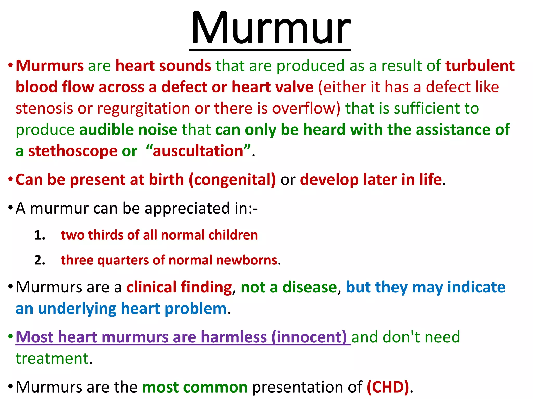
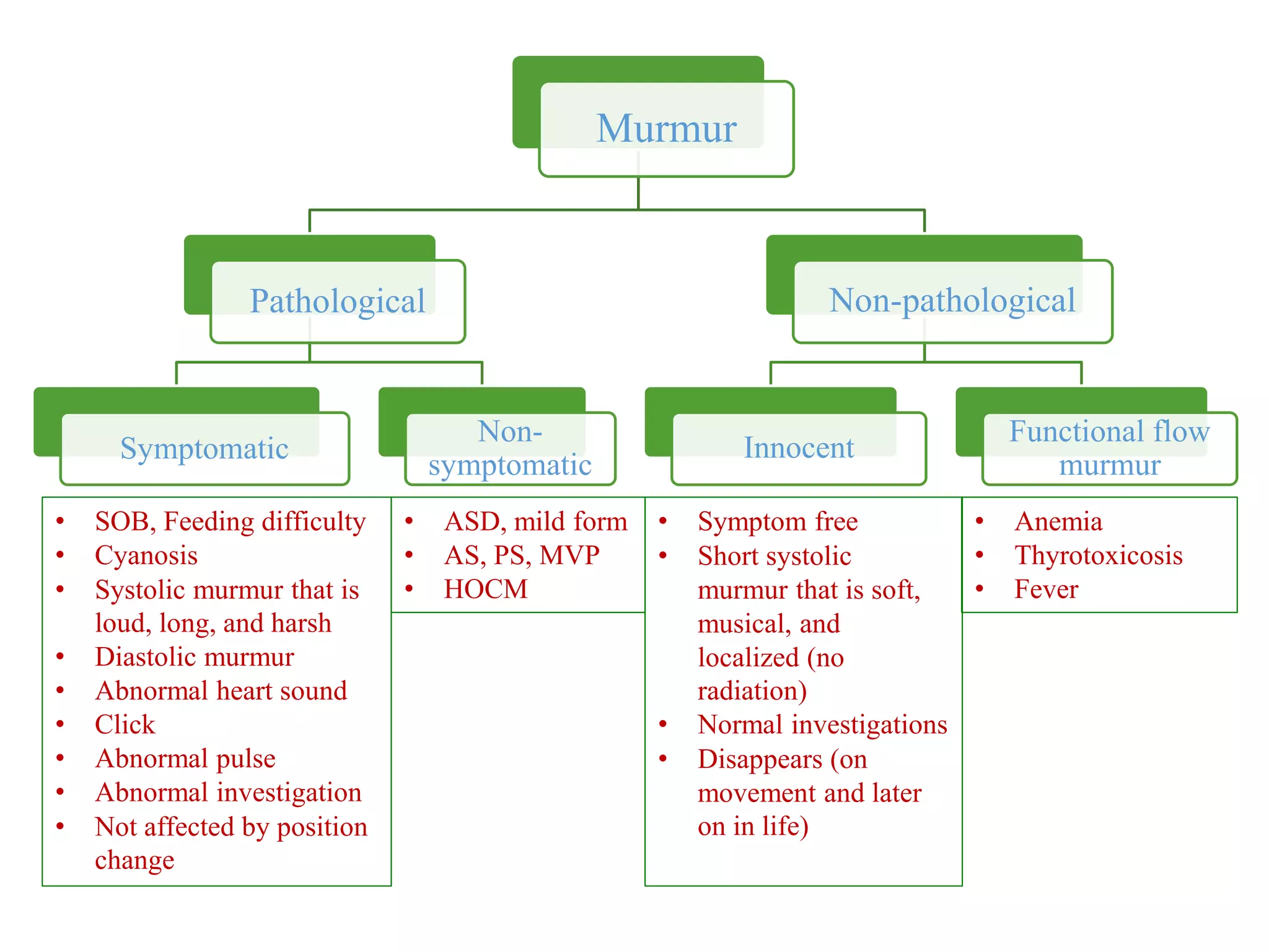
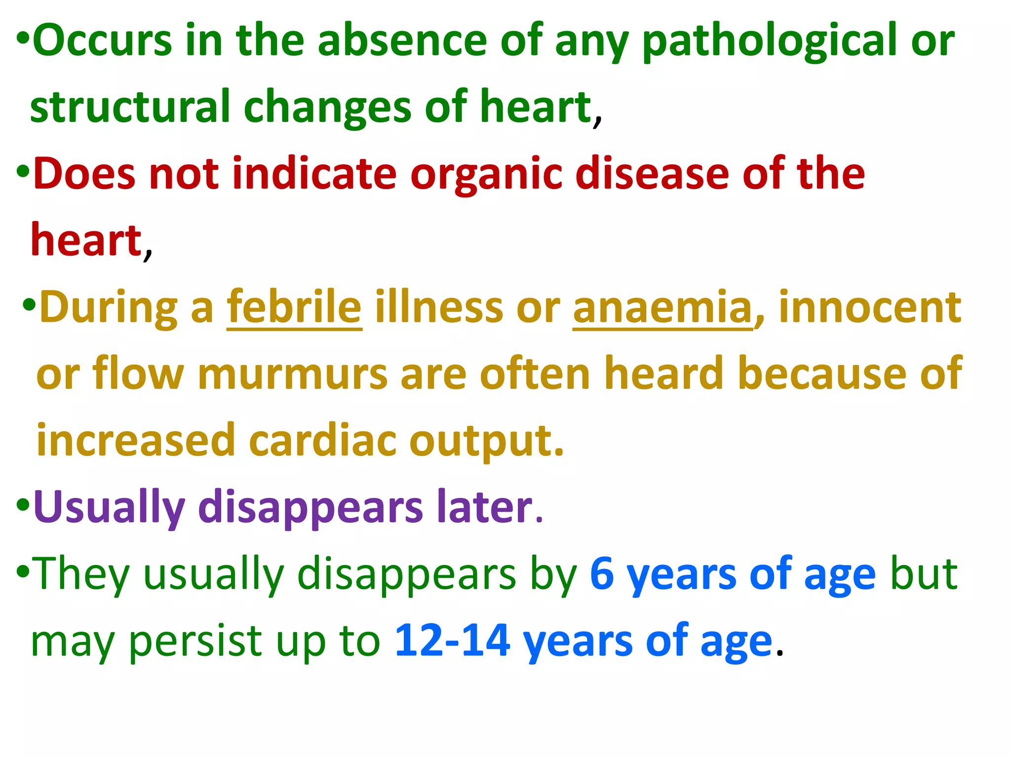
This document discusses the approach to murmurs in pediatrics. It defines a murmur as a heart sound produced by turbulent blood flow across a heart valve or defect. Murmurs can be innocent, pathological, or symptomatic. Innocent murmurs are harmless and common in children. Pathological murmurs indicate an underlying structural issue. Symptomatic murmurs cause issues like shortness of breath. The document outlines characteristics of different types of murmurs and provides guidance on evaluating a child presenting with a murmur through history, physical exam, and potential further workup.
This document discusses various types of arrhythmias that can occur in children. It begins by describing the normal electrical conduction system of the heart and then discusses different types of tachyarrhythmias and bradyarrhythmias. Common pediatric tachyarrhythmias mentioned include supraventricular tachycardia, atrial flutter, atrial fibrillation, ventricular tachycardia, and ventricular fibrillation. Long QT syndrome is also summarized. Treatment options for unstable and stable rhythms are provided.
This document discusses approaches to diagnosing and treating hypertension in children. It defines hypertension and outlines stages of severity. In infants and young children, hypertension is usually secondary to an underlying condition, while adolescents can develop primary or essential hypertension. Evaluation involves measuring blood pressure properly, considering causes of secondary hypertension, assessing for target organ damage like left ventricular hypertrophy, and determining if hypertension is primary or secondary. Treatment involves lifestyle changes, weight management if overweight, and potentially medications to lower blood pressure below guidelines.
This document contains an OSCE (Objective Structured Clinical Examination) practice exam for pediatrics. It includes 10 multiple choice matching questions that pair drugs used in pregnancy with their expected adverse effects on the fetus. It also includes several short clinical vignettes followed by 5 questions each. The vignettes cover topics like interpreting an ABG result, identifying sickle cell anemia from a peripheral smear, making a diagnosis of retropharyngeal abscess from presented symptoms, and more. The goal of the summary is to provide a high-level overview of the content and focus of the practice exam.
This document defines myocarditis as inflammation of the myocardium, outlines its main etiologies as viral and bacterial infections, and describes its pathogenesis. Signs and symptoms range from asymptomatic to cardiogenic shock. Diagnosis involves ECG, chest X-ray, echocardiogram, and endomyocardial biopsy. Treatment focuses on supportive care and conventional heart failure therapies. Prognosis depends on age, with higher mortality in newborns and potential recovery of function in children and adolescents.
The plantar reflex is an important superficial reflex that involves polysynaptic pathways. A normal plantar reflex results in flexion of the toes when the sole is scratched, while an extensor plantar response (Babinski's sign) involves dorsiflexion of the great toe and fanning of the other toes and suggests corticospinal tract dysfunction. There are several methods to elicit the plantar reflex and variations in responses provide information about neurological conditions.
A 15-year-old male presents with concerns of short stature and delayed puberty. Differential diagnoses include gonadotrophin deficiency, gonadal failure, and constitutional delay of growth and puberty. Physical exam and bone age assessment support a diagnosis of constitutional delay of growth and puberty, which is a condition of temporary short stature and delayed puberty but normal expected progression and attainment of full adult height. Reassurance and monitoring are the typical management approach.
Pediatric Acute Liver Failure (PALF) is defined as evidence of liver dysfunction within 8 weeks of symptoms onset in children, with uncorrectable coagulopathy and no evidence of chronic liver disease. Common etiologies include viral hepatitis, drugs, and other metabolic causes. Diagnostic workup involves general and etiology-specific tests. Key parameters to monitor include encephalopathy grade, coagulopathy, electrolytes, and complications. Treatment focuses on supportive care, complication management, and liver transplantation if indicated based on severity scores. Prognosis depends on etiology and degree of encephalopathy.
Croup is a common respiratory illness in young children caused by viruses such as parainfluenza. It causes barking cough, hoarseness, and stridor. Symptoms typically worsen at night. Diagnosis is clinical based on symptoms and appearance of the steeple sign on chest x-ray. Treatment involves corticosteroids which reduce symptoms, and nebulized epinephrine for more severe cases. Most children recover without complications, though a small percentage require hospitalization for respiratory support.
This document discusses different types of heart murmurs, including innocent or benign murmurs versus pathological murmurs. It provides details on specific murmurs such as Still's murmur, pulmonary flow murmur, physiological pulmonary flow murmur in neonates, carotid bruit, and venous hum. Characteristics of different systolic, diastolic, and continuous murmurs are outlined. Nada's criteria for diagnosing the presence of heart disease is also summarized.
Approach to cardiac murmurs and cardiac examination in childrenVarsha Shah
Cardiovascular examination in children for MBBS undergraduate, Residents, Trainees, pediatricians, GP, family physicians, nursing , dental, allied health students
This document discusses supraventricular tachycardia (SVT) in pediatric patients. SVT is the most common abnormal heart rhythm seen in children and the most common arrhythmia requiring treatment. It is usually caused by re-entry mechanisms involving an accessory pathway or the atrioventricular node. Diagnosis involves obtaining an electrocardiogram during episodes to identify P wave patterns. Treatment options include vagal maneuvers, medications like adenosine, calcium channel blockers, or beta blockers, and cardioversion. Radiofrequency ablation can provide a cure for refractory or recurrent cases. Proper diagnosis of the underlying SVT mechanism guides selection of the most appropriate treatment approach.
history and examination in pediatric CVSRaghav Kakar
This document provides guidance on performing a thorough history and physical examination for pediatric patients with suspected cardiovascular disease. Key aspects to assess include symptoms, timing of onset, family history, pre/postnatal history, examination of pulse, blood pressure, jugular venous pressure, precordial examination including auscultation of heart sounds and murmurs. Specific congenital heart defects should be considered based on findings. Investigations are guided by physical exam. A complete cardiovascular exam is essential for accurate diagnosis of heart disease in children.
Basic approach on short stature in childrenAzad Haleem
This document provides an overview of short stature, including definitions, types, diagnostic principles, causes, and management. It defines short stature as height below the 3rd percentile and discusses types such as familial short stature. Diagnosis involves accurate height measurements, bone age assessment, mid-parental height comparison, and medical investigations. Causes include growth hormone deficiency, Turner syndrome, and small size at birth. Management consists of dietary counseling, growth hormone injections, and limb lengthening procedures depending on the underlying cause.
Approach to a child with HepatosplenomegalySunil Agrawal
This document discusses hepatosplenomegaly, or the enlargement of the liver and spleen. It begins with an introduction and overview of hepatomegaly and splenomegaly. It then covers the various causes of hepatosplenomegaly including infections, hematological disorders, vascular congestion, tumors and infiltrations, storage disorders, and miscellaneous causes. The document provides details on evaluating a patient's history, physical examination findings, investigations, and treatment strategies for hepatosplenomegaly in both children and neonates. It concludes with references for further information.
Coma is defined as an unresponsive state with closed eyes lasting less than 24 hours that requires medical intervention. Common causes of pediatric coma include trauma, drowning, infection, metabolic disorders, and lack of oxygen or blood flow to the brain. The initial priorities for treatment are stabilizing breathing, circulation, blood sugar, and other vital functions, as well as identifying and treating any underlying medical issues causing the coma. Outcomes depend on the underlying cause, with complete recovery more likely after toxic or metabolic comas, while severe injuries like trauma or hypoxia often result in long-term neurological impairments.
Central nervous system involvement is a common cause of hypotonia in infants. A thorough history and physical exam seeks to determine if the origin is central or peripheral. Key aspects of the exam include assessing for proximal versus distal weakness, deep tendon reflexes, and distribution of weakness. Investigations such as EMG, nerve conduction studies, muscle biopsy and genetic testing can help characterize disorders of the motor unit to establish a diagnosis. Narrowing the likely etiology is important to guide management and prognostic expectations.
Approach in children with Hepatosplenomegaly
To summarize the key points:
1. A full examination including inspection, palpation, percussion and auscultation of the abdomen should be performed to evaluate for hepatosplenomegaly.
2. Common causes include infections, hematological disorders, vascular congestion, tumors and infiltrations, and storage disorders.
3. Initial investigations should include a complete blood count, liver function tests, ultrasound and further testing based on history and exam findings.
4. Treatment is directed at the underlying cause and may include antibiotics for infections, chemotherapy for tumors, or management of metabolic disorders.
- Pulmonary artery hypertension (PAH) is defined as a mean pulmonary artery pressure of ≥25 mmHg at rest. It is characterized by pre-capillary pulmonary hypertension with a pulmonary wedge pressure <15 mmHg and a pulmonary vascular resistance >3 Wood units.
- The pathophysiology involves sustained vasoconstriction, vascular remodeling, in situ thrombosis, and increased arterial stiffness. Genetic factors like BMPR2 mutations also contribute to PAH development.
- Clinical features range from mild breathlessness to signs of right heart failure. Diagnostic tests include echocardiography, CT scans, V/Q scans, right heart catheterization and lab tests.
- Treatment involves oxygen therapy, diure
This document provides an overview of how to examine the cardiovascular system through inspection, palpation, percussion, and auscultation. Key steps include inspecting the chest shape and size, palpating the jugular venous pulse and apex beat, percussing the heart borders, and auscultating heart sounds and murmurs in various areas. Examination findings are described that may indicate conditions like cardiomegaly, valvular disorders, or pericardial issues. Grading scales help characterize murmur intensity. A thorough cardiovascular exam provides clues to underlying heart and vessel abnormalities.
Paraquat poisoning causes severe oxidative stress and multi-organ failure. Ingestion has a high fatality rate. It is rapidly absorbed and concentrated in lungs, liver, and kidneys, causing cellular damage through redox cycling. Clinical features include oropharyngeal burns, respiratory distress, and acute kidney injury. Diagnosis is confirmed by positive urine dithionite test showing blue color change. Prognosis is poor for those with serum paraquat levels above the Sipp score threshold or rapidly increasing creatinine. Early extracorporeal removal within 4 hours may help severe cases, but long-term outcomes are generally poor with progressive lung fibrosis. Management focuses on supportive care, though antioxidants have been tried with
Human milk fortifiers are products that can be added to expressed breast milk to increase its nutritional content for premature infants. There are three main approaches to fortification - standard fixed dosage, adjustable based on blood urea nitrogen levels, and targeted fortification using human milk analysis. Fortifiers provide additional protein, calories, and minerals to help premature infants achieve adequate growth. While fortification benefits growth, high osmolality from fortifiers can cause feed intolerance and risks like necrotizing enterocolitis. Careful monitoring of infants on fortified breast milk is needed to optimize nutrition and growth.
1. The document provides guidance on evaluating and diagnosing anemia in children. It outlines key signs, symptoms, and pointers that suggest a child may have anemia.
2. Laboratory tests that can help determine the severity and type of anemia include complete blood count, hematocrit, reticulocyte count, blood indices, and peripheral smear.
3. A thorough history, physical exam, and lab work are needed to assess if a child is anemic, determine the severity, and identify the potential cause and type, such as blood loss, decreased red blood cell production, or increased red blood cell destruction.
This document provides an overview of the pediatric examination process. It discusses examining various body systems including:
1. Mental status and neurological assessment including cranial nerves, motor and sensory systems, and reflexes.
2. Assessment of consciousness, behavior, intelligence, memory, and speech.
3. Evaluation of muscle tone, power, involuntary movements, and coordination.
4. Sensory testing including superficial sensations, deep sensations, and cortical sensations.
5. Assessment of various reflexes including superficial, deep, and visceral reflexes.
The summary outlines the key areas addressed in a comprehensive pediatric examination.
Approach to child with generalized body swellingElhadi Hajow
Edema is characterized by swelling caused by excess fluid in the interstitial tissue. It can be localized or generalized. Common causes include cardiac, renal, or hepatic disease which decrease plasma oncotic pressure allowing fluid shift from vessels into tissue. A thorough history, physical exam, and lab tests are needed to determine the underlying cause and guide treatment such as diuretics, dietary changes, or treating the primary disease.
Approach to respiratory distress in childrenWasim Akram
This document provides an overview of pediatric respiratory emergencies. It begins with an introduction to the approach and assessment of a child presenting with breathing difficulties. It then covers the grading of respiratory distress, features of respiratory failure, and the pathophysiology of increased airway resistance and edema. The document further discusses the pathophysiologic approach to various clinical conditions causing respiratory distress. It provides guidance on the initial assessment and immediate care of the child, including airway management, oxygen delivery, circulation support, and diagnostic evaluation. Specific conditions addressed include upper airway obstruction, pneumonia, and wheezing.
The document provides information on assessing cardiovascular health. It defines key terms like systolic and diastolic blood pressure. It describes how to inspect the eyes, skin, chest and edema during assessment. Methods of assessment include measuring blood pressure, taking a health history, and auscultating the heart to identify sounds like S1, S2, murmurs or gallops. A thorough assessment can help identify risk factors for cardiovascular disease.
This document discusses functional or innocent heart murmurs that occur in the absence of cardiac abnormalities. It describes several types of common innocent murmurs including Still's murmur, pulmonary flow murmurs in childhood and infancy, and the venous hum. Still's murmur is the most common, heard as a low frequency systolic murmur. Pulmonary flow murmurs are often heard in adolescents and can increase with fever, anemia or pregnancy. The venous hum is continuous rather than systolic and will cease with neck vein compression. In general, innocent murmurs have normal heart sounds and exam findings.
This document provides an overview of techniques for examining the cardiovascular system in dogs, including:
1. Physical examination techniques like auscultation of heart sounds and palpation of the chest wall to detect murmurs, thrills, and abnormalities.
2. Diagnostic imaging tools like thoracic radiography to evaluate heart size and shape, and look for signs of pulmonary edema or effusion.
3. Electrocardiography to analyze heart rate, rhythm, and intervals to detect arrhythmias or chamber enlargement.
The document describes what each exam can reveal and how findings are interpreted to evaluate for heart disease and abnormalities.
This document discusses continuous murmurs, which are murmurs that begin in systole and continue uninterrupted through diastole. The main causes of continuous murmurs are high to low pressure shunts, such as a patent ductus arteriosus (PDA) or ruptured sinus of valsalva. Continuous murmurs can also be caused by rapid blood flow, such as in hyperthyroidism. The document describes the characteristics, locations, and distinguishing features of continuous murmurs from various underlying conditions.
The document discusses heart sounds and murmurs. It describes the normal first and second heart sounds and variations caused by different cardiac conditions. Abnormal heart sounds like S3, S4, clicks, and pericardial rubs are also outlined. Heart murmurs are classified based on timing, shape, location, intensity, pitch, quality, and radiation. Common causes of systolic, diastolic, and continuous murmurs are provided. Interventions that can change the characteristics of murmurs are summarized.
This document provides information on cardiovascular history taking and physical examination. It discusses important symptoms of heart disease like dyspnea, palpitations, edema, and chest pain. It also outlines the steps for examining arterial pulses, blood pressure, jugular venous pressure, auscultation of heart sounds, and palpation of the precordium. The physical exam aims to evaluate symptoms, risk factors, and detect any abnormalities that could indicate cardiac issues.
This document provides details on performing a cardiovascular examination, including inspection, palpation, percussion, and auscultation. It describes how to evaluate heart sounds and murmurs through auscultation of specific areas of the heart. Key areas covered include the four heart sounds (S1, S2, S3, S4), splitting of S1 and S2, abnormalities in intensity and timing of heart sounds, systolic and diastolic murmurs based on timing and cause, and grading murmurs based on intensity among other characteristics. The goal is to identify any abnormalities that could indicate cardiovascular diseases or conditions.
The document provides information about cardiac auscultation and heart sounds. It discusses the anatomy and function of the heart chambers and valves. Four main heart sounds (S1, S2, S3, S4) are described in detail, including their locations, timing in the cardiac cycle, and pathological variations. Additional extra heart sounds like clicks, murmurs and gallops are also outlined. The importance of assessing six characteristics of heart sounds during auscultation is highlighted. Instructions are given on performing a cardiac exam and auscultating the heart in multiple positions and with different parts of the stethoscope.
This document provides information on assessing the cardiovascular system, including:
- The anatomy of the heart with descriptions of the atria, ventricles, and major vessels.
- Subjective data to collect includes risk factors, symptoms, exercise habits, and pain characteristics.
- Inspection focuses on pulsations, retractions, and apical pulse location.
- Palpation locates pulsations and feels for thrills or abnormalities.
- Auscultation assesses heart sounds, murmurs, and extra sounds at various locations.
- Special maneuvers check for deep phlebitis and signs of arterial/venous insufficiency.
This document discusses heart sounds and murmurs. It describes the four main heart sounds: S1 occurs with mitral/tricuspid closure; S2 with aortic/pulmonic closure; S3 is an early sign of congestive heart failure; S4 indicates pulmonary/aortic stenosis or hypertension. There are three factors that can cause murmurs: high flow, forward flow through a constricted opening, or regurgitant backward flow. Murmurs are classified by timing, loudness, pitch, and location of greatest intensity. Common murmurs include aortic stenosis, mitral regurgitation, pulmonary stenosis, and ventricular septal defect.
Heart sounds are produced by the mechanical activities of the heart during each cardiac cycle, such as the closure of heart valves and the flow of blood through the chambers. The four main heart sounds are the first, second, third, and fourth heart sounds. The first and second heart sounds are the loudest and most commonly heard using a stethoscope. Abnormal heart sounds called murmurs can indicate underlying heart valve problems or other cardiac issues.
Normal and abnormal Heart sounds (Murmurs).pptx
Auscultation of heart sounds
How murmurs are produced
physiology of murmurs
Classification and types of murmurs
causes of murmurs
Unit III. Cardiovascular Disorders B.pptxSani191640
This document provides an outline of disorders of the cardiovascular system (CVS). It begins with an introduction and anatomy/physiology review. It then covers disorders in three sections: disorders of the heart like arrhythmias and coronary heart disease; vascular disorders like DVT and varicose veins; and hematological disorders like anemias and leukemias. Common diagnostic procedures and physical exam techniques are also discussed, including assessing heart sounds, murmurs, and jugular vein pressure. The document provides a comprehensive overview of cardiovascular conditions and their evaluation.
Systolic murmurs can occur in various parts of the cardiac cycle. Ejection systolic murmurs are most common, occurring after some time from S1 and peaking in mid-systole or later. Causes include ventricular outflow obstruction, dilation of the aorta/pulmonary trunk, or accelerated flow. Pan systolic murmurs occur throughout systole due to pressure gradients across valves. Mitral regurgitation and ventricular septal defects cause holosystolic murmurs. Early systolic murmurs begin with S1 and peak in early systole, often caused by small VSDs or innocent murmurs. Late systolic murmurs in mitral valve prolapse occur as leaflets overshoot in late syst
Blood pressure is measured by a sphygmomanometer and stethoscope. It involves two readings: systolic (top number) which is the pressure when the heart contracts, and diastolic (bottom number) which is the pressure when the heart relaxes. Sounds known as Korotkoff sounds are used to determine these readings as the cuff is deflated. Blood pressure is written as the systolic over the diastolic pressure in mmHg and is influenced by factors like age, stress, and medications. Abnormal blood pressures include hypertension (high) and hypotension (low).
The document provides guidance on assessing cardiovascular function through a physical exam. It outlines steps to take a health history, examine vital signs, listen to heart and lung sounds, check the extremities, neck, abdomen, and perform diagnostic tests. The physical exam aims to evaluate symptoms, identify abnormalities, and detect any risks for conditions like heart disease.
1. Aortic stenosis can be caused by rheumatic heart disease, congenital abnormalities of the aortic valve, or age-related degeneration and calcification of the valve.
2. Obstructed left ventricular outflow due to aortic stenosis leads to increased pressure and compensatory hypertrophy of the left ventricle. While cardiac output is maintained at rest, it often fails to rise normally during exertion in severe aortic stenosis.
3. Symptoms of aortic stenosis include angina, syncope, exertional dyspnea, and heart failure, which typically appear when the aortic orifice is reduced to one third of its normal size. Valve replacement surgery is
Similar to Approach to murmur in Paediatrics.pptx (20)
A 3-year-old girl has had loose stools for 2 months that often contain undigested food. She is otherwise well and thriving. The most probable diagnosis is chronic non-specific diarrhea (toddler's diarrhea). Management includes decreasing fluid intake, especially of fruit juice, providing high-fat foods to slow gastric emptying, and increasing fiber intake through bulking agents. Pharmacologic intervention is rarely required as symptoms usually resolve spontaneously by age 3-4 years.
The document discusses infant feeding and nutrition. It covers various topics including the types and definitions of breastfeeding, the physiology of lactation, problems associated with breastfeeding, infant growth phases and their energy requirements, and the importance of proper nutrition. The key components of human milk are discussed, including fat, proteins, carbohydrates, oligosaccharides, prebiotics and probiotics. Guidelines around establishing and maintaining breastfeeding are provided. Common breastfeeding and infant feeding problems are also outlined.
Postpartum hemorrhage (PPH) is excessive bleeding after childbirth, defined as blood loss over 500 ml for vaginal births or 1000 ml for C-sections. The main causes of PPH are uterine atony (failure of the uterus to contract), retained placenta, and trauma to the genital tract. Management involves bimanual uterine massage, uterotonic drugs, vaginal packing, balloon tamponade, and in severe cases surgical interventions like B-Lynch sutures or hysterectomy.
This document discusses the use of various imaging modalities for evaluating neck masses. Ultrasound is useful for differentiating cystic from solid lesions and assessing lymph node size and vascularity. CT provides details of soft tissues and their relationships. MRI is good for lesion detection and involvement of nearby structures but has limitations for nodal assessment. PET/CT is excellent for staging lymphoma and detecting unknown primary cancers. Biopsy is used when malignancy is suspected. The approach depends on whether the mass is in a child or adult, with ultrasound often the initial study. Location provides clues for cystic lesions. Features help characterize solid lesions and lymph nodes. Further tests are guided by ultrasound findings.
This document provides information about fetal cardiotocography (CTG), including:
1. CTG can be performed from 28 weeks of gestation as that is when the fetal autonomic nervous system is mature.
2. Normal CTG findings include a baseline heart rate between 110-160 bpm, variability between 5-25 bpm, and an absence of or early decelerations with at least 2 accelerations in 20 minutes.
3. Abnormal findings include bradycardia (<110 bpm), tachycardia (>160 bpm), decreased variability (<5 bpm), and late or variable decelerations which can indicate fetal hypoxia or distress.
This document discusses the partogram, a tool for recording the progress of labor. It explains that the partogram graphs cervical dilation, fetal descent, and uterine contractions on a chart to allow healthcare providers to monitor labor and identify complications early. The document outlines the components recorded on a partogram, including fetal heart rate, amniotic fluid, maternal vital signs, and medications. It describes how to interpret the alert and action lines plotted on the partogram to determine if labor is progressing normally or requires intervention. The partogram is an important tool that facilitates continuity of care during labor and allows early detection of problems like prolonged or obstructed labor.
Gestational trophoblastic disease (GTD) is a spectrum of tumors caused by abnormal proliferation of placental tissue. It includes hydatidiform moles (complete and partial), which are usually benign, as well as gestational trophoblastic neoplasms like invasive moles, choriocarcinoma, and placental site trophoblastic tumors, which are malignant. GTD is diagnosed using clinical features, ultrasound findings, and elevated human chorionic gonadotropin levels. Treatment may involve D&C for molar pregnancies as well as chemotherapy for malignant or persistent cases. Long term follow up is important to monitor for recurrence or progression to gestational trophoblastic neoplasia due to the
Gametogenesis conversion of germ cells into male and female gametes.pptJwan AlSofi
Gametogenesis refers to the formation of male and female gametes. It begins with primordial germ cells that migrate to the developing gonads. Oogenesis involves the formation of ova through meiotic divisions in females, arresting in prophase I until puberty. Spermatogenesis is the formation of sperm in males through mitotic and meiotic divisions of spermatogonia into spermatids. Spermiogenesis then transforms spermatids into mature spermatozoa through nuclear condensation and tail formation. Abnormal gametes can form with extra nuclei or morphological defects preventing fertilization.
Development of the male& female genital system.pptxJwan AlSofi
The document summarizes the development of the male and female genital systems from an indifferent stage. It describes how in males, the presence of SRY leads testes to develop from indifferent gonads, while in females without SRY ovaries develop. It outlines the development of testes, ovaries, male ducts including epididymis and vas deferens, and female ducts including uterus and vagina from indifferent ducts. External genitalia also develop differently in males under testosterone versus females.
First week of development: Ovulation to Implantation Jwan AlSofi
The document summarizes key aspects of ovulation, fertilization, and early embryonic development. It describes the ovarian and menstrual cycles controlled by hormones like FSH and LH. Ovulation occurs mid-cycle due to an LH surge, releasing an egg. Sperm travel through the reproductive tract while undergoing capacitation. Fertilization typically occurs in the fallopian tubes, involving penetration of the egg's layers and fusion of gametes. This activates the egg and forms pronuclei, leading to cell division and pregnancy if implantation occurs. Otherwise, the corpus luteum regresses and menstruation begins.
Approach to patient with spinal cord lesions & diseases
Localize spinal cord lesions
Determining the Level of the Lesion in Myelopathy
Diseases of spinal cord
Multiple sclerosis is a chronic disease characterized by inflammation, demyelination, and gliosis in the central nervous system. It affects around 5 million people worldwide. The cause is unknown but involves genetic and environmental factors. Symptoms vary widely and can include sensory disturbances, motor symptoms, visual problems, ataxia, and cognitive impairment. Diagnosis involves demonstrating dissemination of lesions in the CNS over time via MRI imaging or evoked potentials testing, and sometimes analysis of cerebrospinal fluid. There are several disease courses including relapsing-remitting MS, primary progressive MS, and secondary progressive MS. Management aims to reduce inflammation and disability progression.
This is a comprehensive approach to a hypertensive patient presenting to the emergency department.
Discussing:-
- Hypertensive emergency
- Hypertensive Urgency
- Hypertensive Crisis
- Hypertensive encephalopathy and retinopathy
- Accelerated Hypertension
- Malignant hypertension
1. Short stature can be caused by familial, constitutional, or pathological factors. Familial short stature runs in families while constitutional short stature involves delayed puberty.
2. Pathological short stature can be disproportionate involving abnormal limb ratios, or proportionate involving prenatal issues like IUGR or postnatal diseases/nutritional disorders.
3. Evaluating a short child involves assessing growth charts, growth velocity, bone age, family history, and screening tests to classify the cause of short stature.
Headache is a common symptom in children and adolescents, with up to 75% experiencing a significant headache by age 15. Headaches can be primary, such as migraines or tension-type headaches, or secondary to other conditions such as viral infections. A thorough history and physical exam are usually sufficient for diagnosis, though imaging may be required if symptoms suggest increased intracranial pressure. Treatment involves acute medication to stop attacks as well as preventive medication and lifestyle modifications if headaches are frequent or disabling.
Neonatal seizures are the most common neurological emergency in newborns. The majority occur within the first day of life, and hypoxic ischemic encephalopathy is the most common cause, especially in term infants. In preterm infants, cerebral vascular events are more often the cause. Neonatal seizures are usually focal and often have identifiable underlying causes, unlike seizures in older children which are often idiophenic. The prognosis depends on the underlying etiology, with hypoxic ischemic encephalopathy carrying the worst prognosis. Phenobarbital remains the first-line treatment, though multiple anticonvulsants may be needed to control seizures.
this is a complete discussion and an approach to a child with febrile seizure / convulsion.
It contains:-
Case scenario
Causes of Seizures in the setting of fever
Definition of Febrile Seizure
Age of Occurrence
Types of Febrile Convulsions
Risks of Recurrent Febrile Seizures
Risk For Developing Epilepsy After Febrile Seizures
Workup for Febrile Seizure
Red Flags in Febrile Seizures
Treatment
Prognosis
Approach to Syncope in Children (Pediatric Syncope).pptxJwan AlSofi
Approach to Syncope in Children (Pediatric Syncope), includes:-
Introduction
Differential diagnosis of syncope
Syncope vs vertigo vs Presyncope vs light-headedness.
Comparison of Clinical Features of Syncope and Seizures
Neurocardiogenic (Vasovagal) syncope
MECHANISMS and Causes of Syncope
Cardiac causes of syncope
Life-threatening causes of syncope
Red Flags in Evaluation of Patients With Syncope
Non-cardiac causes of loss of consciousness.
Noncardiac Causes of Syncope
Differentiating Features for Causes of Syncope
EVALUATION of syncope:- History, Examination,Treatment.
Summary
Co-Chairs, Val J. Lowe, MD, and Cyrus A. Raji, MD, PhD, prepared useful Practice Aids pertaining to Alzheimer’s disease for this CME/AAPA activity titled “Alzheimer’s Disease Case Conference: Gearing Up for the Expanding Role of Neuroradiology in Diagnosis and Treatment.” For the full presentation, downloadable Practice Aids, and complete CME/AAPA information, and to apply for credit, please visit us at https://bit.ly/3PvVY25. CME/AAPA credit will be available until June 28, 2025.
Breast cancer: Post menopausal endocrine therapyDr. Sumit KUMAR
Breast cancer in postmenopausal women with hormone receptor-positive (HR+) status is a common and complex condition that necessitates a multifaceted approach to management. HR+ breast cancer means that the cancer cells grow in response to hormones such as estrogen and progesterone. This subtype is prevalent among postmenopausal women and typically exhibits a more indolent course compared to other forms of breast cancer, which allows for a variety of treatment options.
Diagnosis and Staging
The diagnosis of HR+ breast cancer begins with clinical evaluation, imaging, and biopsy. Imaging modalities such as mammography, ultrasound, and MRI help in assessing the extent of the disease. Histopathological examination and immunohistochemical staining of the biopsy sample confirm the diagnosis and hormone receptor status by identifying the presence of estrogen receptors (ER) and progesterone receptors (PR) on the tumor cells.
Staging involves determining the size of the tumor (T), the involvement of regional lymph nodes (N), and the presence of distant metastasis (M). The American Joint Committee on Cancer (AJCC) staging system is commonly used. Accurate staging is critical as it guides treatment decisions.
Treatment Options
Endocrine Therapy
Endocrine therapy is the cornerstone of treatment for HR+ breast cancer in postmenopausal women. The primary goal is to reduce the levels of estrogen or block its effects on cancer cells. Commonly used agents include:
Selective Estrogen Receptor Modulators (SERMs): Tamoxifen is a SERM that binds to estrogen receptors, blocking estrogen from stimulating breast cancer cells. It is effective but may have side effects such as increased risk of endometrial cancer and thromboembolic events.
Aromatase Inhibitors (AIs): These drugs, including anastrozole, letrozole, and exemestane, lower estrogen levels by inhibiting the aromatase enzyme, which converts androgens to estrogen in peripheral tissues. AIs are generally preferred in postmenopausal women due to their efficacy and safety profile compared to tamoxifen.
Selective Estrogen Receptor Downregulators (SERDs): Fulvestrant is a SERD that degrades estrogen receptors and is used in cases where resistance to other endocrine therapies develops.
Combination Therapies
Combining endocrine therapy with other treatments enhances efficacy. Examples include:
Endocrine Therapy with CDK4/6 Inhibitors: Palbociclib, ribociclib, and abemaciclib are CDK4/6 inhibitors that, when combined with endocrine therapy, significantly improve progression-free survival in advanced HR+ breast cancer.
Endocrine Therapy with mTOR Inhibitors: Everolimus, an mTOR inhibitor, can be added to endocrine therapy for patients who have developed resistance to aromatase inhibitors.
Chemotherapy
Chemotherapy is generally reserved for patients with high-risk features, such as large tumor size, high-grade histology, or extensive lymph node involvement. Regimens often include anthracyclines and taxanes.
Cell Therapy Expansion and Challenges in Autoimmune DiseaseHealth Advances
There is increasing confidence that cell therapies will soon play a role in the treatment of autoimmune disorders, but the extent of this impact remains to be seen. Early readouts on autologous CAR-Ts in lupus are encouraging, but manufacturing and cost limitations are likely to restrict access to highly refractory patients. Allogeneic CAR-Ts have the potential to broaden access to earlier lines of treatment due to their inherent cost benefits, however they will need to demonstrate comparable or improved efficacy to established modalities.
In addition to infrastructure and capacity constraints, CAR-Ts face a very different risk-benefit dynamic in autoimmune compared to oncology, highlighting the need for tolerable therapies with low adverse event risk. CAR-NK and Treg-based therapies are also being developed in certain autoimmune disorders and may demonstrate favorable safety profiles. Several novel non-cell therapies such as bispecific antibodies, nanobodies, and RNAi drugs, may also offer future alternative competitive solutions with variable value propositions.
Widespread adoption of cell therapies will not only require strong efficacy and safety data, but also adapted pricing and access strategies. At oncology-based price points, CAR-Ts are unlikely to achieve broad market access in autoimmune disorders, with eligible patient populations that are potentially orders of magnitude greater than the number of currently addressable cancer patients. Developers have made strides towards reducing cell therapy COGS while improving manufacturing efficiency, but payors will inevitably restrict access until more sustainable pricing is achieved.
Despite these headwinds, industry leaders and investors remain confident that cell therapies are poised to address significant unmet need in patients suffering from autoimmune disorders. However, the extent of this impact on the treatment landscape remains to be seen, as the industry rapidly approaches an inflection point.
Promoting Wellbeing - Applied Social Psychology - Psychology SuperNotesPsychoTech Services
A proprietary approach developed by bringing together the best of learning theories from Psychology, design principles from the world of visualization, and pedagogical methods from over a decade of training experience, that enables you to: Learn better, faster!
8 Surprising Reasons To Meditate 40 Minutes A Day That Can Change Your Life.pptxHolistified Wellness
We’re talking about Vedic Meditation, a form of meditation that has been around for at least 5,000 years. Back then, the people who lived in the Indus Valley, now known as India and Pakistan, practised meditation as a fundamental part of daily life. This knowledge that has given us yoga and Ayurveda, was known as Veda, hence the name Vedic. And though there are some written records, the practice has been passed down verbally from generation to generation.
Nano-gold for Cancer Therapy chemistry investigatory projectSIVAVINAYAKPK
chemistry investigatory project
The development of nanogold-based cancer therapy could revolutionize oncology by providing a more targeted, less invasive treatment option. This project contributes to the growing body of research aimed at harnessing nanotechnology for medical applications, paving the way for future clinical trials and potential commercial applications.
Cancer remains one of the leading causes of death worldwide, prompting the need for innovative treatment methods. Nanotechnology offers promising new approaches, including the use of gold nanoparticles (nanogold) for targeted cancer therapy. Nanogold particles possess unique physical and chemical properties that make them suitable for drug delivery, imaging, and photothermal therapy.
NAVIGATING THE HORIZONS OF TIME LAPSE EMBRYO MONITORING.pdfRahul Sen
Time-lapse embryo monitoring is an advanced imaging technique used in IVF to continuously observe embryo development. It captures high-resolution images at regular intervals, allowing embryologists to select the most viable embryos for transfer based on detailed growth patterns. This technology enhances embryo selection, potentially increasing pregnancy success rates.
Test bank for karp s cell and molecular biology 9th edition by gerald karp.pdfrightmanforbloodline
Test bank for karp s cell and molecular biology 9th edition by gerald karp.pdf
10 Benefits an EPCR Software should Bring to EMS Organizations Traumasoft LLC
The benefits of an ePCR solution should extend to the whole EMS organization, not just certain groups of people or certain departments. It should provide more than just a form for entering and a database for storing information. It should also include a workflow of how information is communicated, used and stored across the entire organization.
5-hydroxytryptamine or 5-HT or Serotonin is a neurotransmitter that serves a range of roles in the human body. It is sometimes referred to as the happy chemical since it promotes overall well-being and happiness.
It is mostly found in the brain, intestines, and blood platelets.
5-HT is utilised to transport messages between nerve cells, is known to be involved in smooth muscle contraction, and adds to overall well-being and pleasure, among other benefits. 5-HT regulates the body's sleep-wake cycles and internal clock by acting as a precursor to melatonin.
It is hypothesised to regulate hunger, emotions, motor, cognitive, and autonomic processes.
Summer is a time for fun in the sun, but the heat and humidity can also wreak havoc on your skin. From itchy rashes to unwanted pigmentation, several skin conditions become more prevalent during these warmer months.
2. Murmur
•Murmurs are heart sounds that are produced as a result of turbulent
blood flow across a defect or heart valve (either it has a defect like
stenosis or regurgitation or there is overflow) that is sufficient to
produce audible noise that can only be heard with the assistance of
a stethoscope or “auscultation”.
•Can be present at birth (congenital) or develop later in life.
•A murmur can be appreciated in:-
1. two thirds of all normal children
2. three quarters of normal newborns.
•Murmurs are a clinical finding, not a disease, but they may indicate
an underlying heart problem.
•Most heart murmurs are harmless (innocent) and don't need
treatment.
•Murmurs are the most common presentation of (CHD).
3. Murmur
Pathological
Symptomatic
Non-
symptomatic
Non-pathological
Innocent
Functional flow
murmur
• SOB, Feeding difficulty
• Cyanosis
• Systolic murmur that is
loud, long, and harsh
• Diastolic murmur
• Abnormal heart sound
• Click
• Abnormal pulse
• Abnormal investigation
• Not affected by position
change
• ASD, mild form
• AS, PS, MVP
• HOCM
• Symptom free
• Short systolic
murmur that is soft,
musical, and
localized (no
radiation)
• Normal investigations
• Disappears (on
movement and later
on in life)
• Anemia
• Thyrotoxicosis
• Fever
4. Clinical Features of murmurs:-
1. Timing
2. Duration
3. Character and pitch
4. Intensity
5. Location
6. Radiation
6. •Occurs in the absence of any pathological or
structural changes of heart,
•Does not indicate organic disease of the
heart,
•During a febrile illness or anaemia, innocent
or flow murmurs are often heard because of
increased cardiac output.
•Usually disappears later.
•They usually disappears by 6 years of age but
may persist up to 12-14 years of age.
7. Criteria of innocent murmur:-
1.Systolic murmur only, not diastolic
2.the quality of the sound - Soft blowing
3.a lack of significant radiation,
4. Left Sternal edge
5.a significant alteration in the intensity of the murmur with
positional changes
6.the cardiovascular history and examination are otherwise
normal.
8.
9.
10. Pulmonary flow murmur of newborn
(or Peripheral pulmonary stenosis or
Pulmonary branch murmur)
(newborn-6months).
- Consequence of flow turbulence made by normal blood flowing from (RV) to (PA)
- Mechanisms:-
1.the pulmonary arteries that had limited blood flow in the uterus, and are therefore
small.
2.increasing cardiac output associated with declining haemoglobin level after birth.
- Features of the murmur of peripheral pulmonary stenosis:-
- heard best in the pulmonary area,
- radiates along the pulmonary arteries (so the murmur can be heard along the axillae and
back- posterior lung fields). What this means is that because the pulmonary arteries go
to each lung, the murmur is often heard in the right and left lateral chest.
11.
12. - A soft systolic at the upper left sternal border
may be because of
1.normal flow across small pulmonary arteries (the
peripheral pulmonary flow murmur)
2.increased blood flow across normal pulmonary
arteries.- e,g, as in ASD.
3.Pulmonic stenosis
13. Still's murmur or Vibratory
murmur (3 - 6 years):-
- is the most common one.
- Due to vibrations in either the right or left ventricle, or ‘‘tendons’’ often
seen in the left ventricle.
- Best heard at the left middle-lower sternal border
- Are loudest when the patient is supine
- Get softer when the patient stands.
(the murmur in HCM, behaves just the opposite of a Still’s murmur- When
any patient stands, gravity takes blood to the lower extremities, and
therefore less blood is in the heart. With less blood filling the ventricle, the
walls of the ventricles get closer together, and in the case of hypertrophic
cardiomyopathy, the obstruction within the cavity of the left ventricle
worsens)
14. Venous hum (3 – 6 years ).
- Is caused by blood flow returning from the child’s head and flowing
from the superior vena cava to the right atrium.
- It is a blowing, continuous murmur, sounding like a soft hum during
both systole and diastole.
- heard at the base of the heart just below the clavicles, mainly on the
right side,
- Best heard while standing
- Disappears while the chest is flat.
- Changes with:-
- Moving the child’s head to either side
- child is lying down
- Light pressure to the right side of the neck, which temporarily stops blood flow through
the right jugular venous system.
15.
16.
17. Carotid bruit (any age).
- Occurs due to normal passing of blood from the
aorta to the carotid, heard best above the clavicle
- Hyperextend the shoulders murmur disappears
18.
19. The Aortic Outflow Murmur
• Heard in adolescents and young adults
• The murmur is usually grade one or two in intensity
• It is different from valvar aortic stenosis in that the
patients do not have an ejection click.
• The murmur is often heard in athletes, who typically
have a low resting heart rate and therefore a large
stroke volume of blood flowing in the left ventricular
outflow tract in systole.
• Standing a patient with hypertrophic cardiomyopathy
should increase the murmur, whereas standing the
patient with the aortic outflow murmur should result in
either a decrease in the murmur, or no significant
change.
20.
21.
22.
23.
24. Pathological Murmurs
•Are sounds produced by turbulent flow due to abnormal intra cardiac
or intravascular obstructions or connections.
•When one or more of the following are present, the murmur is more
likely pathologic and requires cardiac consultation:
1.Abnormal heart sounds S12
2.A systolic murmur that is loud ( grade 3/6 or with a thrill ) long in duration and
transmits well to other parts of the body, of harsh quality
3.holosystolic,
4.late systolic,
5.Diastolic,
6.continuous (except for the venous hum) murmurs
7.presence of a thrill are not normal.
8.Active precordium
9.Abnormal cardiac size or silhouette or abnormal pulmonary vascularity on
chest radiography
10.Abnormal ECG
11.The presence of symptoms, including failure to thrive or dysmorphic features,
12.Associated Cyanosis
25.
26. Innocent Pathological
Symptom free Symptomatic
Short systolic Pansystolic or diastolic
Short, soft, and musical Harsh quality and intensity >grade 3
Localized with no radiation Radiation and posterior propagation
Disappears with changing position Does not disappear
No thrill May be associated with thrill
Normal Investigations Often abnormal
27. Typically – no change with standing/positional changes
ATRIAL SEPTAL DEFECT:
• systolic ejection murmur radiating to axilla &
back
• fixed split second heart sound (S2)
AORTIC VALVE/ PULMONARY VALVE STENOSIS:
•harsh, higher pitched, systolic ejection murmur
•systolic “click”
•AS – radiation RUSB carotids, suprasternal notch thrill
•PS – radiation to axilla and back
Am I missing…
28. VENTRICULAR SEPTAL DEFECT:
• holosystolic murmur
• systolic regurgitant murmur (TR)
• harsh, higher pitched
• large VSDs may not have a loud murmur
PATENT DUCTUS ARTERIOSUS:
• continuous murmur
• left upper sternal border, left infraclavicular area
• murmur does not decrease with head position
changes
Am I missing…
29. HYPERTROPHIC CARDIOMYOPATHY:
• systolic ejection murmur at left sternal border
• does NOT decrease in intensity with standing
(innocent murmur should decrease with
standing)
• may increase in intensity with standing
• may decrease in intensity with squatting
Am I missing …
31. History:
• Pregnancy and birth history
• Intermittent nature
• Normal growth and development
• Negative family history
Physical Examination:
• Characteristic qualities of innocent murmur - practice
• Second heart sound:
o “physiologic splitting” inspiration- splits… expiration- single
o no increased intensity, pounding or loud
• No click, no thrill (grade IV/VI murmur)
• No suprasternal notch thrill
• Positional changes - supine and standing
Innocent Murmurs
What You Can Do
32. • Greater than grade III/VI (thrill)
• Holosystolic
• Diastolic
• Harsh
• Click
• Pulse abnormality
• Failure to thrive
• Significant family history
Murmurs -
When to be concerned?
34. 1. History:
• Feeding history, exercise intolerance. [HF. In <1-year exercise level
is obtained during feeding (an infant with HF can only take small
volumes of milk, develops SOB on sucking, and often perspires)]
• Heavy sweating with minimal or no exertion.
• Poor appetite and failure to grow normally (in infants). [indicates
heart failure (HF)]
• Chronic cough. [indicates lung congestion]
• Swelling or sudden weight gain. [Edema]
• Cyanosis or cyanotic spells ± squatting posture (indicates cyanotic
CHD, classically seen in tetralogy of Fallot).
• Chest pain, prolonged fever. [Endocarditis]
• Dizziness, fainting episodes.
35. • Feeding difficulty:
• When the mother complains that the baby is not able to take
feeds properly (either breast feed or bottle feed), becomes
breathless and has excessive sweating during feeding, the
physician should think of congestive heart failure (CHF) of any
cause.
• If the parents complain that the baby starts crying each time while
taking feeds and if the feed is stopped, feels comfortable, one
should think of a rare possibility of vascular ring malformation.
• Repeated respiratory infection:
• History of repeated cold and cough requiring admission to the
hospital should be noted.
• If the infant is having repeated attacks of breathlessness, rapid
breathing, cough, grunting sounds and restlessness (indicating
repeated lower respiratory tract infection) more than six times
per year, indicates high pulmonary flow due to significant left to
right shunt.
36. • History of blue discoloration of lips, nails especially on crying
indicates the possibility of cyanotic heart disease with decreased
pulmonary blood flow and right to left shunt.
• When the cyanotic infant lies calm and listless having less
physical activity it indicates cyanotic spell or low output state.
• History of squatting after exertion in a cyanosed child indicates:-
1. tetralogy of Fallot (TOF)
2. TOF like physiology and tricuspid atresia (TA).
• History of frequent palpitations in a cyanotic child, one should
think of Ebstein anomaly.
• History of syncope on mild to moderate exertion in an acyanotic
child indicates:-
1. severe aortic stenosis (AS),
2. hypertrophic cardiomyopathy,
3. severe pulmonary hypertension
4. congenitally corrected transposition of great arteries producing
significant bradycardia.
37. • Birth History.
• Was the baby term or preterm (structural abnormalities)?
• Was there asphyxia? (caused by a type of C.M., low O2 to the
heart),
• ask for prolonged labor, history of convulsion, and SGA
• Maternal complications:
• DM leads to HOCM.
• HTN leads to TGA.
• SLE lead to COMPLETE HEART BLOCK.
• TORCH: rubella leads to PDA.
• Drug history:
• Isotretinoin is teratogenic.
• Anti-convulsants.
• Aspirin.
• Is there a family history of congenital heart disease? [There
is a higher risk of heart defects in siblings of children with
congenital heart disease.]
38. 2. Examination
• Appearance
• Posture.
• Color.
• Dysmorphic features.
• Respiratory distress.
• Nutritional assessment.
• Edema.
• Clubbing.
• Vital signs:
• Tachycardia is a sign of cardiac failure.
• The character of the pulse can also give a clue to cardiac pathology.
• Palpate the femoral pulses, as in coarctation of the aorta they are absent or weak
and delayed compared with the radial pulse.
• Take the blood pressure, and if you suspect coarctation you need to do this in
both arms and legs. Normally it is 10-20mmHg higher in the legs.
39. • Other signs of heart failure:
- Tachypnoea, hepatomegaly, and crepitations in the
lungs are the major clinical manifestations of cardiac
failure in childhood.
- Peripheral oedema is rare.
- Cyanosis if present suggests reversal of shunt, urgent
investigation is required.
• Raised JVP.
• Growth parameters: Failure to thrive and poor
growth are important signs of cardiac failure in
childhood and are also important in monitoring
medical management.
43. 4. Investigations
• Chest X-ray:
• size: is not reliable at all in <1-year-olds, so send for echo. Because of thymus
shadow and the heart is initially horizontal then moves down. Cardiothoracic ratio
of up to 55% is normal in infants.
• shape:
• Boot shape: TOF
Truncus arteriosus
• Egg shape: TGA
• Snowman silhouette (double contour of the heart): TAPVR
• vascularity: vascular markings should not exceed 1/3 of the thoracic diameter
normally:
• Increased: plethoric
• Decreased: oligemic
• lung, thoracic abnormalities: rib notching → COA
TAPVR= Total anomalous pulmonary
venous return. Oxygen-rich blood from the
lungs goes to the right atrium instead of the
left atrium.
48. 1. ECG: Rate, Rhythm, axis, (P, QRS, T). gives further
information about ventricular and atrial hypertrophy.
2. Echo is important in evaluating cardiac structure and
performance, gradients across stenotic valves and the
direction of flow across a shunt:
• M-mode
• 2-dimensional
• Doppler
• transesophageal
3. Cardiac Catheterization: is now rarely required for
diagnosis.
4. Angiography (Angiocadiography).
5. MRI.
6. CT.
51. • Which one of the followings is not characteristic
of innocent murmur:
A. Symptom free
B. The murmur is soft in character
C. There is cardiomegaly on chest X-ray film
D. The murmur is a grade I systolic murmur
E. The murmur is heard at the left sternal edge
C.
There
is
cardiomegaly
on
chest
X-ray
film
52. • Alan, a 4-month-old boy, sees his general
practitioner for an ear infection. On listening to his
chest a heart murmur is heard. Which one of the
following features most suggests that it requires
further investigation? Select one answer only.
A. A thrill
B. Disappearance of murmur on lying flat
C. Murmur maximal at the left sternal edge
D. Sinus arrhythmia
E. Systolic murmur
A. A thrill
53. • Nada, a 5 month old female infant has a fever and
runny nose for 2 days. On examination she has a
fever of 38.3° C and a runny nose. Her tongue is
pink. Her breathing is normal. Pulse is 160
beats/min. Her heart sounds are normal but she
has a soft systolic murmur at the left sternal edge.
Pulses are normal. Diagnosis???
Innocent murmur