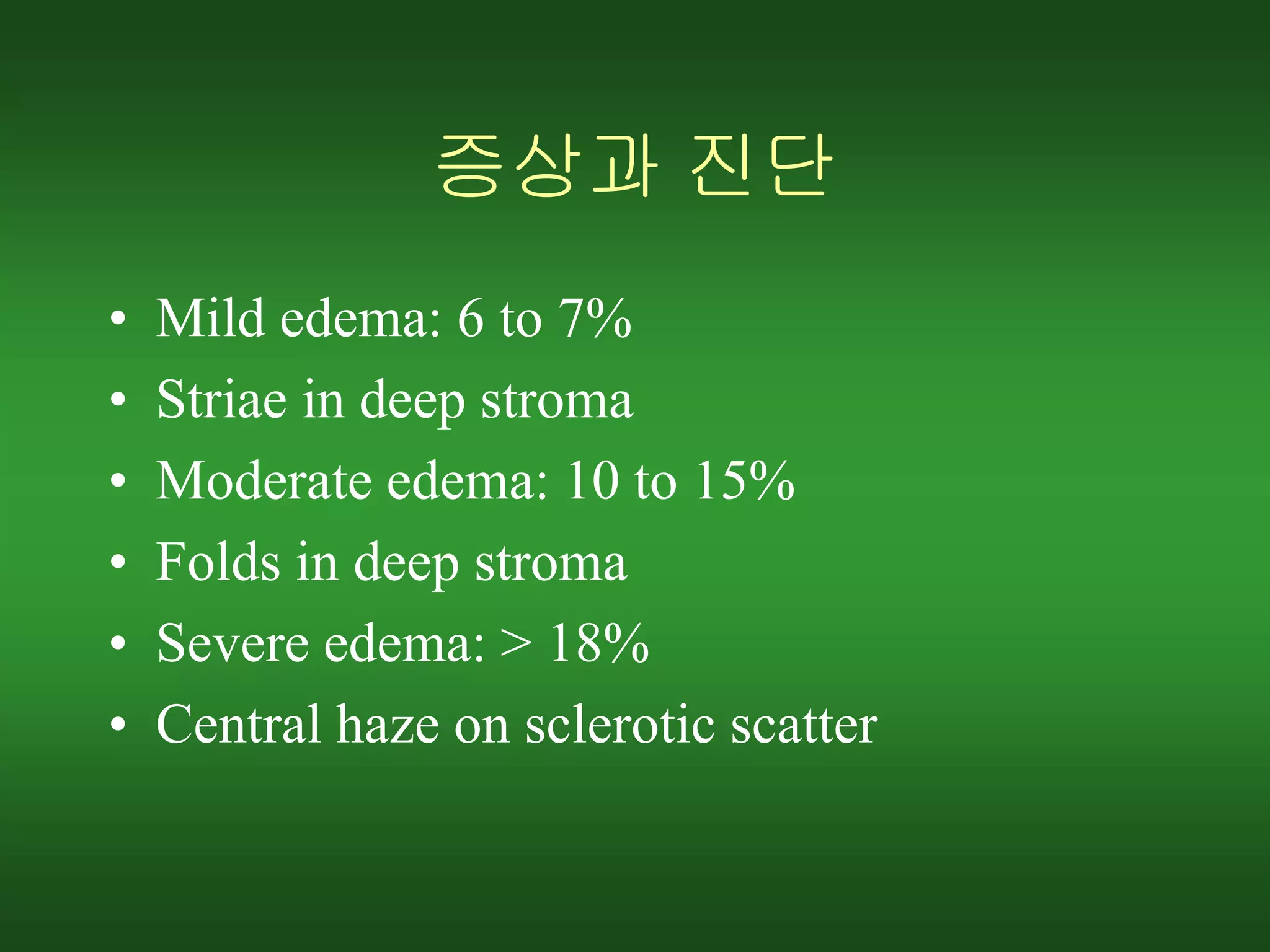
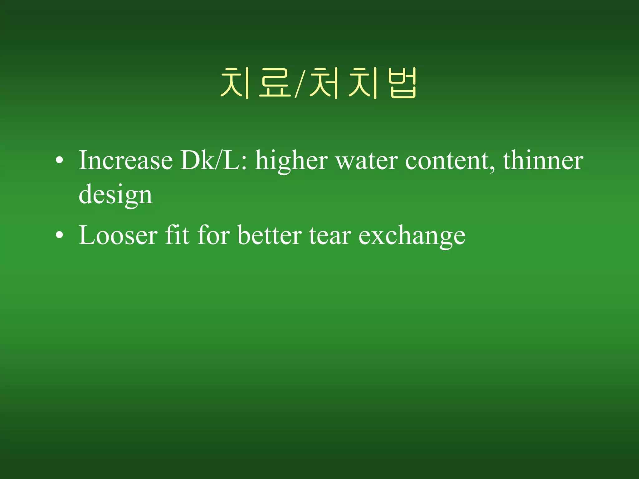
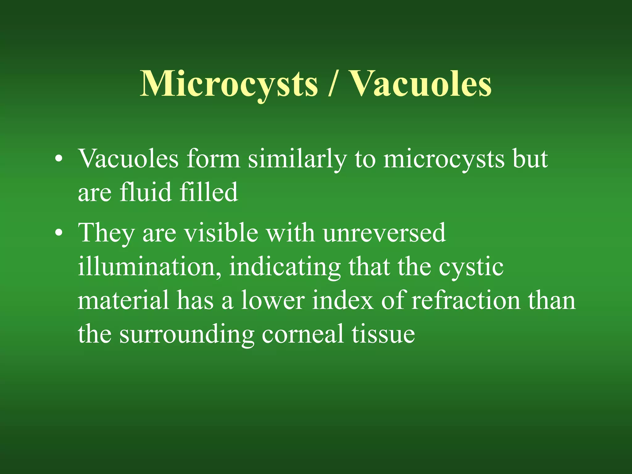
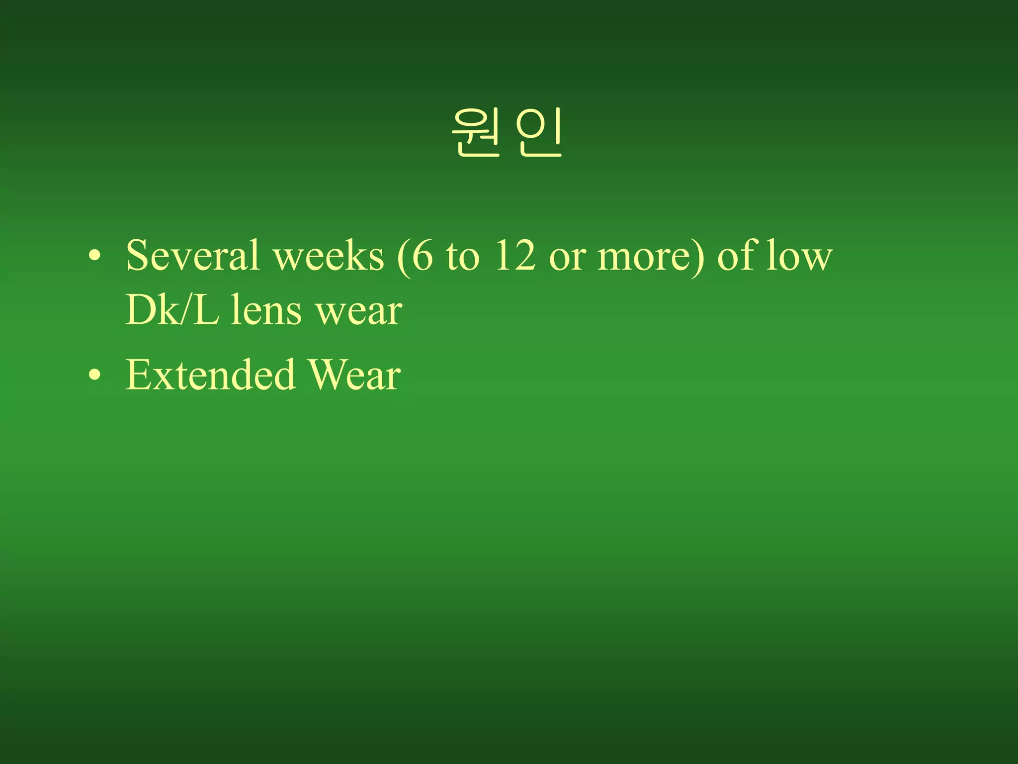
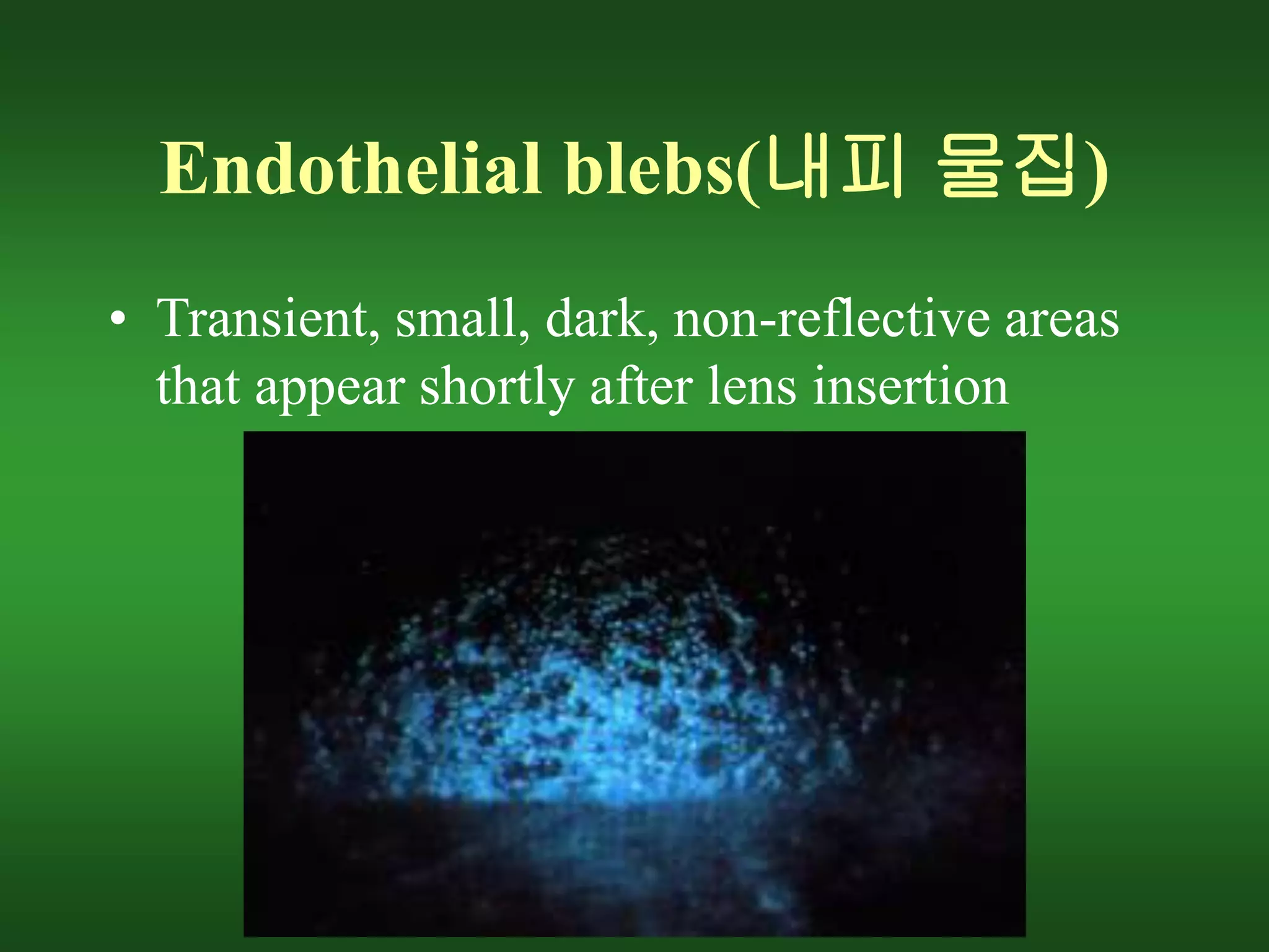
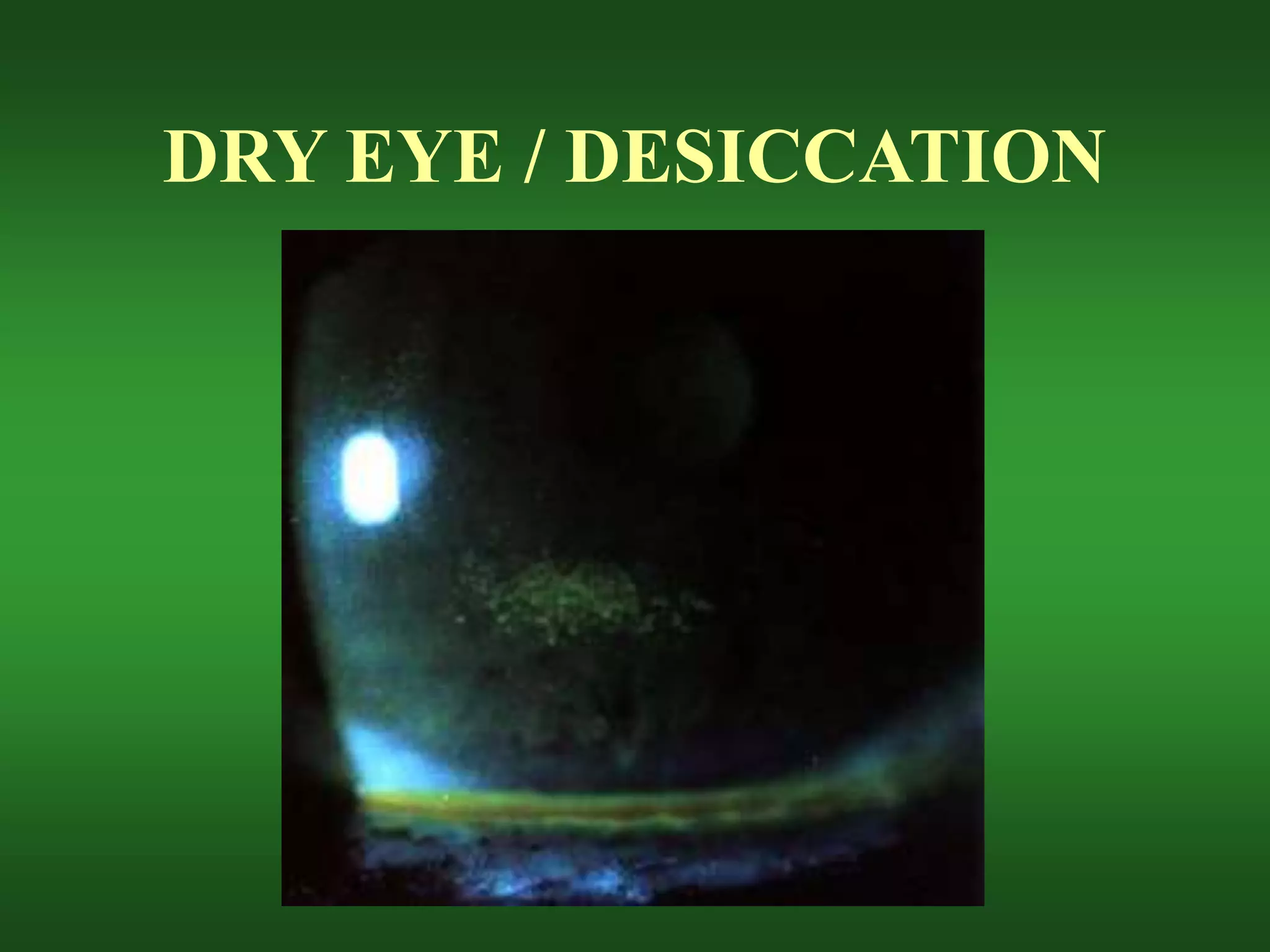
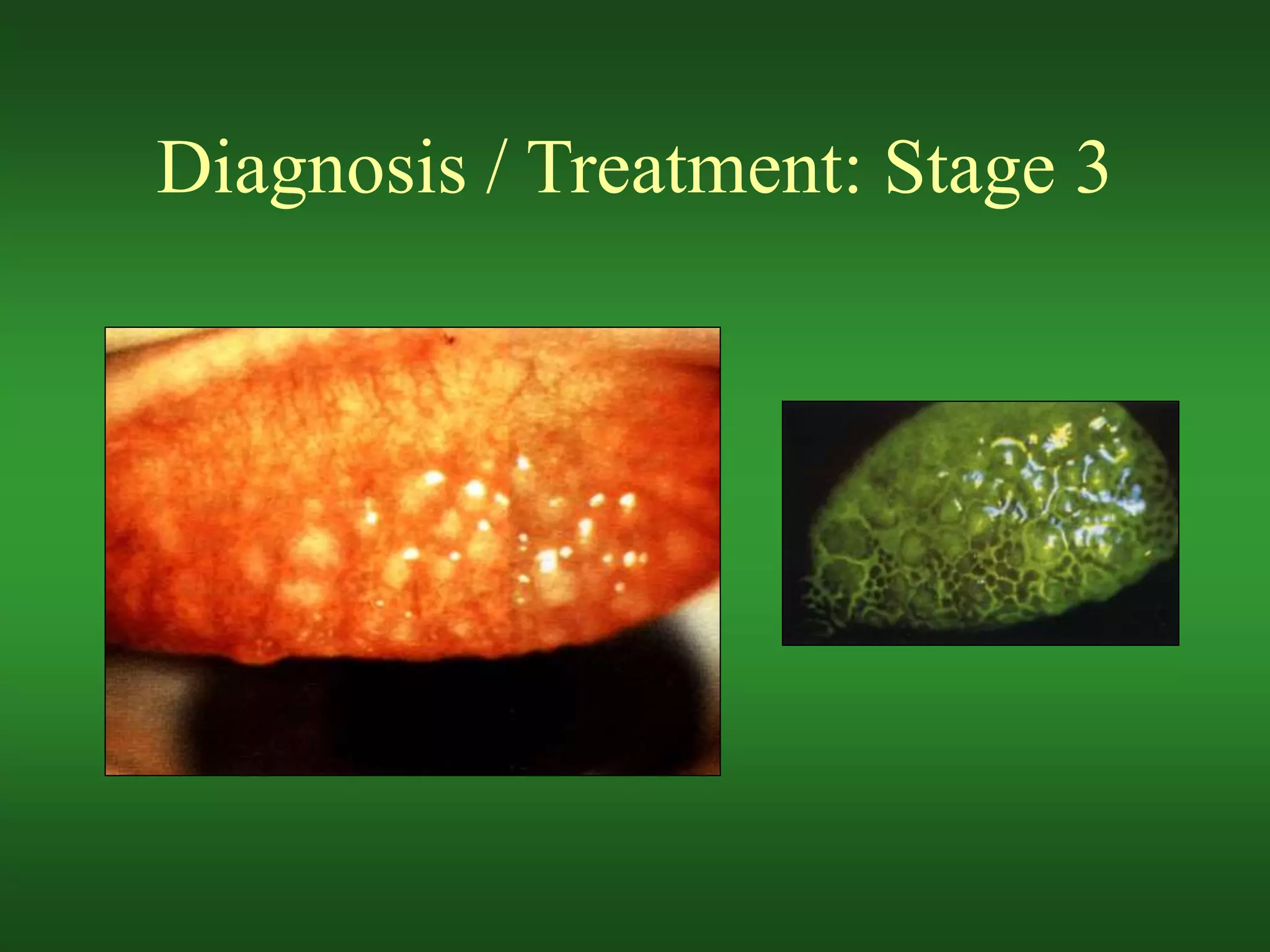
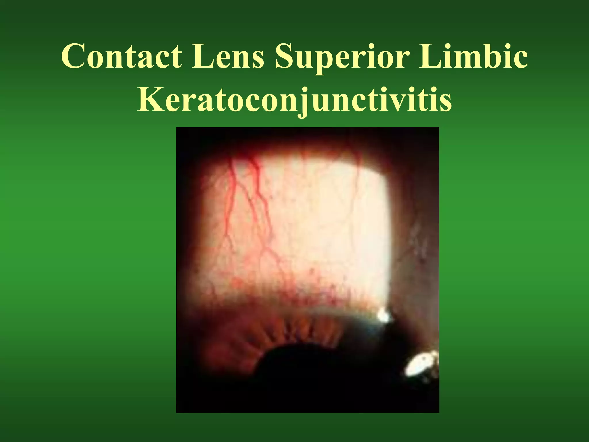
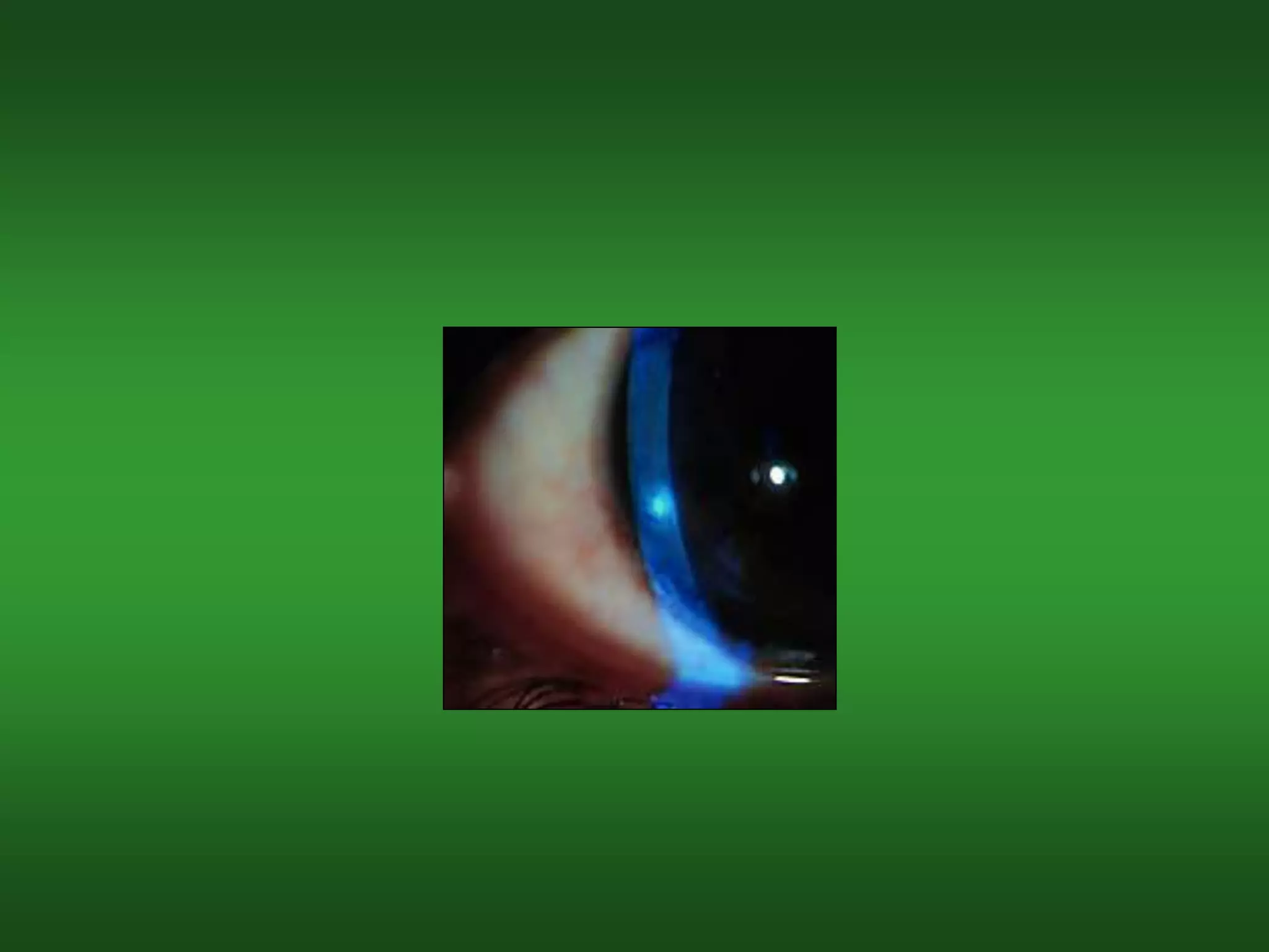
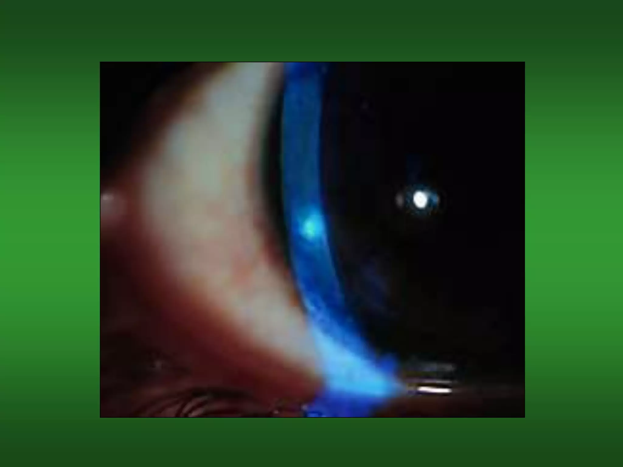
The document discusses various risks and complications that can arise from soft contact lens wear, including hypoxia, desiccation, deposit buildup, mechanical issues, and inflammatory responses. It covers specific conditions like epithelial edema, stromal edema, microcysts, vascularization, polymegethism, endothelial blebs, dry eye, contact lens papillary conjunctivitis, contact lens superior limbic keratoconjunctivitis, acute red eye, sterile infiltrates and ulcers, and preservative allergies and sensitivities. Potential causes, symptoms, diagnoses, and treatment approaches are provided for each condition.
Contact lenses can cause or exacerbate dry eye symptoms due to changes in the pre-corneal tear film and decreased tear production. Several factors influence contact lens-related dry eye, including the water content and material of the contact lens. Higher water content lenses dehydrate more, reducing oxygen transmission and sensitivity of the cornea, leading to less tear production. Symptoms include dryness, irritation, and red eyes. Signs include corneal staining and lens surface dehydration. Management involves addressing any underlying conditions, optimizing the contact lens parameters like water content and material, using artificial tears, and punctal plugs for more severe cases.
Common complications from rigid gas permeable (RGP) contact lens wear include inflammation and staining, oedema and hypoxia, and mechanical and pressure issues. Inflammation and staining complications include papillary conjunctivitis, 3 and 9 o'clock staining, corneal dellen, and vascularized limbal keratitis. Oedema and hypoxia complications include corneal oedema. Mechanical and pressure complications involve lens adherence, corneal warpage, and incomplete blinking leading to peripheral corneal desiccation and staining. Managing complications requires determining the underlying cause, such as lens design or fitting issues, and taking steps to improve lens physiology and ocular health.
This document provides an overview of common eye conditions and disorders organized by anatomical structure of the eye. Key points covered include:
- Dry eye, conjunctivitis, keratoconus, refractive errors, uveitis, color blindness, retinal detachment, diabetic retinopathy, age-related macular degeneration, glaucoma, cataracts, and floaters.
- Causes, symptoms, and treatment options are described for each condition.
- Additional details are given on types of conjunctivitis, refractive errors, retinal detachments, diabetic retinopathy, age-related macular degeneration, glaucoma, and surgical procedures for cataracts and retinal detachment.
most common ophthalmic disorder seen in all over world. in India 2015 incidence of cataract patient was 62.6 % (9 million). so the awareness and the management is very important for this disease condition. i hope this presentation is very helpful to all the student and people to understanding the cataract refractive ophthalmic disease
Keratoconus is a degenerative eye disorder where the cornea thins and changes to a conical shape, causing irregular astigmatism and vision problems. The exact cause is unknown but factors like eye rubbing, genetics, contact lens overuse, and UV exposure may play a role. It is diagnosed based on symptoms like blurred vision, history, and signs seen on exam like scissoring reflex and cone formation. Treatment options depend on the stage and severity but include glasses, rigid gas permeable contact lenses, collagen cross-linking to stop progression, and surgery like INTACS or corneal transplantation for advanced cases. Regular follow up is important as the condition progresses over time.
This document discusses dry eyes, also known as aqueous tear deficiency. It defines dry eyes as a non-infectious ocular surface disorder caused by a lack of tear fluid. The three layers of the normal tear film are described: the lipid layer from meibomian glands, the aqueous layer from lacrimal glands, and the mucin layer from goblet cells. Various causes of dry eyes are classified, including deficiencies in the aqueous, lipid, or mucin layers. Signs, symptoms, and diagnostic tests are outlined. Management involves tear supplementation, preservation, treatment of underlying conditions, punctal plugs, anti-inflammatories, and surgery in severe cases.
Dry eye, also known as keratoconjunctivitis sicca, is a condition caused by inadequate tear production or unstable tear film. It results in ocular irritation, redness, and visual disturbance. The tear film consists of an outer lipid layer, middle aqueous layer, and inner mucin layer. In chronic dry eye, tears have increased salts and decreased proteins and lipocalins. Treatment focuses on lubricating the eyes, managing underlying conditions like blepharitis, and occasionally punctal plugs or anti-inflammatory drugs. Diagnosis involves tests of tear production, stability, and ocular surface staining to determine the best lubricating and anti-inflammatory treatments.
Conventional contact lenses and its common problems
How to manage contact lens in covid 19 situation
Basic care & maintenance
In which aspect conventional Cl are using
Hydrogel contact lens
The cornea has five layers - epithelium, Bowman's layer, stroma, Descemet's membrane, and endothelium. It is avascular and transparent. The epithelium regenerates every 7 days. The endothelium maintains dehydration via ion pumps. Diseases like keratoconus cause thinning. Examination uses a slit lamp to assess size, shape, surface, transparency, and vascularization. Stains like fluorescein detect erosions while rose bengal finds filaments. The cornea has high innervation and refractive power of 45 diopters.
This document provides an overview of dry eye disease. It defines dry eye as a multifactorial disease resulting from tear deficiency or excess evaporation, causing symptoms like eye discomfort. Diagnostic tests evaluate tear secretion, stability, and damage to the ocular surface. Clinical features include irritation, redness, blurred vision, and staining of the cornea or conjunctiva. Dry eye exists on a spectrum of severity and has many predisposing factors like age, gender, medication use, contact lens wear, surgery, and autoimmune diseases.
This document discusses retinal detachment, including its causes, symptoms, diagnosis, and treatment options. The retina is the light-sensitive layer of tissue in the back of the eye. Retinal detachment occurs when the retina separates from its underlying layer, and fluid accumulates underneath. Common causes are trauma, advanced diabetes, high nearsightedness, and vitreous shrinkage. Symptoms include floaters, light flashes, and blurred or lost vision. Treatment options aim to reattach the retina through procedures like laser photocoagulation, cryopexy, pneumatic retinopexy, scleral buckling, or vitrectomy surgery.
This document discusses keratoconus, a progressive eye disorder causing corneal thinning and irregular astigmatism. Key signs include an irregular red reflex seen with an ophthalmoscope, Vogt striae seen on slit lamp exam, and a protruding cone shape of the cornea. Diagnosis involves examining the cornea and using tools like keratometry and OCT. Treatment focuses on rigid contact lenses to correct vision and corneal collagen cross-linking to slow progression. Surgical options like intrastromal corneal ring segments or keratoplasty may be considered for more advanced cases.
Dry eye is a multifactorial disease characterized by loss of tear film homeostasis and accompanied by ocular symptoms. It involves tear film instability, hyperosmolarity, ocular surface inflammation, and neurosensory abnormalities. It can be caused by aqueous deficiency, evaporative loss, or external diseases like Sjogren's syndrome. Diagnosis involves assessing risk factors, tear film parameters, ocular staining, and questionnaires. Treatment is staged based on disease severity and may include lifestyle modifications, lubricants, anti-inflammatories, punctal plugs, and in severe cases amniotic membrane grafts or surgery.
Therapeutic contact lenses are used for therapeutic, diagnostic and cosmetic purposes to treat various ocular surface diseases and conditions. They provide mechanical protection and support to the cornea, maintain corneal epithelial hydration, and can be used to deliver medications to the eye. The type of therapeutic contact lens chosen depends on the specific condition being treated and should aim to maximize oxygen to the cornea unless the eye has no vision. Common complications include ocular redness, minor corneal edema and lens deposits which require regular follow up visits.
L11-1434 vision L1 .pdf special senses.luckymbasela
This document provides an overview of the physiology of the special senses, with a focus on vision. It describes the anatomy of the eye, including the layers, tissues, humors, and role of each component. Key points covered include refraction of light as it passes through the eye, accommodation through the lens, and the roles of the retina, fovea and optic nerve in visual processing. Common vision conditions like glaucoma, cataracts and errors of refraction are explained. The document also discusses binocular vision and its advantages over monocular vision.
This document discusses ocular involvement in leprosy. It notes that 70-75% of leprosy patients experience ocular involvement, with 10-50% experiencing severe symptoms and 5% becoming blind. The major parts of the eye affected include the eyelids, cornea, iris, ciliary body, conjunctiva, and nerves. Common complications include blepharochalasis, madarosis, trichiasis, lagophthalmos, dacryocystitis, punctate keratitis, pannus, iridocyclitis, glaucoma, and cataract. Modes of infection include direct invasion via blood or nerves. Management involves antibiotics, steroids, surgery, and
Dry Eye Disease is a multifactorial disease that results in discomfort and visual disturbances due to tear film instability and inflammation of the ocular surface. It is caused by a disturbance in the lacrimal functional unit involving tear production, distribution, and clearance. Diagnosis involves evaluating tear secretion, volume, and stability as well as assessing ocular surface damage. Management focuses on eliminating exacerbating factors, supplementing tears, anti-inflammatory therapy, punctal plugs, and newer treatments targeting tear stimulation and mucous secretion when needed.
Lacrimal system ii,03.08.2016, a.r.rajalakshmiSrikanth K
The document discusses the lacrimal system and dry eye disease. It describes the structure and functions of the normal tear film and its three layers: lipid, aqueous, and mucin. Signs and symptoms of dry eye include dryness, irritation, blurry vision, redness, and corneal staining. Investigations include tear breakup time, Schirmer test, and ocular surface staining. Treatment is based on disease severity and may include artificial tears, anti-inflammatories, punctal plugs, and surgery for advanced cases.
This presentation describes all the clinical aspects of keratoconus management
You can watch the illustrated presentation in this link :
https://www.youtube.com/watch?v=pYxwZPGm7e4&list=PLZ_mM13I_TrhWavjTmE9NjW1O5bGxkONO&index=13
Mr. Jaydip J. Ninama is a lecturer in the department of medical surgical nursing. The document discusses the anatomy and physiology of the lens, causes and types of cataracts, signs and symptoms, diagnosis, and treatment. The main types of cataracts discussed are nuclear, cortical, subcapsular, and age-related cataracts. Treatment involves removing the cloudy lens surgically, usually through phacoemulsification. Post-operative care and potential complications are also outlined.
corneal response to anoxia stress from contact lens wearTheeratep
Contact lens wear can lead to corneal hypoxia due to the lens acting as a barrier to oxygen supply. Prolonged hypoxia causes changes to the tear film, epithelium, stroma, and endothelium. Symptoms include red eyes, irritation, tearing, and vision changes. Management involves discontinuing lens use, refitting with a higher oxygen permeability lens, or reducing hours of wear. Choosing a lens material with good wettability, oxygen permeability, and low deposition can reduce risks. Immune events like allergic conjunctivitis or mechanical issues like abrasions may also occur but can often be managed by changing lens parameters or care.
The document summarizes rhegmatogenous retinal detachment (RRD), which occurs when the retina separates from the retinal pigment epithelium due to a break or tear, allowing fluid from the vitreous cavity to enter the subretinal space. It describes the anatomy of the retina, risk factors for RRD like vitreous liquefaction and posterior vitreous detachment, signs and symptoms, examination techniques, and management options like laser retinopexy, pneumatic retinopexy, scleral buckling, and vitrectomy. Rhegmatogenous retinal detachment is the most common type of retinal detachment and can lead to vision loss if not treated.
Fuchs endothelial corneal dystrophy is a slowly progressive, bilateral inherited condition affecting the corneal endothelium. It presents as cloudy spots on the cornea called guttae and can lead to corneal edema. Recent advances in treatment include Descemet's stripping only (DSO), which removes the dysfunctional central endothelial cells to allow peripheral cells to migrate in, reducing the need for transplantation. Injection of cultured endothelial cells combined with rho kinase inhibitors may also help treat Fuchs' by promoting cell growth to replace dysfunctional cells. While these methods show promise, transplantation is still needed for advanced cases where not enough healthy cells remain.
History & materials of conatct lens by pushkar dhirPushkar Dhir
This document provides a history of contact lenses, including early sketches by Leonardo Da Vinci and Rene Descartes, and developments made by Adolf Fick, Fa Muller and Sons, and Carl Zeiss in the late 19th century. It discusses the evolution of materials from PMMA to silicone, hydrogel, and various surface treatments. Contact lenses are classified based on purpose, anatomical location, material, water content, and wearing schedule. Parameters like base curve, diameter, power, and various lens designs are also outlined.
1. Cataracts are opacities or cloudiness in the lens of the eye that impair vision. They are most commonly age-related but can be caused by eye injuries, medical conditions like diabetes, or exposure to radiation.
2. Symptoms include reduced visual acuity, glare, distorted vision, and difficulty seeing colors. Examination reveals a dim red reflex and opacity visible through a slit lamp.
3. Treatment is usually surgical removal of the cloudy lens, most commonly through phacoemulsification which uses ultrasound to break up the lens for removal. Post-operative care involves steroid and antibiotic eye drops. Potential complications include infection, bleeding, inflammation, and refractive errors.
Cataract is a clouding of the normally clear lens of the eye. It occurs when the lens proteins clump together, preventing clear images from reaching the retina. Risk factors include increasing age, diabetes, smoking, obesity, and corticosteroid use. Symptoms include blurred vision, sensitivity to light, and fading colors. Cataract surgery involves removing the cloudy lens and replacing it with an artificial intraocular lens, improving vision. Complications may include infection, swelling, bleeding or retinal detachment.
Dry eye, also known as dysfunctional tear syndrome, is a multifactorial disease of the tears and ocular surface. It results in symptoms of discomfort, visual disturbance, and tear film instability, and can potentially damage the ocular surface. The document discusses the prevalence, classification, signs, symptoms, and diagnostic tests and tools for dry eye. The prevalence increases significantly with age and is higher in women. Diagnosis involves evaluating symptoms, performing a slit lamp exam to check for signs of damage, and conducting tests to assess tear film stability, tear secretion, and ocular surface damage. Management involves classifying dry eye severity levels based on signs and symptoms to determine appropriate treatment.
Lens insertion for basic optical dispensingmikaelgirum
The document provides instructions for inserting lenses into plastic frames. It discusses heating the frame if needed, pre-shaping the eye wire to the lens curve, and inserting the lens by snapping the temporal edge in first and then the nasal edge. It describes checking that the lens is fully inserted and not twisted, as well as techniques for adjusting improperly inserted lenses.
standard frame alignment for basic frame adgustmentmikaelgirum
The document provides guidelines for standardizing the alignment of different types of eyewear frames, including adjusting the bridge, temples, and nose pads to be properly aligned based on impersonal standards. It describes checking frames for horizontal and vertical alignment and adjusting plastic, metal, and rimless frames, noting that plastic frames require heating during adjustment while metal frames can often be adjusted without heating. The goal of standard alignment is to configure eyewear to fit comfortably and symmetrically on a variety of face shapes.
The document discusses various parts and types of eyeglass frames. It describes the basic components of frames including the front, hinges, temples, bridges and end pieces. It outlines different frame constructions such as plastic, metal, nylon cord and combination frames. It also discusses specialized frame types like half-eyes, rimless and semi-rimless frames. The document provides details on materials used in frames like cellulose acetate, propionate, nylon and carbon fiber.
The document discusses factors to consider when selecting frames for a patient's prescription lenses. It describes how frame selection can affect the apparent length and width of the face, as well as facial balance and features. Frame shape, size, color, thickness, and bridge design were some of the key factors discussed in relation to complementing different facial structures. The document emphasizes matching frames to the individual's facial characteristics for optimal aesthetic results.
This document discusses eye optics and refractive errors. It provides 3 case studies: a 14-year-old boy unable to see the blackboard, a 51-year-old man with difficulty reading the newspaper, and a 6-year-old girl who squints when looking farther than 2 feet away. The objectives are to understand eye optics, visual acuity assessment, common refractive errors, and color perception assessment. Key refractive errors like myopia, hyperopia, presbyopia, and astigmatism are explained in terms of etiology, presentation, diagnosis, and treatment.
Keratometry is a technique used to measure the curvature of the cornea. It works by analyzing the reflection of light off the cornea's convex surface. Keratometry can determine the radius of curvature and refractive power of the cornea. This information is useful for contact lens fitting and calculating intraocular lens power for cataract surgery. Modern keratometers automate the measurement process for improved accuracy and efficiency.
This document discusses cataracts, including their classification, causes, symptoms, diagnosis, and treatment. It notes that cataracts are the opacification and clouding of the lens of the eye. They are classified morphologically or etiologically. Senile cataracts related to aging affect over 90% of people by age 70 and are the most common type. Examination involves assessing visual acuity, eye pressure, and examining the anterior segment and fundus. Treatment options include glasses initially, but surgery such as phacoemulsification is often needed for more advanced cases. Complications of surgery can include inflammation, edema, and retinal detachment.
Myopia, also known as nearsightedness, is a refractive error where light rays focus in front of the retina rather than directly on it. There are several types of myopia including simple/developmental myopia, pathological myopia, and acquired myopia. Treatment options include optical correction using concave lenses, surgical options like LASIK, and general measures like visual hygiene and a balanced diet. Prognosis depends on the type and severity of myopia.
Refractive error is a condition where the eye fails to form a clear image on the retina when light enters from infinity and accommodation is relaxed. The major types of refractive error are myopia, hyperopia, and astigmatism. Uncorrected refractive error is a leading cause of blindness worldwide, affecting over 2 billion people with 88.4 million having uncorrected refractive error. Children in urban areas and people over age 50 are most affected. The main reasons for high rates of uncorrected refractive error are lack of vision testing, limited refractive services, and affordability and cultural issues regarding corrective devices. Comprehensive community-level refractive services and school vision testing programs are important strategies to address this major global cause of visual impairment.
This document discusses visual impairment and blindness in Ethiopia. It identifies the main causes of visual impairment as cataract, uncorrected refractive error, glaucoma, corneal opacities, and diabetic retinopathy. For childhood blindness, the leading causes are uncorrected refractive error, cataract, retinopathy of prematurity, trauma, trachoma, and xerophthalmia. The document also explains how visual acuity is measured using a Snellen chart and expressing the results as a fraction where the top number indicates the test distance and the bottom number is the equivalent distance for a person with normal vision.
This document discusses anti-reflective coatings. It begins by describing their applications in corrective lenses and photolithography. It then outlines different types of coatings, including index-matching, single-layer interference, multi-layer interference, absorbing, moth eye, and circular polarizers. The document also covers the theory behind how reflection and interference coatings work to reduce reflection at surfaces. Key points include using layers of different refractive indices to destructively interfere reflected beams while constructively interfering transmitted beams.
This document discusses adjusting eyeglass frames to properly fit a patient's face. It covers adjusting the temple spread, positioning nose pads, checking the frame height and pantoscopic tilt. Temples should exert minimal pressure and have the bend just above the ear. Nose pads should rest halfway between the nose crest and inner eye, with the long diameter perpendicular to the floor. Proper adjustment ensures comfort and correct lens positioning.
1. The document discusses properties of light including its nature as electromagnetic radiation, its movement as waves, and its interaction with materials through transmission, reflection, absorption, and scattering.
2. Visual functions like visual acuity, dark adaptation, visual fields, and color vision are assessed clinically and their development and measurement are described.
3. Methods for measuring visual acuity both qualitatively and quantitatively are outlined, including grating acuity, Vernier acuity, and various acuity tests appropriate for different populations. Factors that affect acuity measurements like crowding effects are also addressed.
This document provides guidelines for routine refraction procedures and prescription writing. It discusses evaluating the patient history and vision, performing subjective and objective refraction tests, and guidelines for prescribing corrections for myopia, hyperopia, astigmatism, and presbyopia. It also addresses some common refraction cases involving these conditions and provides guidance on determining the appropriate correction or next steps.
1) Eye movements can be translatory (sideways, up/down, forward/backward) or rotary (around vertical, horizontal, or anteroposterior axes). Rotary movements occur around a center of rotation within the eyeball, which does not remain fixed.
2) Eye movements are governed by Donders' Law, Listing's Laws, Sherrington's Law of Reciprocal Innervation, and Hering's Law of Equal Innervation. These laws describe how eye movements maintain a consistent retinal image orientation and involve coordinated contraction/relaxation of agonist and antagonist muscles between the two eyes.
3) Eye movements are categorized as versions (simultaneous movements of both eyes),
Vision disorders are common in children and can negatively impact development if left untreated. A comprehensive eye exam evaluates vision, eye alignment and tracking, focusing ability, and screens for conditions like nearsightedness and lazy eye. Exams are important because many issues have no obvious symptoms. Screenings miss up to 1/3 of problems and may lead to misdiagnoses. Comprehensive exams are recommended at ages 6 months, 3 years, before school, and every 2 years thereafter to catch issues early for best outcomes. Bringing an infant for an exam requires preparing them to be well-rested and not distressed.
This document discusses vision syndrome, also known as binocular, accommodative, and oculomotor dysfunction (BAOD). BAOD is a common condition that affects reading and learning and often goes undetected on standard vision screenings. It involves dysfunctions in binocular control, accommodation, and oculomotor control. The document outlines signs and symptoms of BAOD, provides prevalence data, and recommends doctors evaluate for BAOD in patients with learning difficulties and refer for office-based vision therapy if needed.
Binocular vision develops gradually in infants, from following light monocularly at 2-3 weeks to binocularly at 6 weeks to 6 months. By 6 months, accommodation has developed but lags behind convergence. Sensory fusion blends monocular images into a single image, while motor fusion maintains eye alignment. Binocular vision provides advantages like single vision, stereopsis, and an enlarged visual field. Normal binocular vision requires clear visual axes, sensory fusion, and motor fusion.
These lecture slides, by Dr Sidra Arshad, offer a quick overview of the physiological basis of a normal electrocardiogram.
Learning objectives:
1. Define an electrocardiogram (ECG) and electrocardiography
2. Describe how dipoles generated by the heart produce the waveforms of the ECG
3. Describe the components of a normal electrocardiogram of a typical bipolar lead (limb II)
4. Differentiate between intervals and segments
5. Enlist some common indications for obtaining an ECG
6. Describe the flow of current around the heart during the cardiac cycle
7. Discuss the placement and polarity of the leads of electrocardiograph
8. Describe the normal electrocardiograms recorded from the limb leads and explain the physiological basis of the different records that are obtained
9. Define mean electrical vector (axis) of the heart and give the normal range
10. Define the mean QRS vector
11. Describe the axes of leads (hexagonal reference system)
12. Comprehend the vectorial analysis of the normal ECG
13. Determine the mean electrical axis of the ventricular QRS and appreciate the mean axis deviation
14. Explain the concepts of current of injury, J point, and their significance
Study Resources:
1. Chapter 11, Guyton and Hall Textbook of Medical Physiology, 14th edition
2. Chapter 9, Human Physiology - From Cells to Systems, Lauralee Sherwood, 9th edition
3. Chapter 29, Ganong’s Review of Medical Physiology, 26th edition
4. Electrocardiogram, StatPearls - https://www.ncbi.nlm.nih.gov/books/NBK549803/
5. ECG in Medical Practice by ABM Abdullah, 4th edition
6. Chapter 3, Cardiology Explained, https://www.ncbi.nlm.nih.gov/books/NBK2214/
7. ECG Basics, http://www.nataliescasebook.com/tag/e-c-g-basics
Adhd Medication Shortage Uk - trinexpharmacy.comreignlana06
The UK is currently facing a Adhd Medication Shortage Uk, which has left many patients and their families grappling with uncertainty and frustration. ADHD, or Attention Deficit Hyperactivity Disorder, is a chronic condition that requires consistent medication to manage effectively. This shortage has highlighted the critical role these medications play in the daily lives of those affected by ADHD. Contact : +1 (747) 209 – 3649 E-mail : sales@trinexpharmacy.com
Clinic ^%[+27633867063*Abortion Pills For Sale In Tembisa Central19various
Clinic ^%[+27633867063*Abortion Pills For Sale In Tembisa Central Clinic ^%[+27633867063*Abortion Pills For Sale In Tembisa CentralClinic ^%[+27633867063*Abortion Pills For Sale In Tembisa CentralClinic ^%[+27633867063*Abortion Pills For Sale In Tembisa CentralClinic ^%[+27633867063*Abortion Pills For Sale In Tembisa Central
Integrating Ayurveda into Parkinson’s Management: A Holistic ApproachAyurveda ForAll
Explore the benefits of combining Ayurveda with conventional Parkinson's treatments. Learn how a holistic approach can manage symptoms, enhance well-being, and balance body energies. Discover the steps to safely integrate Ayurvedic practices into your Parkinson’s care plan, including expert guidance on diet, herbal remedies, and lifestyle modifications.
Muktapishti is a traditional Ayurvedic preparation made from Shoditha Mukta (Purified Pearl), is believed to help regulate thyroid function and reduce symptoms of hyperthyroidism due to its cooling and balancing properties. Clinical evidence on its efficacy remains limited, necessitating further research to validate its therapeutic benefits.
Cell Therapy Expansion and Challenges in Autoimmune DiseaseHealth Advances
There is increasing confidence that cell therapies will soon play a role in the treatment of autoimmune disorders, but the extent of this impact remains to be seen. Early readouts on autologous CAR-Ts in lupus are encouraging, but manufacturing and cost limitations are likely to restrict access to highly refractory patients. Allogeneic CAR-Ts have the potential to broaden access to earlier lines of treatment due to their inherent cost benefits, however they will need to demonstrate comparable or improved efficacy to established modalities.
In addition to infrastructure and capacity constraints, CAR-Ts face a very different risk-benefit dynamic in autoimmune compared to oncology, highlighting the need for tolerable therapies with low adverse event risk. CAR-NK and Treg-based therapies are also being developed in certain autoimmune disorders and may demonstrate favorable safety profiles. Several novel non-cell therapies such as bispecific antibodies, nanobodies, and RNAi drugs, may also offer future alternative competitive solutions with variable value propositions.
Widespread adoption of cell therapies will not only require strong efficacy and safety data, but also adapted pricing and access strategies. At oncology-based price points, CAR-Ts are unlikely to achieve broad market access in autoimmune disorders, with eligible patient populations that are potentially orders of magnitude greater than the number of currently addressable cancer patients. Developers have made strides towards reducing cell therapy COGS while improving manufacturing efficiency, but payors will inevitably restrict access until more sustainable pricing is achieved.
Despite these headwinds, industry leaders and investors remain confident that cell therapies are poised to address significant unmet need in patients suffering from autoimmune disorders. However, the extent of this impact on the treatment landscape remains to be seen, as the industry rapidly approaches an inflection point.
Cell Therapy Expansion and Challenges in Autoimmune Disease
ContactLensComplications[1].ppt
1. 소프트 콘택트렌즈 착용에 관한
부작용
Peter B. Lee, OD
121st General Hospital
Seoul, Korea
2. CL 부작용의 위험 요소
• Hypoxia (저산소증)
– virtually all CLs reduce O2 supply to cornea
• Desiccation / Alteration of tear
film(렌즈탈수/각막건조)
– CLs are much thicker than tear film
– SCLs evaporate to the atmosphere
• Deposit build-up(침전)
– SCL>RGP, but all develop deposits made of
proteins, mucous, lipids, dirt, and microbes
3. CL 부작용의 위험 요소
• Mechanical(물리적 요소)
– Tightness & looseness
– Abrasions & friction
• Inflammatory response(염증)
– Proteins and other surface deposits
– Microbes and their toxins
– Solution preservatives and other components
4. Contributory Factors
(문제가 될 수 있는 요소)
• Lens material
– Dk(산소 침투성)
– Wettability and deposit attraction
– Stiffness and other physical characteristics
• Lens design and fitting
– Lens-to-cornea relationship
– Position and movement
– Thickness profile
5. • Wearing time(착용 시간)
– DW vs. EW
– Daily vs. part-time
– Patient’s physiology
• Patient compliance(착용자의 순응도)
– Wear time
– Cleaning and disinfection
– Lens replacements
– Follow-up visits
7. Effects of Hypoxia
1. Epithelial and stromal edema(각막부종)
2. Decrease in epithelial mitosis(상피 분열)
3. Increase in stromal lactate
accumulation(유산 축적 in anterior
chamber)
4. Decrease in corneal sensitivity
8. Corneal Edema(각막부종)
• The cornea requires a certain amount of
oxygen to prevent swelling, or edema
• During sleep, the oxygen supply is reduced
to approx. 8%, compared to about 21%
during waking hours
• The cornea swells 3% to 4% overnight
• Therefore, it appears the cornea needs more
than 8% O2 to prevent swelling
9. Corneal sensitivity(각막 민감도)
» Millodot and O'Leary (1980) found that the cornea
needs at least 8% oxygen to prevent a reduction in
corneal sensitivity
10. Oxygen Requirements
• The cornea requires 8% to 13% O2 to
maintain normal corneal thickness,
metabolism, and sensitivity.
• What are today’s contact lenses able to
deliver?
11. Oxygen Requirements
• CLs are not rated by % O2 delivered
• Transmissibility (Dk/L) is a rough indicator
of how well a CL transmits oxygen
• Dk/L is the most common measure to rate
how well contact lenses deliver oxygen to
the cornea
• Other methods have also been developed
but are not in widespread use
13. • Epithelial edema manifests as clouding of
the epithelial tissue, usually centrally, with
associated punctate epithelial staining,
steepening of the corneal curvature and
possible irregularity of the corneal surface
Epithelial Edema(각막 부종)
14. 원인
• Low Dk(산소 침투성) contact lens wear
(PMMA more likely than soft lenses)
• Long-term, extended hours or extended
wear(연속 착용)
15. 증상과 진단
• Poor endpoint visual acuity
• Distorted keratometry mires, steepening of
K's
• Central circular clouding (CCC)
• Punctate epithelial staining centrally
• Increased minus on refraction
16. 치료/처치법
• Discontinue lens wear or immediate refit to
higher Dk material
• Refit to higher Dk material
• Decrease wear time
• Looser fit for better tear exchange
• Monitor K and SR changes
– Prescribe new spectacles when stable to allow
decreased CL wear time
17. Stromal Edema(고유층 부종)
• Stromal edema manifests as fine wrinkling
(striae) or folds in Descemet's membrane
• Striae and folds appear with approx. 6%
stromal edema
• Stromal clouding requires extensive edema,
at least 15%, and is usually not induced by
contact lens wear
19. 원인
• Thick lens design (aphakic, prism ballast),
low Dk material or EW
20. 증상과 진단
• Mild edema: 6 to 7%
• Striae in deep stroma
• Moderate edema: 10 to 15%
• Folds in deep stroma
• Severe edema: > 18%
• Central haze on sclerotic scatter
21. 치료/처치법
• Increase Dk/L: higher water content, thinner
design
• Looser fit for better tear exchange
23. Microcysts(상피 미세낭종) /
Vacuoles(엑포)
• Microcysts are tiny cystic bodies containing
cellular debris
• They form at the basal cells and slowly
migrate towards the corneal surface
• They are visible with reversed illumination,
indicating that the cystic material has a
higher index of refraction than the
surrounding corneal tissue
25. Microcysts / Vacuoles
• Vacuoles form similarly to microcysts but
are fluid filled
• They are visible with unreversed
illumination, indicating that the cystic
material has a lower index of refraction than
the surrounding corneal tissue
26. Microcysts / Vacuoles
• Although they do not induce any
symptomalogy, the presence of microcysts
and vacuoles indicate a chronic hypoxic
condition and a resulting alteration of
corneal metabolism
• Removal of the offending contact lens will
result in their disappearance over the course
of several weeks
27. 원인
• Several weeks (6 to 12 or more) of low
Dk/L lens wear
• Extended Wear
28. 증상 및 진단
• Small cysts in epithelium visible with
retroillumination
• Microcysts: reversed illumination (higher
index than cornea)
• Vacuoles: unreversed illumination (lower
index than cornea)
29.
30. 치료/처치법
• Increase Dk/L
• D/C extended wear
• D/C lens wear if more than 50 are observed
• Monitor for 3 to 6 months for resolution
– # may increase after d/c lens wear due to
abnormal processing of epithelial cells
31. Vascularization(혈관신생)
• Cellular debris and other metabolic by-
products may be other stimuli to vessel
growth
• Almost all soft lens patients will
demonstrate some amount of
vascularization
33. Vascularization(혈관신생)
• In most cases, vascularization is
asymptomatic, but encroachment into the
visual axis may result in a reduction in
vision
• Vascularization can be graded according to
the number of quadrants and the extent to
which the vessels have grown into the
cornea
34. 원인
• Long-term wear of any soft lens, but
particularly lenses with low Dk/L (aphakic,
prism ballasted, etc.)
• Extended wear
35. 진단
• Varying degrees of vessels encroaching into
previously avascular cornea
– Grade 1: Sectorial or circumlimbal vessel
extension less than 0.5 mm, or 1 or 2 vessels
extending less than 1.5 mm into cornea.
– Grade 2: Sectorial or circumlimbal vessel
extension 0.5 to 1.5 mm into cornea.
36.
37. 치료/처치법
• Grade 1 or 2: monitor Q6mos
• Grade 3: decrease wear time, increase Dk/L
or refit to RGP material
• Grade 4: refit to RGP material or D/C lens
wear
• Though vasc may regress, vessel walls do
not disappear
– Watch for development of ghost vessels
38. Polymegethism(폴리메가티즘)
• A permanent condition of endothelial
cell(내피) change such that the cells are no
longer uniform but are of varying sizes
39. Endothelial blebs(내피 물집)
• Transient, small, dark, non-reflective areas
that appear shortly after lens insertion
40. 원인
• Long-term wear of any soft lens, but
particularly lenses with low Dk/L (aphakic,
prism ballasted, etc.), extended wear
41. 치료/처치법
• Change to higher Dk or RGP
• Monitor for further cellular changes,
corneal edema
43. Soft Lens Desiccation(탈수)
• Soft lenses dehydrate when exposed to the
atmosphere
• As the lens dehydrates, it draws water from
the post-lens tear film, resulting in dryness
of the cornea in these areas
• Tear flow dynamics from tight or high
modulus soft lenses may contribute to this
staining pattern
44. 원인
• Thin, high water soft lens
• Borderline dry eye
• Extended wear
• Incomplete / infrequent blink
• Dry environment
• Heavy protein deposition
52. 진단
• Large papillae on upper tarsal conjunctiva
– SCL: diffuse distribution
– RGP: more common along lid margin (zone 3)
• Heavily protein coated contact lens
• Poorly designed lens edge
• White, ropy mucous discharge
53. Diagnosis / Treatment: Stage 1
• Itching
• Mucous discharge
• Upper tarsus
hyperemic but
relatively normal
• Discontinue lens wear
or immediate refit in
new lens
• Unpreserved lubricants
54. Diagnosis / Treatment: Stage 2
• Enlarged papillae on
upper tarsus
• Hyperemic and
edematous upper tarsal
conjunctiva
• Increased itching and
mucous
• Mild blurring of vision
• Reduced wear time
• Discontinue lens wear
• Unpreserved lubricants
55. Diagnosis / Treatment: Stage 3
• Large, clover-like
papillae that may stain
with fluorescein
• Increase in tarsal
hyperemia
• Increased itching and
mucous
• Lens discomfort
• Discontinue lens wear
• Topical antihistamine
• Topical vasoconstrictor
• Topical NSAID
• Refit to disposables or
RGPs
60. Contact Lens Superior Limbic
Keratoconjunctivitis
(상윤부각결막염)
• Immunological response to lens surface
deposits and solution preservatives
• Its superior location suggests that there may
be a hypoxia component
• May also have a mechanical component
61. 원인
• Soft contact lens wear
• Protein deposition
• Use of solutions containing thimerosal
64. 치료/처치법
• Discontinue lens wear until resolution
• Lubricants
• Topical steroid if severe
• Refit to different material
• Patient education on compliance with lens care
• Discontinue use of thimerosal preserved
solutions
66. 원인
• Extended wear of soft lenses, particularly
tightly fitting lenses
• Extended wear of rigid lens with lens
adherence
67. 증상
• Awaken with unilateral discomfort or pain
• Redness
• Tearing
• Photophobia
68. 진단
• Subepithelial infiltrates
• Conjunctival and limbal hyperemia
• Minimal epithelial staining
• Debris trapped between lens and cornea
69. 치료/처치법
• Discontinue lens wear until infiltrates resolve
• Lubricants
• Prophylactic broad spectrum antibiotic drops
if epithelial defect or infiltrates are present
(fluoroquinolones, Polytrim)
• Change wearing schedule to daily wear
70. Case example
• 26 세, 남
• Sx of moderate irritation and localized
redness OD for 4 days
• No pain, discharge, photophobia, blurry
vision
• SCL wearer for 10 years
– Focus Monthly: replaces Q2mos
– 16 hrs/day
– Renu MPS
71.
72. Sterile Infiltrate / Ulcer
(무감염성 각막궤양)
• An infiltrate is a localized collection of
white blood cells
• An ulcer is an excavated area of the cornea
that has undergone necrosis due to
immunological or microbial invasion
73. 원인
• Extended wear, long wear times
• Low Dk material
• Preserved solutions
• Poor compliance with care regimen
75. 진단
• Peripheral, small diameter white
subepithelial fuzzy lesions
• Overlying epithelial defect / disruption may
be present
• Mild to moderate sectoral injection
• None to moderate anterior chamber reaction
76.
77. 치료/처치법
• Discontinue lens wear
• Culture if epithelial defect present: cannot
diagnose as sterile until culture is done
• Artificial tears if no epithelial defect present
• Topical broad spectrum antibiotic if
epithelial defect present
– Fluoroquinolone (Ciloxan, Ocuflox)
– Fortified aminoglycoside
78. • Continue antibiotic therapy until culture
results are in, or until re-epithelialization
– qh for 24 hours, then taper
• Topical steroid possible after
re-epithelialization if infiltrates are
central and persistent
80. Preservative Allergies
and Sensitivities (방부제에
의한 앨러지)
• Chemical care systems provide convenient
disinfection with little damage to the
contact lenses. However, many of the
preservatives in use today are capable of
inducing a Type IV delayed hypersensitivity
response.
81. 원인
• Long-term use of preserved soaking/rinsing
solution
• Potential offending agents:
– Polyquaternium-1 (Alcon)
– Polyaminopropyl biguanide (B&L)
– Polyhexamethylene biguanide (Allergan)
– Polyhexanide (Ciba)
– Chlorhexidine (Boston, Claris)
– Benzalkonium chloride (Allergan Wet n Soak)
– Thimerosal
84. 치료/처치법
• Discontinue lens wear if infiltrates or
moderate to severe SPK are present
• Change preservative system or change to
unpreserved care system
• If infiltrates persist after 2 weeks, consider a
steroid
• Steroid Tx should be aggressive and of
sufficient duration