SlideShare a Scribd company logo
DORV
Dr. PRAMOD KUMAR
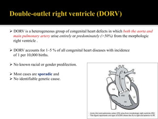
 DORV is a heterogeneous group of congenital heart defects in which both the aorta and
main pulmonary artery arise entirely or predominately (>50%) from the morphologic
right ventricle .
 DORV accounts for 1–5 % of all congenital heart diseases with incidence
of 1 per 10,000 births.
 No known racial or gender predilection.
 Most cases are sporadic and
 No identifiable genetic cause.
DORV is the spectrum of conotruncal defects.
Conus: is a circular tube of muscle beneath the valves of each great artery in equal
distribution, creating fibrous discontinuity with the atrioventricular valves.
Normally, the muscle under the aortic valve resorbs whereas the subpulmonary conus
continues to grow.
This therefore produces a right and posterior position of the aortic valve with mitro-
aortic fibrous continuity and Left & anterior position of the pulmonary valve with a
fibrous discontinuity with the tricuspid valve .
In conotruncal defects, there is a spectrum between heart anomaly, in which minimal
conus exists beneath the aorta, as seen in tetralogy of Fallot, and no conus exists under
the pulmonary valve, as with transposition of the great arteries .
DORV falls in the middle of the spectrum, that have variable amounts of conus under each
semilunar valve.
One end of spectrum is TOF type DORV:
“TOF type”: Near-normal length of conus beneath the pulmonary valve and minimal conus
beneath the aortic valve. Consequently, there is No aorto-mitral continuity, and the
pulmonary valve is left, anterior and superior.
Other end of spectrum is TGA type DORV:
“Transposition type”: Large amount of conus under the aortic valve with relatively little
under the P-Valve. Aorta is pushed anteriorly and superiorly, resulting in rightward
positioning of the aorta relative to the pulmonary artery. No pulmonary-mitral continuity.
Between the TOF type and TGA type:
There are many variants with bilateral conus that have neither aorto-mitral , nor pulmonary-
mitral continuity and have variable distribution of conal septum.
Solitus (Normal)
Inversus
Side by side
D-Malposition
(D-MAG)
Side by side
L-Malposition
(L-MAG)
L-Transposition
(L-TGA/CC-TGA)
D-Transposition
(D-TGA)
Spatial Relationship Of Great Arteries
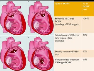
Van Praagh developed the most widely accepted classification of DORV, which included
the following:
• DORV with a subaortic VSD
• DORV with a subpulmonic VSD
• DORV with a doubly committed (both subaortic and subpulmonic) VSD
• DORV with a remote VSD (noncommitted)
Type of DORV % of
DORV
case
Subaortic VSD-type
DORV
(tetralogy of Fallot-type)
>50 %
Subpulmonary VSD-type
(k/a Taussig–Bing
anomaly)
30%
Doubly committed VSD-
type
10%
Noncommitted or remote
VSD-type DORV
10%
Four types of great artery relationships at the level of the semilunar valves have been
described in DORV:
• Aorta right and posterior to the pulmonary artery (normal)
• Aorta right and lateral to the pulmonary artery (side by side)
• Aorta right and anterior to the pulmonary artery (dextro-malposition)
• Aorta left and anterior to the pulmonary artery (levo-malposition)
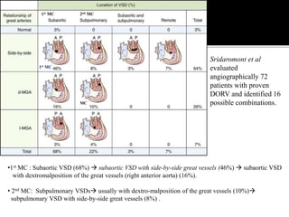
Sridaromont et al
evaluated
angiographically 72
patients with proven
DORV and identified 16
possible combinations.
•1st MC : Subaortic VSD (68%)  subaortic VSD with side-by-side great vessels (46%)  subaortic VSD
with dextromalposition of the great vessels (right anterior aorta) (16%).
• 2nd MC: Subpulmonary VSDs usually with dextro-malposition of the great vessels (10%)
subpulmonary VSD with side-by-side great vessels (8%) .
1st MC
1st MC 2nd MC
MC
Sridaromont et al
evaluated
angiographically 72
patients with proven
DORV and identified 16
possible combinations.
• Doubly committed VSDs (3%)VSD is usually large and in the superior position, closely
related to both semilunar valves.
• Remote or noncommitted VSD (7%) with muscular VSD.
Pulmonary Stenosis (PS) is the most common lesion associated with DORV.
Occurs in ~50% of patients and may be valvular or subvalvar.
Secundum-ASD are seen in 25% of all types, whereas primum-ASD are seen in the 8% of
DORV patients .
Coronary arterial anomalies occur in about 10% of patients—most commonly
anomalous origin of the LAD from the RCA. It is of surgical importance, because they
may alter considerations for surgical repair due to their effect on feasibility of conduit
placement or coronary arterial transfer .
Associated aortic arch coarctation, hypoplasia, or interruption—also found in about 10%
of patients.
Rarely, the VSD will be absent, and it is accompanied by a small ASD and hypoplasia of
the mitral valve and hypoplastic left ventricle .
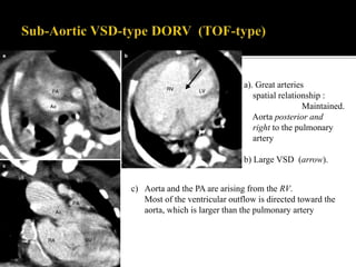
> 50 % of DORV cases .
Great arteries spatial relationship : Maintained, Aortic origin posterior and to the right
of the pulmonary origin .
Pulmonary stenosis is present in up to 50 % of patients. In these patients, the
physiology resembles that of tetralogy of Fallot, where the aorta completely overrides
the right ventricle.
In the absence of pulmonary stenosis, the physiology resembles that of a large isolated
VSD . This anatomy may result in congestive heart failure.
Subaortic VSD is associated with L-transposition of the great arteries and an
anomalous course of the right coronary artery, which crosses the pulmonary outflow
tract.
c) Aorta and the PA are arising from the RV.
Most of the ventricular outflow is directed toward the
aorta, which is larger than the pulmonary artery
a). Great arteries
spatial relationship :
Maintained.
Aorta posterior and
right to the pulmonary
artery
b) Large VSD (arrow).
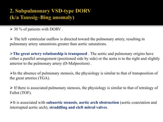
 30 % of patients with DORV .
 The left ventricular outflow is directed toward the pulmonary artery, resulting in
pulmonary artery saturations greater than aortic saturations.
The great artery relationship is transposed . The aortic and pulmonary origins have
either a parallel arrangement (positioned side by side) or the aorta is to the right and slightly
anterior to the pulmonary artery (D-Malposition) .
In the absence of pulmonary stenosis, the physiology is similar to that of transposition of
the great arteries (TGA).
 If there is associated pulmonary stenosis, the physiology is similar to that of tetralogy of
Fallot (TOF).
It is associated with subaortic stenosis, aortic arch obstruction (aortic coarctation and
interrupted aortic arch), straddling and cleft mitral valves.
a) Aorta and PA in a parallel arrangement (positioned side by side).
b) Aorta and PA are arising from the RV.
Most of the ventricular outflow is directed toward the pulmonary artery
a) Aorta anterior and right to the PA (D-transposition).
b/c) Aorta and PA are arising from the dilated RV.
(c) Very large VSD.
 10 % of cases of DORV.
The left ventricular outflow is equally directed to the aorta and pulmonary artery.
The great arteries are normally related .
Since the left ventricular outflow is equally shared by the aorta and pulmonary
artery, the pathophysiology resembles that of a VSD.
4. Non-committed or remote VSD-type DORV
10 % of DORV cases.
 Most commonly the great arteries are normally related .
The anatomy and physiology is similar to that of an isolated VSD .
 Patients present by age range 1 day–4 years.
Presentation is variable and is dependent on the type of DORV and associated cardiac
anomalies.
DORV with subaortic VSD +PS (TOF type):
C/F indistinguishible from classic Tetrology of Fallot.
Cyanosis develops and progresses during the early month of life..
Hypoxic spell may occur.
DORV with subaortic VSD without PS:
In absence of PS Increased flow to pulmonary circulation PAH.
C/F resembles that of large isolated VSD.
DORV with subpulmonary VSD ( Taussing Bing malformation):
C/F similar as TGA with VSD.
Cyanosis in newborn.
Early development of Heart failure.
Increasing breathlessness, poor feeding and slow weight gain are prominent feature.
Associated coarctation of aorta is frequent and leads to early (1st week of life) onset of HF.
Most patients with DORV are diagnosed in the first month of life and undergo
Palliative repair (pulmonary artery banding or Blalock–Taussig shunt) or Surgical repair.
The surgical interventions depend on the location of the VSD, the size of the left ventricle,
type of ventriculo-arterial connection, and the type of pulmonary blood flow (restricted on
unrestricted) .
Definitive repairs vary and include :
Arterial switch operation to connect the left ventricle to the neo-aorta in combination with
VSD closure. The subpulmonic or “Taussig–Bing” anomaly with physiology similar to
transposition may be treated with an arterial switch operation.
Bidirectional Glenn shunt (superior vena cava to pulmonary artery) .
Intraventricular repair: for the subaortic and doubly committed VSDs .
Fontan procedure: For directing the systemic flow of venous blood to the lungs without
passing through a ventricle.
Fig: Original Fontan procedure,
SVC was connected to the RPA and the
RA to MPA.
Fig: Modified Fontan procedure.
RA was connected to MPA.
SCA was connected to PA
Potential complications after surgical repair include :
 Residual or recurrent VSD,
 Residual or recurrent outflow tract obstructions,
 Atrioventricular valve regurgitation
stenosis/narrowing of the Glenn shunt or Fontan procedure.
THANK YOU

More Related Content

What's hot

Congenitally corrected transposition of great arteries
Congenitally corrected transposition of great arteriesCongenitally corrected transposition of great arteries
Congenitally corrected transposition of great arteriesDheeraj Sharma
 
single ventricle physiology
single ventricle physiologysingle ventricle physiology
single ventricle physiology
richamalik99
 
ECHO ASSESSMENT OF ASD FOR DEVICE CLOSURE
ECHO ASSESSMENT OF ASD FOR DEVICE CLOSURE ECHO ASSESSMENT OF ASD FOR DEVICE CLOSURE
ECHO ASSESSMENT OF ASD FOR DEVICE CLOSURE
SR,CARDIOLOGY,JIPMER,PUDUCHERRY
 
Segmental approach to Congenital Heart Disease
Segmental approach to Congenital Heart DiseaseSegmental approach to Congenital Heart Disease
Segmental approach to Congenital Heart Disease
Tanat Tabtieang
 
ASD devices
ASD devicesASD devices
ASD devices
Navin Agrawal
 
Single ventricle presentation for pediatrician
Single ventricle presentation for pediatricianSingle ventricle presentation for pediatrician
Single ventricle presentation for pediatrician
Laxmi Ghimire
 
Low flow low gradient aortic stenosis
Low flow low gradient aortic stenosisLow flow low gradient aortic stenosis
Low flow low gradient aortic stenosis
Malleswara rao Dangeti
 
Dorv thab
Dorv thab Dorv thab
Dorv thab
Thabit Ahmed
 
Echocardiographic Evaluation of Hypertrophic Cardiomyopathy
Echocardiographic Evaluation of Hypertrophic CardiomyopathyEchocardiographic Evaluation of Hypertrophic Cardiomyopathy
Echocardiographic Evaluation of Hypertrophic Cardiomyopathy
Hatem Soliman Aboumarie
 
Transposition of the great arteries(TGA)
Transposition of the great arteries(TGA)Transposition of the great arteries(TGA)
Transposition of the great arteries(TGA)
Sid Kaithakkoden
 
Pulmonary atresia with intact ventricular septum
Pulmonary atresia with intact ventricular septumPulmonary atresia with intact ventricular septum
Pulmonary atresia with intact ventricular septumRamachandra Barik
 
Dr ranjith mp av canal defect
Dr ranjith mp av canal defectDr ranjith mp av canal defect
Dr ranjith mp av canal defectdrranjithmp
 
Double outlet right ventricle
Double outlet right ventricleDouble outlet right ventricle
Double outlet right ventricle
Ramachandra Barik
 
Speckle Tracking Echocardiography
Speckle Tracking EchocardiographySpeckle Tracking Echocardiography
Speckle Tracking Echocardiography
MADHURJAIN78
 
D TGA
D TGAD TGA
Atrioventricular canal defect, Firas Aljanadi,MD
Atrioventricular canal defect, Firas Aljanadi,MDAtrioventricular canal defect, Firas Aljanadi,MD
Atrioventricular canal defect, Firas Aljanadi,MD
FIRAS ALJANADI
 
Dobutamine stress echocardiography
Dobutamine stress echocardiographyDobutamine stress echocardiography
Dobutamine stress echocardiography
Himanshu Rana
 
Transposition of Great Arteries
Transposition of Great ArteriesTransposition of Great Arteries
Transposition of Great Arteries
Dr. Harshil Joshi
 
How to echo... tricuspid regurgitation.ppt
How to echo... tricuspid regurgitation.pptHow to echo... tricuspid regurgitation.ppt
How to echo... tricuspid regurgitation.ppt
Vinayak Vadgaonkar
 

What's hot (20)

D o r v
D o r vD o r v
D o r v
 
Congenitally corrected transposition of great arteries
Congenitally corrected transposition of great arteriesCongenitally corrected transposition of great arteries
Congenitally corrected transposition of great arteries
 
single ventricle physiology
single ventricle physiologysingle ventricle physiology
single ventricle physiology
 
ECHO ASSESSMENT OF ASD FOR DEVICE CLOSURE
ECHO ASSESSMENT OF ASD FOR DEVICE CLOSURE ECHO ASSESSMENT OF ASD FOR DEVICE CLOSURE
ECHO ASSESSMENT OF ASD FOR DEVICE CLOSURE
 
Segmental approach to Congenital Heart Disease
Segmental approach to Congenital Heart DiseaseSegmental approach to Congenital Heart Disease
Segmental approach to Congenital Heart Disease
 
ASD devices
ASD devicesASD devices
ASD devices
 
Single ventricle presentation for pediatrician
Single ventricle presentation for pediatricianSingle ventricle presentation for pediatrician
Single ventricle presentation for pediatrician
 
Low flow low gradient aortic stenosis
Low flow low gradient aortic stenosisLow flow low gradient aortic stenosis
Low flow low gradient aortic stenosis
 
Dorv thab
Dorv thab Dorv thab
Dorv thab
 
Echocardiographic Evaluation of Hypertrophic Cardiomyopathy
Echocardiographic Evaluation of Hypertrophic CardiomyopathyEchocardiographic Evaluation of Hypertrophic Cardiomyopathy
Echocardiographic Evaluation of Hypertrophic Cardiomyopathy
 
Transposition of the great arteries(TGA)
Transposition of the great arteries(TGA)Transposition of the great arteries(TGA)
Transposition of the great arteries(TGA)
 
Pulmonary atresia with intact ventricular septum
Pulmonary atresia with intact ventricular septumPulmonary atresia with intact ventricular septum
Pulmonary atresia with intact ventricular septum
 
Dr ranjith mp av canal defect
Dr ranjith mp av canal defectDr ranjith mp av canal defect
Dr ranjith mp av canal defect
 
Double outlet right ventricle
Double outlet right ventricleDouble outlet right ventricle
Double outlet right ventricle
 
Speckle Tracking Echocardiography
Speckle Tracking EchocardiographySpeckle Tracking Echocardiography
Speckle Tracking Echocardiography
 
D TGA
D TGAD TGA
D TGA
 
Atrioventricular canal defect, Firas Aljanadi,MD
Atrioventricular canal defect, Firas Aljanadi,MDAtrioventricular canal defect, Firas Aljanadi,MD
Atrioventricular canal defect, Firas Aljanadi,MD
 
Dobutamine stress echocardiography
Dobutamine stress echocardiographyDobutamine stress echocardiography
Dobutamine stress echocardiography
 
Transposition of Great Arteries
Transposition of Great ArteriesTransposition of Great Arteries
Transposition of Great Arteries
 
How to echo... tricuspid regurgitation.ppt
How to echo... tricuspid regurgitation.pptHow to echo... tricuspid regurgitation.ppt
How to echo... tricuspid regurgitation.ppt
 

Similar to Dorv ppt

Transposition of Great Arteries;TGA,Firas Aljanadi,MD
Transposition of Great Arteries;TGA,Firas Aljanadi,MDTransposition of Great Arteries;TGA,Firas Aljanadi,MD
Transposition of Great Arteries;TGA,Firas Aljanadi,MD
FIRAS ALJANADI
 
Tetrology of Fallot (TOF) - A Review
Tetrology of Fallot (TOF) - A ReviewTetrology of Fallot (TOF) - A Review
Tetrology of Fallot (TOF) - A Review
Vivek Rana
 
Tetralogy of Fallot : Dr. Akif Baig
Tetralogy of Fallot : Dr. Akif BaigTetralogy of Fallot : Dr. Akif Baig
Tetralogy of Fallot : Dr. Akif Baig
akifab93
 
TOF.pptx
TOF.pptxTOF.pptx
TOF.pptx
akifab93
 
Antenatal Diagnosis of Fetal Heart Disease
Antenatal Diagnosis of Fetal Heart DiseaseAntenatal Diagnosis of Fetal Heart Disease
Antenatal Diagnosis of Fetal Heart Disease
Tarique Ajij
 
Single ventricle
Single ventricleSingle ventricle
Single ventricle
Nazmun Ara
 
Single ventricle
Single ventricleSingle ventricle
Single ventricle
Nazmun Ara
 
Single ventricle
Single ventricleSingle ventricle
Single ventricle
Nazmun Ara
 
Tga rakesh edited ppt
Tga rakesh edited pptTga rakesh edited ppt
Tga rakesh edited ppt
drrakesh choudhary
 
Tga rakesh edited ppt
Tga rakesh edited pptTga rakesh edited ppt
Tga rakesh edited ppt
drrakesh choudhary
 
Atrial septal defect
Atrial septal defectAtrial septal defect
Atrial septal defect
Dhanesh Bhardwaj
 
Complete transposition of great arteries
Complete transposition of great arteriesComplete transposition of great arteries
Complete transposition of great arteriesDheeraj Sharma
 
Endocardial Cushion Defect / AVSD
Endocardial Cushion Defect / AVSDEndocardial Cushion Defect / AVSD
Endocardial Cushion Defect / AVSD
Harshitha
 
ACHD Guidelines part II
ACHD Guidelines part II ACHD Guidelines part II
ACHD Guidelines part II
Praveen Nagula
 
A v canal defect may 2021
A v canal defect  may 2021A v canal defect  may 2021
A v canal defect may 2021
rajasthan govt
 
Truncus arteriosus
Truncus arteriosusTruncus arteriosus
Truncus arteriosusgisa_legal
 
Truncus arteriosus
Truncus arteriosusTruncus arteriosus
Truncus arteriosus
gisa_legal
 
seminar ASD
seminar ASDseminar ASD
seminar ASD
Dr. Habibur Rahim
 

Similar to Dorv ppt (20)

Transposition of Great Arteries;TGA,Firas Aljanadi,MD
Transposition of Great Arteries;TGA,Firas Aljanadi,MDTransposition of Great Arteries;TGA,Firas Aljanadi,MD
Transposition of Great Arteries;TGA,Firas Aljanadi,MD
 
Tetrology of Fallot (TOF) - A Review
Tetrology of Fallot (TOF) - A ReviewTetrology of Fallot (TOF) - A Review
Tetrology of Fallot (TOF) - A Review
 
Tetralogy of Fallot : Dr. Akif Baig
Tetralogy of Fallot : Dr. Akif BaigTetralogy of Fallot : Dr. Akif Baig
Tetralogy of Fallot : Dr. Akif Baig
 
TOF.pptx
TOF.pptxTOF.pptx
TOF.pptx
 
Congenital heart diseases 1
Congenital heart diseases 1Congenital heart diseases 1
Congenital heart diseases 1
 
Antenatal Diagnosis of Fetal Heart Disease
Antenatal Diagnosis of Fetal Heart DiseaseAntenatal Diagnosis of Fetal Heart Disease
Antenatal Diagnosis of Fetal Heart Disease
 
Single ventricle
Single ventricleSingle ventricle
Single ventricle
 
Single ventricle
Single ventricleSingle ventricle
Single ventricle
 
Single ventricle
Single ventricleSingle ventricle
Single ventricle
 
Tga rakesh edited ppt
Tga rakesh edited pptTga rakesh edited ppt
Tga rakesh edited ppt
 
Tga rakesh edited ppt
Tga rakesh edited pptTga rakesh edited ppt
Tga rakesh edited ppt
 
Atrial septal defect
Atrial septal defectAtrial septal defect
Atrial septal defect
 
Complete transposition of great arteries
Complete transposition of great arteriesComplete transposition of great arteries
Complete transposition of great arteries
 
Endocardial Cushion Defect / AVSD
Endocardial Cushion Defect / AVSDEndocardial Cushion Defect / AVSD
Endocardial Cushion Defect / AVSD
 
Cyanotic Heart Disease
Cyanotic Heart DiseaseCyanotic Heart Disease
Cyanotic Heart Disease
 
ACHD Guidelines part II
ACHD Guidelines part II ACHD Guidelines part II
ACHD Guidelines part II
 
A v canal defect may 2021
A v canal defect  may 2021A v canal defect  may 2021
A v canal defect may 2021
 
Truncus arteriosus
Truncus arteriosusTruncus arteriosus
Truncus arteriosus
 
Truncus arteriosus
Truncus arteriosusTruncus arteriosus
Truncus arteriosus
 
seminar ASD
seminar ASDseminar ASD
seminar ASD
 

Recently uploaded

Pharynx and Clinical Correlations BY Dr.Rabia Inam Gandapore.pptx
Pharynx and Clinical Correlations BY Dr.Rabia Inam Gandapore.pptxPharynx and Clinical Correlations BY Dr.Rabia Inam Gandapore.pptx
Pharynx and Clinical Correlations BY Dr.Rabia Inam Gandapore.pptx
Dr. Rabia Inam Gandapore
 
Evaluation of antidepressant activity of clitoris ternatea in animals
Evaluation of antidepressant activity of clitoris ternatea in animalsEvaluation of antidepressant activity of clitoris ternatea in animals
Evaluation of antidepressant activity of clitoris ternatea in animals
Shweta
 
For Better Surat #ℂall #Girl Service ❤85270-49040❤ Surat #ℂall #Girls
For Better Surat #ℂall #Girl Service ❤85270-49040❤ Surat #ℂall #GirlsFor Better Surat #ℂall #Girl Service ❤85270-49040❤ Surat #ℂall #Girls
For Better Surat #ℂall #Girl Service ❤85270-49040❤ Surat #ℂall #Girls
Savita Shen $i11
 
Ozempic: Preoperative Management of Patients on GLP-1 Receptor Agonists
Ozempic: Preoperative Management of Patients on GLP-1 Receptor Agonists  Ozempic: Preoperative Management of Patients on GLP-1 Receptor Agonists
Ozempic: Preoperative Management of Patients on GLP-1 Receptor Agonists
Saeid Safari
 
BRACHYTHERAPY OVERVIEW AND APPLICATORS
BRACHYTHERAPY OVERVIEW  AND  APPLICATORSBRACHYTHERAPY OVERVIEW  AND  APPLICATORS
BRACHYTHERAPY OVERVIEW AND APPLICATORS
Krishan Murari
 
micro teaching on communication m.sc nursing.pdf
micro teaching on communication m.sc nursing.pdfmicro teaching on communication m.sc nursing.pdf
micro teaching on communication m.sc nursing.pdf
Anurag Sharma
 
Charaka Samhita Sutra sthana Chapter 15 Upakalpaniyaadhyaya
Charaka Samhita Sutra sthana Chapter 15 UpakalpaniyaadhyayaCharaka Samhita Sutra sthana Chapter 15 Upakalpaniyaadhyaya
Charaka Samhita Sutra sthana Chapter 15 Upakalpaniyaadhyaya
Dr KHALID B.M
 
ACUTE SCROTUM.....pdf. ACUTE SCROTAL CONDITIOND
ACUTE SCROTUM.....pdf. ACUTE SCROTAL CONDITIONDACUTE SCROTUM.....pdf. ACUTE SCROTAL CONDITIOND
ACUTE SCROTUM.....pdf. ACUTE SCROTAL CONDITIOND
DR SETH JOTHAM
 
ARTIFICIAL INTELLIGENCE IN HEALTHCARE.pdf
ARTIFICIAL INTELLIGENCE IN  HEALTHCARE.pdfARTIFICIAL INTELLIGENCE IN  HEALTHCARE.pdf
ARTIFICIAL INTELLIGENCE IN HEALTHCARE.pdf
Anujkumaranit
 
The Normal Electrocardiogram - Part I of II
The Normal Electrocardiogram - Part I of IIThe Normal Electrocardiogram - Part I of II
The Normal Electrocardiogram - Part I of II
MedicoseAcademics
 
Triangles of Neck and Clinical Correlation by Dr. RIG.pptx
Triangles of Neck and Clinical Correlation by Dr. RIG.pptxTriangles of Neck and Clinical Correlation by Dr. RIG.pptx
Triangles of Neck and Clinical Correlation by Dr. RIG.pptx
Dr. Rabia Inam Gandapore
 
Ophthalmology Clinical Tests for OSCE exam
Ophthalmology Clinical Tests for OSCE examOphthalmology Clinical Tests for OSCE exam
Ophthalmology Clinical Tests for OSCE exam
KafrELShiekh University
 
ANATOMY AND PHYSIOLOGY OF URINARY SYSTEM.pptx
ANATOMY AND PHYSIOLOGY OF URINARY SYSTEM.pptxANATOMY AND PHYSIOLOGY OF URINARY SYSTEM.pptx
ANATOMY AND PHYSIOLOGY OF URINARY SYSTEM.pptx
Swetaba Besh
 
24 Upakrama.pptx class ppt useful in all
24 Upakrama.pptx class ppt useful in all24 Upakrama.pptx class ppt useful in all
24 Upakrama.pptx class ppt useful in all
DrSathishMS1
 
Flu Vaccine Alert in Bangalore Karnataka
Flu Vaccine Alert in Bangalore KarnatakaFlu Vaccine Alert in Bangalore Karnataka
Flu Vaccine Alert in Bangalore Karnataka
addon Scans
 
How to Give Better Lectures: Some Tips for Doctors
How to Give Better Lectures: Some Tips for DoctorsHow to Give Better Lectures: Some Tips for Doctors
How to Give Better Lectures: Some Tips for Doctors
LanceCatedral
 
Ocular injury ppt Upendra pal optometrist upums saifai etawah
Ocular injury  ppt  Upendra pal  optometrist upums saifai etawahOcular injury  ppt  Upendra pal  optometrist upums saifai etawah
Ocular injury ppt Upendra pal optometrist upums saifai etawah
pal078100
 
Cervical & Brachial Plexus By Dr. RIG.pptx
Cervical & Brachial Plexus By Dr. RIG.pptxCervical & Brachial Plexus By Dr. RIG.pptx
Cervical & Brachial Plexus By Dr. RIG.pptx
Dr. Rabia Inam Gandapore
 
Physiology of Special Chemical Sensation of Taste
Physiology of Special Chemical Sensation of TastePhysiology of Special Chemical Sensation of Taste
Physiology of Special Chemical Sensation of Taste
MedicoseAcademics
 
Knee anatomy and clinical tests 2024.pdf
Knee anatomy and clinical tests 2024.pdfKnee anatomy and clinical tests 2024.pdf
Knee anatomy and clinical tests 2024.pdf
vimalpl1234
 

Recently uploaded (20)

Pharynx and Clinical Correlations BY Dr.Rabia Inam Gandapore.pptx
Pharynx and Clinical Correlations BY Dr.Rabia Inam Gandapore.pptxPharynx and Clinical Correlations BY Dr.Rabia Inam Gandapore.pptx
Pharynx and Clinical Correlations BY Dr.Rabia Inam Gandapore.pptx
 
Evaluation of antidepressant activity of clitoris ternatea in animals
Evaluation of antidepressant activity of clitoris ternatea in animalsEvaluation of antidepressant activity of clitoris ternatea in animals
Evaluation of antidepressant activity of clitoris ternatea in animals
 
For Better Surat #ℂall #Girl Service ❤85270-49040❤ Surat #ℂall #Girls
For Better Surat #ℂall #Girl Service ❤85270-49040❤ Surat #ℂall #GirlsFor Better Surat #ℂall #Girl Service ❤85270-49040❤ Surat #ℂall #Girls
For Better Surat #ℂall #Girl Service ❤85270-49040❤ Surat #ℂall #Girls
 
Ozempic: Preoperative Management of Patients on GLP-1 Receptor Agonists
Ozempic: Preoperative Management of Patients on GLP-1 Receptor Agonists  Ozempic: Preoperative Management of Patients on GLP-1 Receptor Agonists
Ozempic: Preoperative Management of Patients on GLP-1 Receptor Agonists
 
BRACHYTHERAPY OVERVIEW AND APPLICATORS
BRACHYTHERAPY OVERVIEW  AND  APPLICATORSBRACHYTHERAPY OVERVIEW  AND  APPLICATORS
BRACHYTHERAPY OVERVIEW AND APPLICATORS
 
micro teaching on communication m.sc nursing.pdf
micro teaching on communication m.sc nursing.pdfmicro teaching on communication m.sc nursing.pdf
micro teaching on communication m.sc nursing.pdf
 
Charaka Samhita Sutra sthana Chapter 15 Upakalpaniyaadhyaya
Charaka Samhita Sutra sthana Chapter 15 UpakalpaniyaadhyayaCharaka Samhita Sutra sthana Chapter 15 Upakalpaniyaadhyaya
Charaka Samhita Sutra sthana Chapter 15 Upakalpaniyaadhyaya
 
ACUTE SCROTUM.....pdf. ACUTE SCROTAL CONDITIOND
ACUTE SCROTUM.....pdf. ACUTE SCROTAL CONDITIONDACUTE SCROTUM.....pdf. ACUTE SCROTAL CONDITIOND
ACUTE SCROTUM.....pdf. ACUTE SCROTAL CONDITIOND
 
ARTIFICIAL INTELLIGENCE IN HEALTHCARE.pdf
ARTIFICIAL INTELLIGENCE IN  HEALTHCARE.pdfARTIFICIAL INTELLIGENCE IN  HEALTHCARE.pdf
ARTIFICIAL INTELLIGENCE IN HEALTHCARE.pdf
 
The Normal Electrocardiogram - Part I of II
The Normal Electrocardiogram - Part I of IIThe Normal Electrocardiogram - Part I of II
The Normal Electrocardiogram - Part I of II
 
Triangles of Neck and Clinical Correlation by Dr. RIG.pptx
Triangles of Neck and Clinical Correlation by Dr. RIG.pptxTriangles of Neck and Clinical Correlation by Dr. RIG.pptx
Triangles of Neck and Clinical Correlation by Dr. RIG.pptx
 
Ophthalmology Clinical Tests for OSCE exam
Ophthalmology Clinical Tests for OSCE examOphthalmology Clinical Tests for OSCE exam
Ophthalmology Clinical Tests for OSCE exam
 
ANATOMY AND PHYSIOLOGY OF URINARY SYSTEM.pptx
ANATOMY AND PHYSIOLOGY OF URINARY SYSTEM.pptxANATOMY AND PHYSIOLOGY OF URINARY SYSTEM.pptx
ANATOMY AND PHYSIOLOGY OF URINARY SYSTEM.pptx
 
24 Upakrama.pptx class ppt useful in all
24 Upakrama.pptx class ppt useful in all24 Upakrama.pptx class ppt useful in all
24 Upakrama.pptx class ppt useful in all
 
Flu Vaccine Alert in Bangalore Karnataka
Flu Vaccine Alert in Bangalore KarnatakaFlu Vaccine Alert in Bangalore Karnataka
Flu Vaccine Alert in Bangalore Karnataka
 
How to Give Better Lectures: Some Tips for Doctors
How to Give Better Lectures: Some Tips for DoctorsHow to Give Better Lectures: Some Tips for Doctors
How to Give Better Lectures: Some Tips for Doctors
 
Ocular injury ppt Upendra pal optometrist upums saifai etawah
Ocular injury  ppt  Upendra pal  optometrist upums saifai etawahOcular injury  ppt  Upendra pal  optometrist upums saifai etawah
Ocular injury ppt Upendra pal optometrist upums saifai etawah
 
Cervical & Brachial Plexus By Dr. RIG.pptx
Cervical & Brachial Plexus By Dr. RIG.pptxCervical & Brachial Plexus By Dr. RIG.pptx
Cervical & Brachial Plexus By Dr. RIG.pptx
 
Physiology of Special Chemical Sensation of Taste
Physiology of Special Chemical Sensation of TastePhysiology of Special Chemical Sensation of Taste
Physiology of Special Chemical Sensation of Taste
 
Knee anatomy and clinical tests 2024.pdf
Knee anatomy and clinical tests 2024.pdfKnee anatomy and clinical tests 2024.pdf
Knee anatomy and clinical tests 2024.pdf
 

Dorv ppt

  • 2.  DORV is a heterogeneous group of congenital heart defects in which both the aorta and main pulmonary artery arise entirely or predominately (>50%) from the morphologic right ventricle .  DORV accounts for 1–5 % of all congenital heart diseases with incidence of 1 per 10,000 births.  No known racial or gender predilection.  Most cases are sporadic and  No identifiable genetic cause.
  • 3. DORV is the spectrum of conotruncal defects. Conus: is a circular tube of muscle beneath the valves of each great artery in equal distribution, creating fibrous discontinuity with the atrioventricular valves. Normally, the muscle under the aortic valve resorbs whereas the subpulmonary conus continues to grow. This therefore produces a right and posterior position of the aortic valve with mitro- aortic fibrous continuity and Left & anterior position of the pulmonary valve with a fibrous discontinuity with the tricuspid valve . In conotruncal defects, there is a spectrum between heart anomaly, in which minimal conus exists beneath the aorta, as seen in tetralogy of Fallot, and no conus exists under the pulmonary valve, as with transposition of the great arteries .
  • 4. DORV falls in the middle of the spectrum, that have variable amounts of conus under each semilunar valve. One end of spectrum is TOF type DORV: “TOF type”: Near-normal length of conus beneath the pulmonary valve and minimal conus beneath the aortic valve. Consequently, there is No aorto-mitral continuity, and the pulmonary valve is left, anterior and superior. Other end of spectrum is TGA type DORV: “Transposition type”: Large amount of conus under the aortic valve with relatively little under the P-Valve. Aorta is pushed anteriorly and superiorly, resulting in rightward positioning of the aorta relative to the pulmonary artery. No pulmonary-mitral continuity. Between the TOF type and TGA type: There are many variants with bilateral conus that have neither aorto-mitral , nor pulmonary- mitral continuity and have variable distribution of conal septum.
  • 5.
  • 6. Solitus (Normal) Inversus Side by side D-Malposition (D-MAG) Side by side L-Malposition (L-MAG) L-Transposition (L-TGA/CC-TGA) D-Transposition (D-TGA) Spatial Relationship Of Great Arteries
  • 7. Van Praagh developed the most widely accepted classification of DORV, which included the following: • DORV with a subaortic VSD • DORV with a subpulmonic VSD • DORV with a doubly committed (both subaortic and subpulmonic) VSD • DORV with a remote VSD (noncommitted)
  • 8. Type of DORV % of DORV case Subaortic VSD-type DORV (tetralogy of Fallot-type) >50 % Subpulmonary VSD-type (k/a Taussig–Bing anomaly) 30% Doubly committed VSD- type 10% Noncommitted or remote VSD-type DORV 10%
  • 9. Four types of great artery relationships at the level of the semilunar valves have been described in DORV: • Aorta right and posterior to the pulmonary artery (normal) • Aorta right and lateral to the pulmonary artery (side by side) • Aorta right and anterior to the pulmonary artery (dextro-malposition) • Aorta left and anterior to the pulmonary artery (levo-malposition)
  • 10. Sridaromont et al evaluated angiographically 72 patients with proven DORV and identified 16 possible combinations. •1st MC : Subaortic VSD (68%)  subaortic VSD with side-by-side great vessels (46%)  subaortic VSD with dextromalposition of the great vessels (right anterior aorta) (16%). • 2nd MC: Subpulmonary VSDs usually with dextro-malposition of the great vessels (10%) subpulmonary VSD with side-by-side great vessels (8%) . 1st MC 1st MC 2nd MC MC
  • 11. Sridaromont et al evaluated angiographically 72 patients with proven DORV and identified 16 possible combinations. • Doubly committed VSDs (3%)VSD is usually large and in the superior position, closely related to both semilunar valves. • Remote or noncommitted VSD (7%) with muscular VSD.
  • 12. Pulmonary Stenosis (PS) is the most common lesion associated with DORV. Occurs in ~50% of patients and may be valvular or subvalvar. Secundum-ASD are seen in 25% of all types, whereas primum-ASD are seen in the 8% of DORV patients . Coronary arterial anomalies occur in about 10% of patients—most commonly anomalous origin of the LAD from the RCA. It is of surgical importance, because they may alter considerations for surgical repair due to their effect on feasibility of conduit placement or coronary arterial transfer . Associated aortic arch coarctation, hypoplasia, or interruption—also found in about 10% of patients. Rarely, the VSD will be absent, and it is accompanied by a small ASD and hypoplasia of the mitral valve and hypoplastic left ventricle .
  • 13. > 50 % of DORV cases . Great arteries spatial relationship : Maintained, Aortic origin posterior and to the right of the pulmonary origin . Pulmonary stenosis is present in up to 50 % of patients. In these patients, the physiology resembles that of tetralogy of Fallot, where the aorta completely overrides the right ventricle. In the absence of pulmonary stenosis, the physiology resembles that of a large isolated VSD . This anatomy may result in congestive heart failure. Subaortic VSD is associated with L-transposition of the great arteries and an anomalous course of the right coronary artery, which crosses the pulmonary outflow tract.
  • 14. c) Aorta and the PA are arising from the RV. Most of the ventricular outflow is directed toward the aorta, which is larger than the pulmonary artery a). Great arteries spatial relationship : Maintained. Aorta posterior and right to the pulmonary artery b) Large VSD (arrow).
  • 15.  30 % of patients with DORV .  The left ventricular outflow is directed toward the pulmonary artery, resulting in pulmonary artery saturations greater than aortic saturations. The great artery relationship is transposed . The aortic and pulmonary origins have either a parallel arrangement (positioned side by side) or the aorta is to the right and slightly anterior to the pulmonary artery (D-Malposition) . In the absence of pulmonary stenosis, the physiology is similar to that of transposition of the great arteries (TGA).  If there is associated pulmonary stenosis, the physiology is similar to that of tetralogy of Fallot (TOF). It is associated with subaortic stenosis, aortic arch obstruction (aortic coarctation and interrupted aortic arch), straddling and cleft mitral valves.
  • 16. a) Aorta and PA in a parallel arrangement (positioned side by side). b) Aorta and PA are arising from the RV. Most of the ventricular outflow is directed toward the pulmonary artery
  • 17. a) Aorta anterior and right to the PA (D-transposition). b/c) Aorta and PA are arising from the dilated RV. (c) Very large VSD.
  • 18.  10 % of cases of DORV. The left ventricular outflow is equally directed to the aorta and pulmonary artery. The great arteries are normally related . Since the left ventricular outflow is equally shared by the aorta and pulmonary artery, the pathophysiology resembles that of a VSD. 4. Non-committed or remote VSD-type DORV 10 % of DORV cases.  Most commonly the great arteries are normally related . The anatomy and physiology is similar to that of an isolated VSD .
  • 19.  Patients present by age range 1 day–4 years. Presentation is variable and is dependent on the type of DORV and associated cardiac anomalies. DORV with subaortic VSD +PS (TOF type): C/F indistinguishible from classic Tetrology of Fallot. Cyanosis develops and progresses during the early month of life.. Hypoxic spell may occur.
  • 20. DORV with subaortic VSD without PS: In absence of PS Increased flow to pulmonary circulation PAH. C/F resembles that of large isolated VSD. DORV with subpulmonary VSD ( Taussing Bing malformation): C/F similar as TGA with VSD. Cyanosis in newborn. Early development of Heart failure. Increasing breathlessness, poor feeding and slow weight gain are prominent feature. Associated coarctation of aorta is frequent and leads to early (1st week of life) onset of HF.
  • 21. Most patients with DORV are diagnosed in the first month of life and undergo Palliative repair (pulmonary artery banding or Blalock–Taussig shunt) or Surgical repair. The surgical interventions depend on the location of the VSD, the size of the left ventricle, type of ventriculo-arterial connection, and the type of pulmonary blood flow (restricted on unrestricted) . Definitive repairs vary and include : Arterial switch operation to connect the left ventricle to the neo-aorta in combination with VSD closure. The subpulmonic or “Taussig–Bing” anomaly with physiology similar to transposition may be treated with an arterial switch operation. Bidirectional Glenn shunt (superior vena cava to pulmonary artery) . Intraventricular repair: for the subaortic and doubly committed VSDs . Fontan procedure: For directing the systemic flow of venous blood to the lungs without passing through a ventricle.
  • 22. Fig: Original Fontan procedure, SVC was connected to the RPA and the RA to MPA. Fig: Modified Fontan procedure. RA was connected to MPA.
  • 23.
  • 25. Potential complications after surgical repair include :  Residual or recurrent VSD,  Residual or recurrent outflow tract obstructions,  Atrioventricular valve regurgitation stenosis/narrowing of the Glenn shunt or Fontan procedure.