Download to read offline
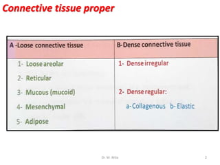
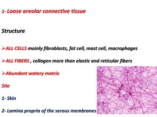
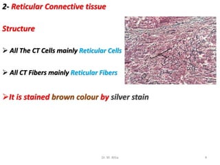
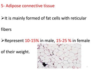
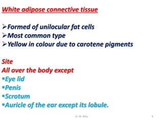
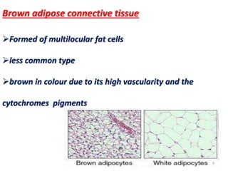
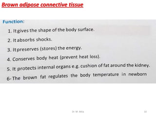
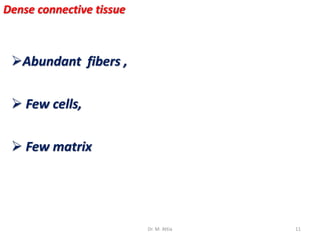
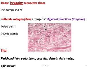
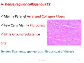
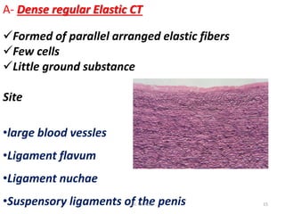
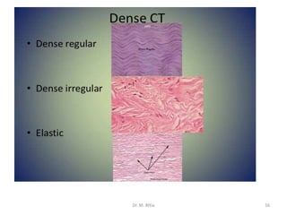


This document describes the different types of connective tissue proper, including loose areolar connective tissue found in the skin and serous membranes, reticular connective tissue stained brown by silver, mucous connective tissue in the umbilical cord and developing teeth, and mesenchymal connective tissue present during embryonic life. It also discusses adipose connective tissue, including white adipose tissue formed of unilocular fat cells found all over the body except certain areas, and brown adipose tissue formed of multilocular fat cells and brown in color. Finally, it covers dense connective tissue with abundant fibers and few cells and matrix, including dense irregular connective tissue in capsules and dermis, and dense regular


