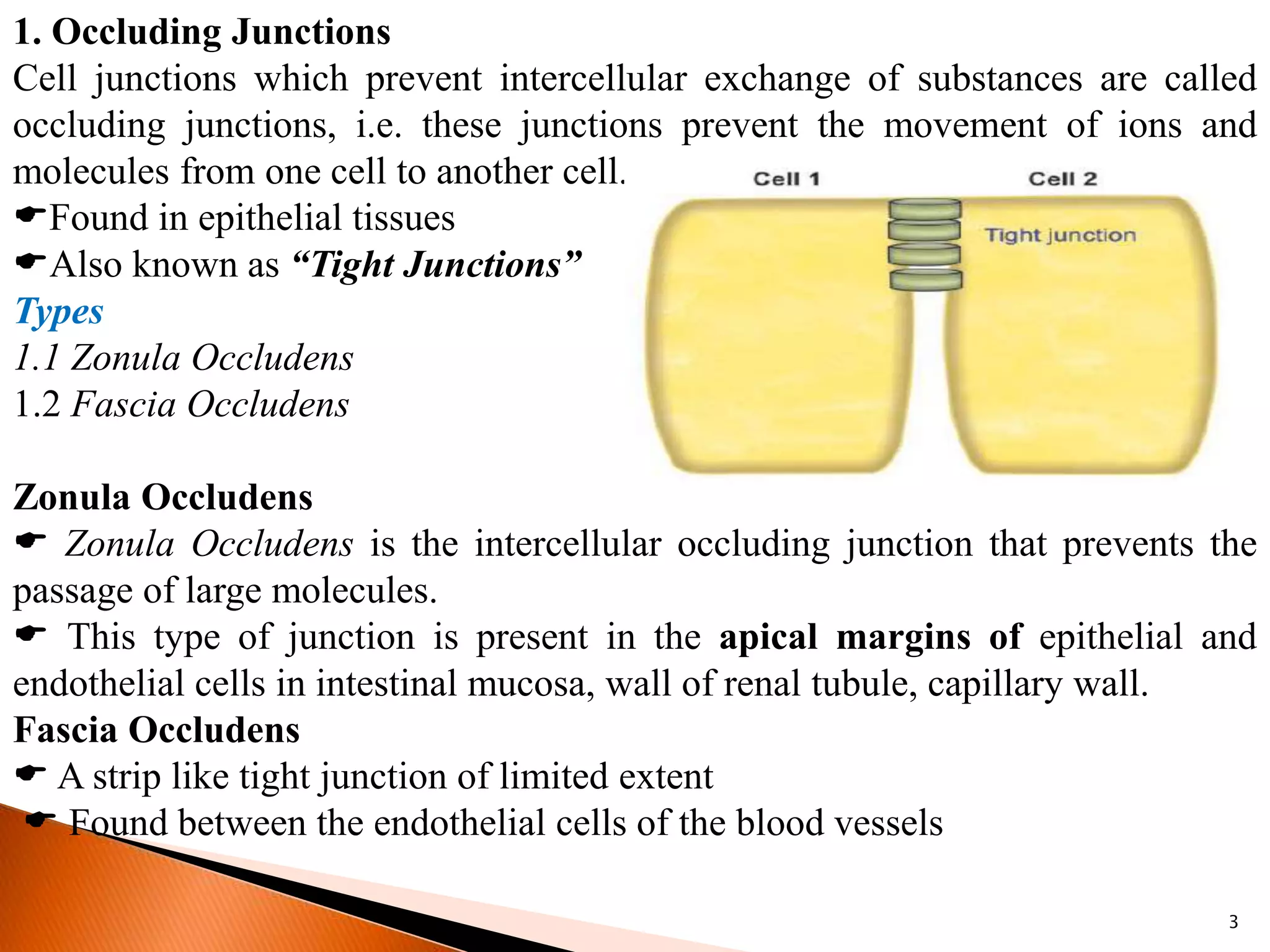
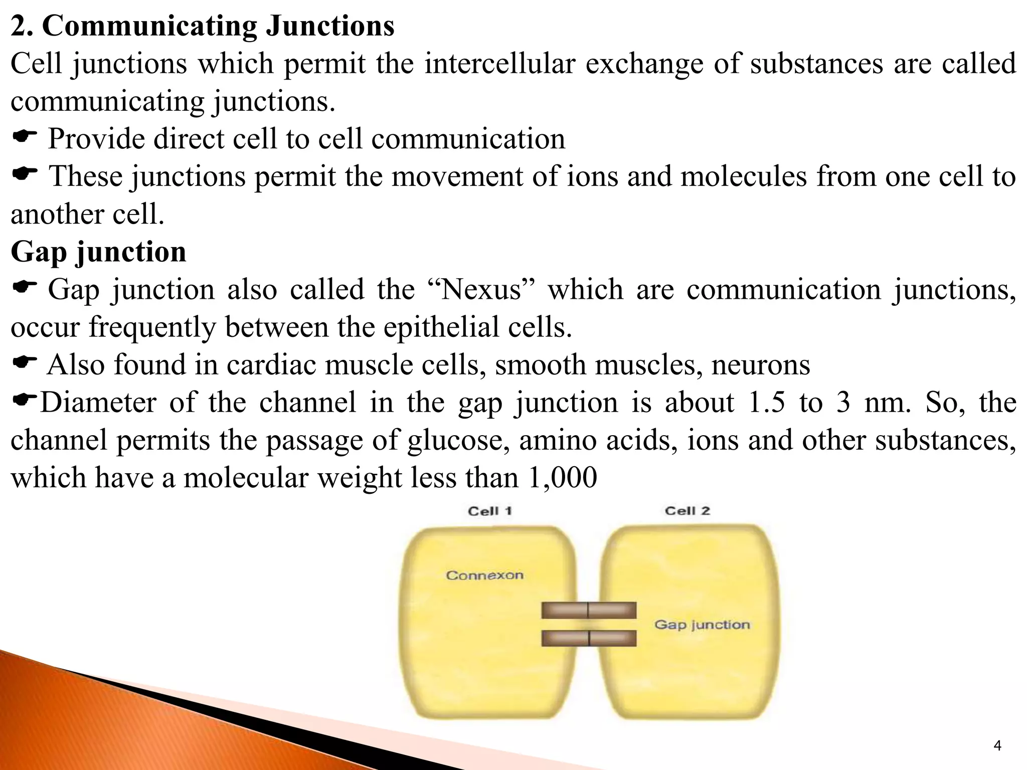
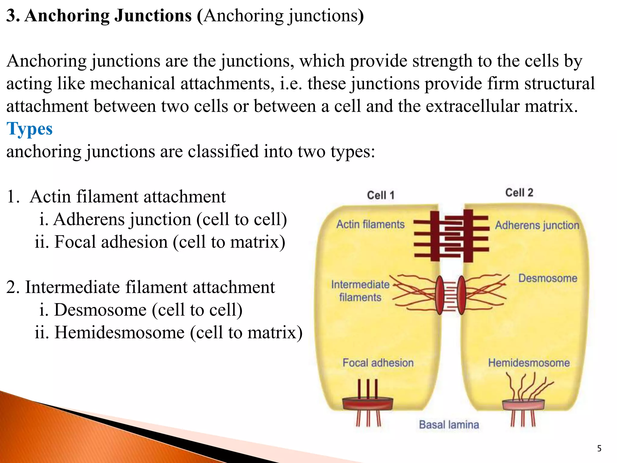
CELL
(Cell Junctions, Transport Through
Cell Membranes)
Shaibal Chandra
Assistant Professor
Pharmacology
Bengal School Of Technology
1
Cell Junctions
Cell junction is the connection between the neighboring cells or the contact
between the cell and extracellular matrix. It is also called membrane junction.
 Intercellular space in closely packed tissue is about 20nm.
 The cells are bound together by the specific adhesive glycoprotein
(Cadherins).
Types of Cell Junctions
Cell junctions are classified into three types:
1. Occluding junctions
2. Communicating junctions
3. Anchoring junctions.
2
1. Occluding Junctions
Cell junctions which prevent intercellular exchange of substances are called
occluding junctions, i.e. these junctions prevent the movement of ions and
molecules from one cell to another cell.
Found in epithelial tissues
Also known as “Tight Junctions”
Types
1.1 Zonula Occludens
1.2 Fascia Occludens
Zonula Occludens
 Zonula Occludens is the intercellular occluding junction that prevents the
passage of large molecules.
 This type of junction is present in the apical margins of epithelial and
endothelial cells in intestinal mucosa, wall of renal tubule, capillary wall.
Fascia Occludens
 A strip like tight junction of limited extent
 Found between the endothelial cells of the blood vessels
3
2. Communicating Junctions
Cell junctions which permit the intercellular exchange of substances are called
communicating junctions.
 Provide direct cell to cell communication
 These junctions permit the movement of ions and molecules from one cell to
another cell.
Gap junction
 Gap junction also called the “Nexus” which are communication junctions,
occur frequently between the epithelial cells.
 Also found in cardiac muscle cells, smooth muscles, neurons
Diameter of the channel in the gap junction is about 1.5 to 3 nm. So, the
channel permits the passage of glucose, amino acids, ions and other substances,
which have a molecular weight less than 1,000
4
3. Anchoring Junctions (Anchoring junctions)
Anchoring junctions are the junctions, which provide strength to the cells by
acting like mechanical attachments, i.e. these junctions provide firm structural
attachment between two cells or between a cell and the extracellular matrix.
Types
anchoring junctions are classified into two types:
1. Actin filament attachment
i. Adherens junction (cell to cell)
ii. Focal adhesion (cell to matrix)
2. Intermediate filament attachment
i. Desmosome (cell to cell)
ii. Hemidesmosome (cell to matrix)
5
Adherens Junction
Adherens junction is the cell to cell junction, which connects the actin filaments
of one cell to those of another cell.
In some places like epithelial linings, this junction forms a continuous
adhesion (zonula adherens)
Adherens junction provides strong mechanical attachments of the adjacent
cells
Focal Adhesion
Focal adhesion is the cell to matrix junctions, which connects the actin
filaments of the cell to the extracellular matrix.
Mainly found in Cardiac Muscle
6
Desmosome
Desmosome is a cell to cell junction, where the intermediatefilaments connect
two adjacent cells. Desmosome is also called macula adherens
The membranes of two adjacent cells, which oppose each other, are
thickened and become spotlike patches.
Desmosomes function like tight junctions.
It is also found in cardiac muscle cells.
Hemidesmosome
Hemidesmosome is a cell to matrix junction, which connects the intermediate
filaments of the cell to the extracellular matrix.
 These junctions serve to anchor the epithelial cells to the basal lamina
 The proteins involved in this are integrins
7
8
9
CELLADHESION MOLECULES
Cell adhesion molecules (CAMs) or cell adhesion proteins are the protein
molecules, which are responsible for the attachment of cells to their neighbors
or to basal lamina (or basal membrane).
TYPES OF CELLADHESION MOLECULES
Cell adhesion molecules are classified into four types:
1. Cadherins, which form the molecular limbs between neighboring cells. These
CAMs form adherens junction and desomosome.
2. Integrins, which form the focal adhesion and hemidesmosome
3. IgG super family, which form the cell adhesion molecules in nervous system
4. Selectins, which act as receptors for carbohydrates (ligand or mucin) and are
found in platelets and endothelial cells.
10
TRANSPORT THROUGH CELL MEMBRANE
All the cells in the body must be supplied with essential substances like
nutrients, water, electrolytes, etc. Cells also must get rid of many unwanted
substances like waste materials, carbon dioxide, etc. The cells achieve these by
means of transport mechanisms across the cell membrane.
BASIC MECHANISM OF TRANSPORT
Two types of basic mechanisms are involved in the transport of substances
across the cell membrane:
1. Passive transport mechanism
2. Active transport mechanism.
Passive Transport
Passive transport is the transport of substances along the concentration gradient
or electrical gradient or both (electrochemical gradient). It is also known as
diffusion or downhill movement. It does not need energy.
11
Passive diffusion types
1. Simple diffusion through lipid layer
2. Simple diffusion through protein layer
3. Facilitated or carrier-mediated diffusion
SIMPLE DIFFUSION THROUGH LIPID LAYER
Lipid layer of the cell membrane is permeable only to lipid-soluble substances
like oxygen, carbon dioxide and alcohol.
SIMPLE DIFFUSION THROUGH PROTEIN LAYER
Protein layer of the cell membrane is permeable to water-soluble substances.
Mainly, electrolytes diffuse through the protein layer.
FACILITATED OR CARRIER MEDIATED DIFFUSION
Facilitated or carrier-mediated diffusion is the type of diffusion by which the
water-soluble substances having larger molecules are transported through the
cell membrane with the help of a carrier protein. Glucose and amino acids are
transported by facilitated diffusion.
12
Hypothetical diagram of simple diffusion through the cell membrane.
A. Diffusion through lipid layer; B. Diffusion through ungated channel;
C. Diffusion through gated channel
13
FACILITATED OR CARRIER MEDIATED DIFFUSION
Hypothetical diagram of facilitated diffusion from higher concentration (ECF)
to lower concentration (ICF).
Stage 1. Glucose binds with carrier protein. Stage 2. Conformational change
occurs in the carrier protein and glucose is released into ICF.
14
ACTIVE TRANSPORT
 Active transport is the movement of substances against the chemical or
electrical or electrochemical gradient.
 It is like swimming against the water tide in a river.
 It is also called uphill transport.
Active transport requires energy, which is obtained mainly by breakdown
of high energy compounds like adenosine triphosphate (ATP).
CARRIER PROTEINS OF ACTIVE TRANSPORT
Carrier proteins involved in active transport are of two types:
1. Uniport
2. Symport or antiport.
1. Uniport
Carrier protein that carries only one substance in a single direction is called
uniport. It is also known as uniport pump.
2. Symport or Antiport
Symport or antiport is the carrier protein that transports two substances at a
time.
15
TYPES OF ACTIVE TRANSPORT
Active transport is of two types:
1. Primary active transport
2. Secondary active transport.
PRIMARY ACTIVE TRANSPORT
Primary active transport is the type of transport mechanism in which the energy
is liberated directly from the breakdown of ATP.
SECONDARY ACTIVE TRANSPORT
Secondary active transport is the transport of a substance with sodium ion, by
means of a common carrier protein.
16

Cell Junctions & Transport Through Cell Membranes

  • 1.
    CELL (Cell Junctions, TransportThrough Cell Membranes) Shaibal Chandra Assistant Professor Pharmacology Bengal School Of Technology 1
  • 2.
    Cell Junctions Cell junctionis the connection between the neighboring cells or the contact between the cell and extracellular matrix. It is also called membrane junction.  Intercellular space in closely packed tissue is about 20nm.  The cells are bound together by the specific adhesive glycoprotein (Cadherins). Types of Cell Junctions Cell junctions are classified into three types: 1. Occluding junctions 2. Communicating junctions 3. Anchoring junctions. 2
  • 3.
    1. Occluding Junctions Celljunctions which prevent intercellular exchange of substances are called occluding junctions, i.e. these junctions prevent the movement of ions and molecules from one cell to another cell. Found in epithelial tissues Also known as “Tight Junctions” Types 1.1 Zonula Occludens 1.2 Fascia Occludens Zonula Occludens  Zonula Occludens is the intercellular occluding junction that prevents the passage of large molecules.  This type of junction is present in the apical margins of epithelial and endothelial cells in intestinal mucosa, wall of renal tubule, capillary wall. Fascia Occludens  A strip like tight junction of limited extent  Found between the endothelial cells of the blood vessels 3
  • 4.
    2. Communicating Junctions Celljunctions which permit the intercellular exchange of substances are called communicating junctions.  Provide direct cell to cell communication  These junctions permit the movement of ions and molecules from one cell to another cell. Gap junction  Gap junction also called the “Nexus” which are communication junctions, occur frequently between the epithelial cells.  Also found in cardiac muscle cells, smooth muscles, neurons Diameter of the channel in the gap junction is about 1.5 to 3 nm. So, the channel permits the passage of glucose, amino acids, ions and other substances, which have a molecular weight less than 1,000 4
  • 5.
    3. Anchoring Junctions(Anchoring junctions) Anchoring junctions are the junctions, which provide strength to the cells by acting like mechanical attachments, i.e. these junctions provide firm structural attachment between two cells or between a cell and the extracellular matrix. Types anchoring junctions are classified into two types: 1. Actin filament attachment i. Adherens junction (cell to cell) ii. Focal adhesion (cell to matrix) 2. Intermediate filament attachment i. Desmosome (cell to cell) ii. Hemidesmosome (cell to matrix) 5
  • 6.
    Adherens Junction Adherens junctionis the cell to cell junction, which connects the actin filaments of one cell to those of another cell. In some places like epithelial linings, this junction forms a continuous adhesion (zonula adherens) Adherens junction provides strong mechanical attachments of the adjacent cells Focal Adhesion Focal adhesion is the cell to matrix junctions, which connects the actin filaments of the cell to the extracellular matrix. Mainly found in Cardiac Muscle 6
  • 7.
    Desmosome Desmosome is acell to cell junction, where the intermediatefilaments connect two adjacent cells. Desmosome is also called macula adherens The membranes of two adjacent cells, which oppose each other, are thickened and become spotlike patches. Desmosomes function like tight junctions. It is also found in cardiac muscle cells. Hemidesmosome Hemidesmosome is a cell to matrix junction, which connects the intermediate filaments of the cell to the extracellular matrix.  These junctions serve to anchor the epithelial cells to the basal lamina  The proteins involved in this are integrins 7
  • 8.
  • 9.
    9 CELLADHESION MOLECULES Cell adhesionmolecules (CAMs) or cell adhesion proteins are the protein molecules, which are responsible for the attachment of cells to their neighbors or to basal lamina (or basal membrane). TYPES OF CELLADHESION MOLECULES Cell adhesion molecules are classified into four types: 1. Cadherins, which form the molecular limbs between neighboring cells. These CAMs form adherens junction and desomosome. 2. Integrins, which form the focal adhesion and hemidesmosome 3. IgG super family, which form the cell adhesion molecules in nervous system 4. Selectins, which act as receptors for carbohydrates (ligand or mucin) and are found in platelets and endothelial cells.
  • 10.
    10 TRANSPORT THROUGH CELLMEMBRANE All the cells in the body must be supplied with essential substances like nutrients, water, electrolytes, etc. Cells also must get rid of many unwanted substances like waste materials, carbon dioxide, etc. The cells achieve these by means of transport mechanisms across the cell membrane. BASIC MECHANISM OF TRANSPORT Two types of basic mechanisms are involved in the transport of substances across the cell membrane: 1. Passive transport mechanism 2. Active transport mechanism. Passive Transport Passive transport is the transport of substances along the concentration gradient or electrical gradient or both (electrochemical gradient). It is also known as diffusion or downhill movement. It does not need energy.
  • 11.
    11 Passive diffusion types 1.Simple diffusion through lipid layer 2. Simple diffusion through protein layer 3. Facilitated or carrier-mediated diffusion SIMPLE DIFFUSION THROUGH LIPID LAYER Lipid layer of the cell membrane is permeable only to lipid-soluble substances like oxygen, carbon dioxide and alcohol. SIMPLE DIFFUSION THROUGH PROTEIN LAYER Protein layer of the cell membrane is permeable to water-soluble substances. Mainly, electrolytes diffuse through the protein layer. FACILITATED OR CARRIER MEDIATED DIFFUSION Facilitated or carrier-mediated diffusion is the type of diffusion by which the water-soluble substances having larger molecules are transported through the cell membrane with the help of a carrier protein. Glucose and amino acids are transported by facilitated diffusion.
  • 12.
    12 Hypothetical diagram ofsimple diffusion through the cell membrane. A. Diffusion through lipid layer; B. Diffusion through ungated channel; C. Diffusion through gated channel
  • 13.
    13 FACILITATED OR CARRIERMEDIATED DIFFUSION Hypothetical diagram of facilitated diffusion from higher concentration (ECF) to lower concentration (ICF). Stage 1. Glucose binds with carrier protein. Stage 2. Conformational change occurs in the carrier protein and glucose is released into ICF.
  • 14.
    14 ACTIVE TRANSPORT  Activetransport is the movement of substances against the chemical or electrical or electrochemical gradient.  It is like swimming against the water tide in a river.  It is also called uphill transport. Active transport requires energy, which is obtained mainly by breakdown of high energy compounds like adenosine triphosphate (ATP). CARRIER PROTEINS OF ACTIVE TRANSPORT Carrier proteins involved in active transport are of two types: 1. Uniport 2. Symport or antiport. 1. Uniport Carrier protein that carries only one substance in a single direction is called uniport. It is also known as uniport pump. 2. Symport or Antiport Symport or antiport is the carrier protein that transports two substances at a time.
  • 15.
    15 TYPES OF ACTIVETRANSPORT Active transport is of two types: 1. Primary active transport 2. Secondary active transport. PRIMARY ACTIVE TRANSPORT Primary active transport is the type of transport mechanism in which the energy is liberated directly from the breakdown of ATP. SECONDARY ACTIVE TRANSPORT Secondary active transport is the transport of a substance with sodium ion, by means of a common carrier protein.
  • 16.