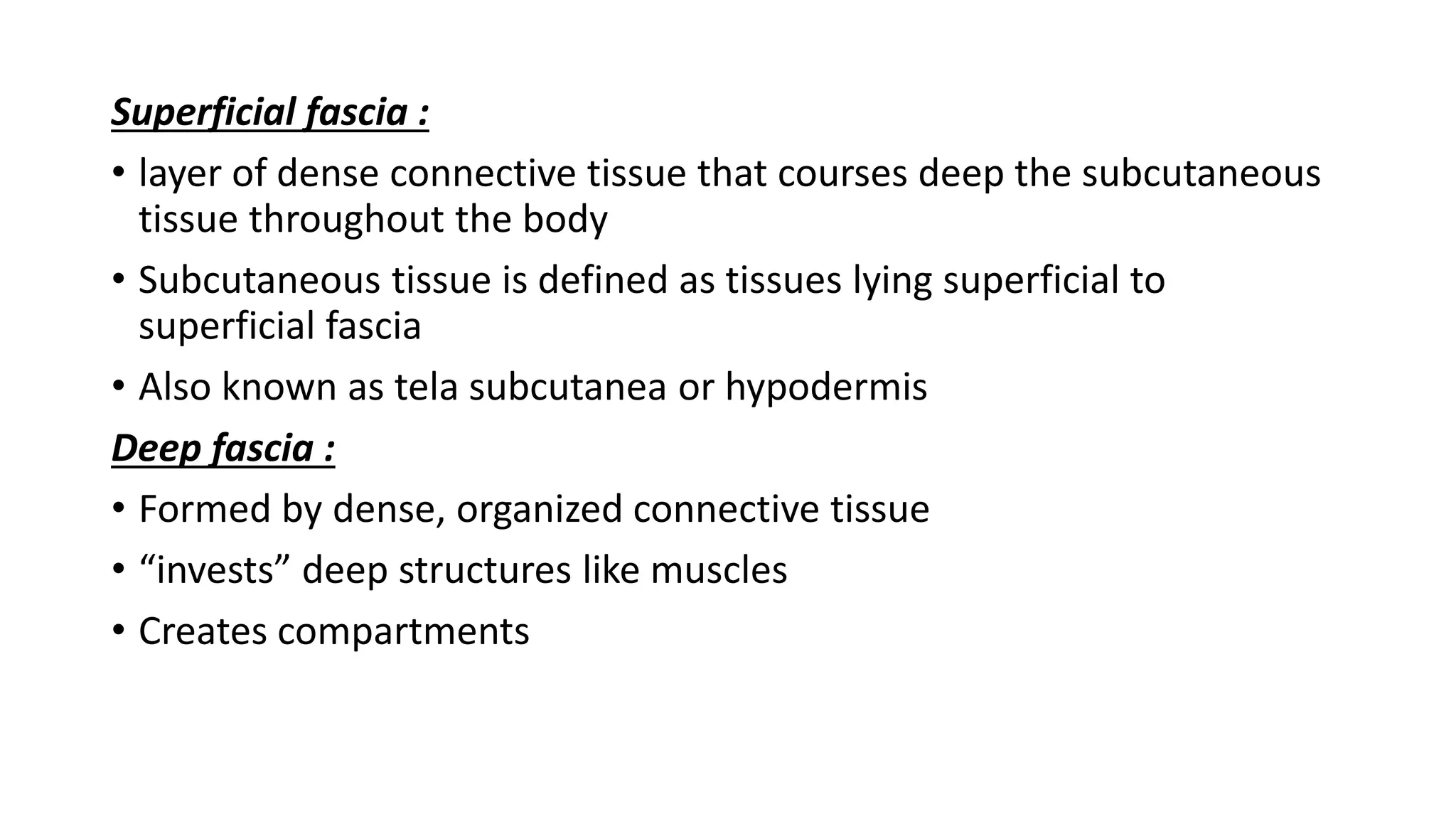
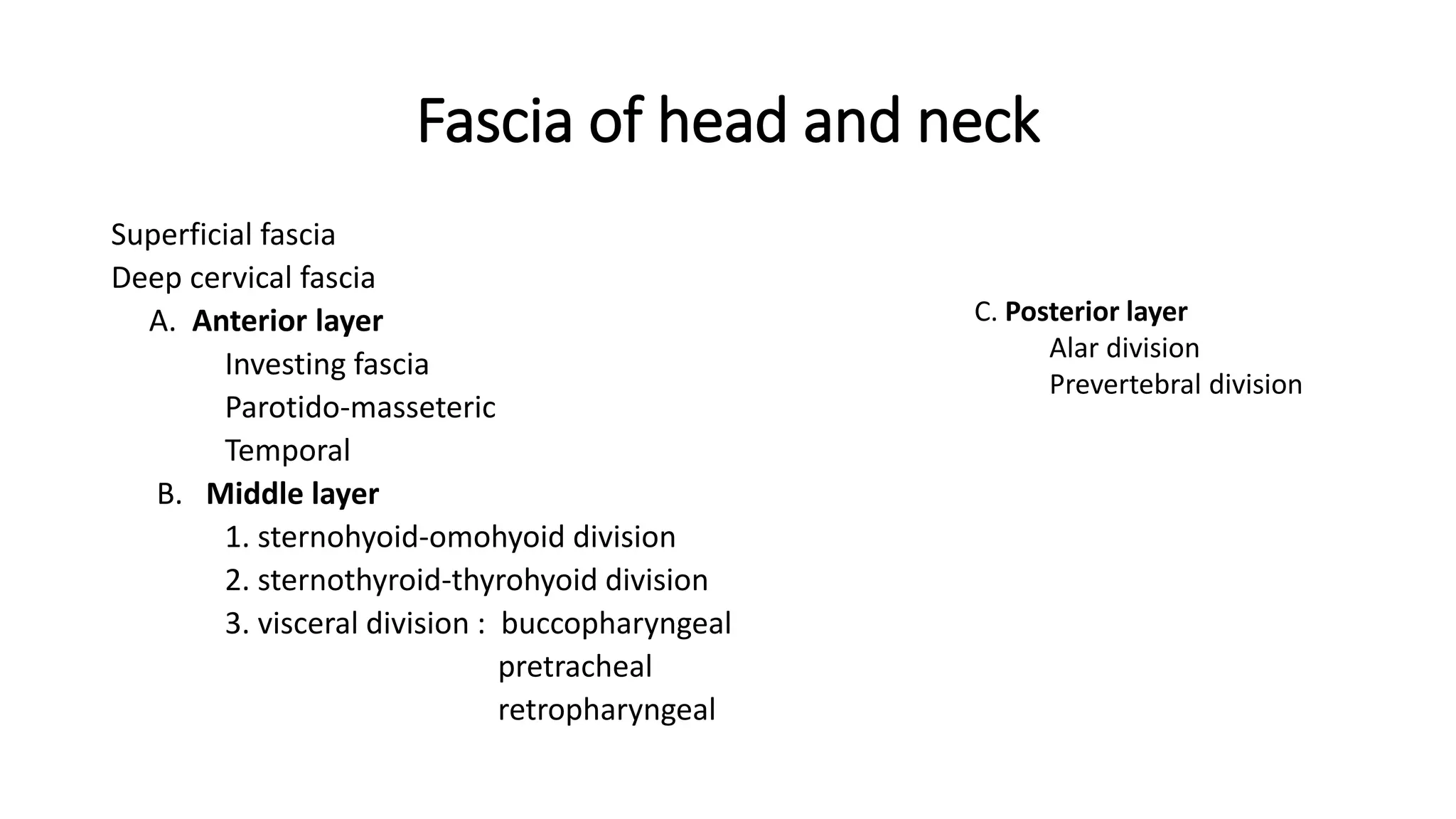
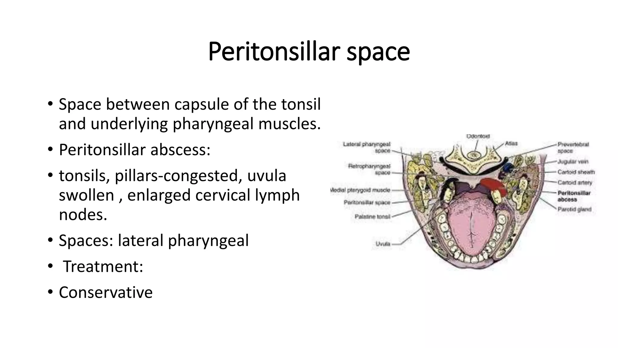
Fascial spaces
By Dr Rayan.M
Guided by Dr Archana
Contents
• Introduction
• Classification
• Fascial spaces
• Infections of spaces
• Treatment
Fascial spaces
Definition
The fascial spaces in the head and neck are the potential spaces
between the various layers of fascia normally filled with loose
connective tissue and bounded by anatomical barriers, usually of bone,
muscle or fascial layers
Moore 1975
Shapiro defined fascial spaces as potential spaces between layers of fascia.
It is filled with loose connective tissues and various anatomical structures.
Fascia
The term fascia is used to
describe broad sheets of dense
connective tissue whose
function is to separate
structures that must pass over
each other during movement,
covers muscles and glands, and
serves as a pathway for the
course of vascular and neural
structures
• Fascia of head and neck can be described under
 Superficial fascia
 Deep fascia
Superficial fascia :
• layer of dense connective tissue that courses deep the subcutaneous
tissue throughout the body
• Subcutaneous tissue is defined as tissues lying superficial to
superficial fascia
• Also known as tela subcutanea or hypodermis
Deep fascia :
• Formed by dense, organized connective tissue
• “invests” deep structures like muscles
• Creates compartments
Fascia of head and neck
Superficial fascia
Deep cervical fascia
A. Anterior layer
Investing fascia
Parotido-masseteric
Temporal
B. Middle layer
1. sternohyoid-omohyoid division
2. sternothyroid-thyrohyoid division
3. visceral division : buccopharyngeal
pretracheal
retropharyngeal
C. Posterior layer
Alar division
Prevertebral division
Superficial fascia
• Layer of dense connective tissue that courses deep to subcutaneous
tissue through the body
• Subcutaneous space mainly contains variable amount of fat and
areolar tissue.
• In the lower third of the face, muscles of facial expression lie deep to
the superficial fascia, where as in the upper face the muscles of facial
expression are positioned superficial to this layer
Deep Fascia
Investing layer
Investing layer of deep cervical fascia
• It lies beneath the platysma
• Superior: External occipital protuberance & superior nuchal lines, mastoid
process, EAM, lower margin of zygomatic arch, lower border of body of
mandible
• Inferiorly : Spine & acromial process of scapula, upper surface of clavicle,
suprasternal notch
• Posterior: Ligamentum nuchae and cervical spine of vertebrae
• Anterior : Continuous with opposite side
• EXTENT
• Horizontal: ligamentum nuchae, trapezius, posterior triangle, SCM,
anterior triangle.
• Vertical : Traced above:from anterior border of SCM, across muscles
of floor of the mouth, attach to mandible, fascia splits to enclose the
submandibular and parotid gland
• Behind - parotidomasseteric fascia
stylomandibular ligament
Traced below: between 2 heads of SCM, splits above sternum- supra
sternal space and supraclavicular space
• Just above the sternum fascia splits to form the suprasternal space or
the space of Burns
Middle layer of deep cervical fascia
• Sternohyoid-omohyoid
• Sternothyroid-thyrohyoid
• Visceral :Often broken down into Retropharyngeal, Lateral
Pharyngeal and Buccopharyngeal components as it
posteriorly to anteriorly envelops the pharynx
• Buccopharyngeal fascia:
• Wraps around posterior and lateral side of the pharynx,
superficial side of constrictors.
• Continuous above with buccinator
• Below with visceral fascia on oesophagus
Surround the corresponding strap muscles of the neck
between the hyoid bone and clavicle
Primary surgical significance of these layers is that they must
be divided in the midline in a surgical approach to the
trachea or thyroid gland
Pretracheal fascia
Extent:
• Horizontal: behind the strap
muscles, undercover of SCM,
passes medially forming anterior
wall of carotid sheath,encloses
thyroid gland,passes in front of
trachea.
• Vertical :above: hyoid bone,
oblique line of thyroid cartilage.
• Below: enters thorax, blends
with apex of fibrous pericardium.
Prevertebral fascia
Extent :
• Horizontal: Beneath SCM, passes
medially forming the posterior wall
of carotid sheath, covers the
cervical vertebrae and prevertebral
muscles
• Vertical: Above to base of skull,
Below: Anterior layer is alar fascia ,
Posterior layer enters the thorax
and blends with anterior
longitudinal ligament.
Carotid sheath
• Formed by all three layers of deep fascia
• Anterior wall: Pretracheal fascia
• Posterior wall: Prevertebral fascia.
• The sheath extends downwards along
the vessels from base of skull to arch of
aorta
• Lincoln highway – coined by Mosher,
extending from jugular foramen and
carotid canal at the base of skull to the
middle mediastinum
Fascial spaces
Most of the anatomic spaces in head and neck communicate with each
other either directly or indirectly, and, when the balance between
bacterial virulence and patient resistance favors extension of the
infection, spread can occur from one region to another
Classification of Fascial Spaces
• Primary Fascial spaces
• Maxillary
• Canine
• Buccal
• Infratemporal
• Mandibular
• Submental
• Buccal
• Sublingual
• Submandibular
Secondary Fascial Spaces
Pterygomandibular
Retropharyngeal
Lateral pharyngeal
Prevertebral
Classification Grondinsky & Holyoke 1938
Deep neck spaces
• Space 1: superficial to the superficial fascia – subcutaneous space
• Space 2: group of spaces surrounding the strap muscles lying superficial to
the first 2 divisions of middle layer
• Space 3: lying superficial to the visceral division – contains pretracheal,
retropharyngeal & lateral pharyngeal
• Space 3A: is the carotid sheath
• Space 4: lies between the alar & prevertebral division – danger space
• Space 4A: is in posterior triangle of the neck, posterior to carotid sheath
• Space 5: prevertebral space
• Space 5A: enclosed by the prevertebral fascia, posterior to the transverse
processes of the vertebrae, as it surrounds the scalene and spinal postural
muscles.
Vestibular space
• Space between oral vestibular mucosa and nearby muscles of facial
expression.
• Anterior -intrinsic muscles of lip
• Posterior- buccinator
• Contents- long buccal, mental nerve.
• Dentoalveolar abscess occupy a portion of this space
• Clinical features:
• Swelling of mucosa
• Treatment:
• Incision on most dependent part.
Subcutaneous space
• The potential space between the
superficial fascia, along with muscles
of facial expression, and the skin
• Infection in any deep fascial space
may point through the subcutaneous
space to the skin
Infraorbital space/Canine space
• Anterior: nasal cartilages
• Posterior: Anterior Maxilla
• Superior: Levator labi superioris
• Inferior:oral mucosa
• Medial:quadratus labi superioris
• Lateral: levator anguli oris
• Contents:
• Angular vein, infraorbital nerve.
• Space: buccal
• Infection from canines can involve
this space
• Clinical features: marked cellulitis of
the lower eyelids, obliteration of
the naso labial fold.
• Treatment: incision given high in
maxillary labial vestibule
• Complications: cavernous sinus
thrombosis
Buccal space
• Boundaries
• Anterior : posterior border of
zygomaticus major above and below
depressor anguli oris
• Posterior: anterior edge masseter
• Superior : zygomatic arch
• Inferior: lower border of mandible
• Medial : buccinator and its covering
buccopharengeal fascia.
• Lateral: skin and subcutaneous tissue
• Contents :parotid duct, transverse
facial artery, buccal pad of fat
• Infections from upper molars and lower
1st and 2nd molars
• Clinical features: dome shaped swelling
and considerable edema
Communications
• Superficially – submassetric space
• infra orbital
• Medially – pterygomandibular,
• Superficial temporal and infratemporal
• Lateral pharengeal space
• Treatment:
• extra oral approach
• intraoral approach
Submandibular space
• Boundaries
• Laterally: skin superficial fascia, platysma & superficial layer of deep
cervical fascia
• Medial: mylohyiod, hyoglossus and styloglossus
• Superior: medial surface of mandible and attachment of Mylohyoid
• Inferior: anterior and posterior bellies of digastric muscle
• Contents: submandibular gland, branches of facial artery, lymph
nodes
• Spaces: sublingual, submental, lateral pharyngeal, buccal
• Infections from mandibular 2nd
and 3rd molars, submental space
infection
• Clinical features:
• Firm swelling , tenderness,
redness, tooth sensitive to
percussion
• Treatment: incision and drainage
Sublingual space
• Boundaries
• Anterior: lingual surface of mandible
• Posterior: body of hyoid
• Superior: oral mucosa
• Inferior: mylohyoid muscle
• Medial: geniohyoid genioglossus and styloglossus
• Lateral: lingual surface of mandible
• Contents: sublingual gland, Wharton’s duct, lingual nerve, sublingual
vessels
• Spaces: submandibular, lateral pharyngeal.
• Infections from
mandibular pre molars,
mandibular 1st molars and
2nd molars
• Features:
• Little swelling, firm
painful swelling in floor of
the mouth
• Treatment:
• Incised intraorally at base
of alveolar process in
lingual sulcus.
Submental space
• Anterior: Inferior border of mandible
• Superior: mylohyoid muscle
• Inferior: skin, superficial fascia, platysma
and deep cervical fascia
• Medial: investing fascia
• Lateral: anterior belly of diagastric
• Contents:anterior jugular vein, lymph
nodes
• Space:submandibular
• Causes: Mandibular anteriors
• Clinical features:
• Midline swelling below the
chin
• intra oral-tender tooth.
• Treatment:
• Bilaterally through skin.
Masticator spaces
• Submassetric space
• Pterygomandibular space
• Superficial temporal
• Deep temporal
Submassetric space
• Anterior : facial extension of
parotideo-masseteric fascia
• Posterior: parotid gland
• Superior:zygomatic arch
• Inferior:inferior border of
mandible
• Medial:ascending ramus of
mandible
• Lateral: masseter
• Contents: massetric vessels
• Spaces: buccal, pterygomandibular,
superficial temporal, parotid
• Causes:
• Mandibular 3rd molar, trauma
• Clinical features:
trismus,pain,dysphagia,
• Brawny induration
• Treatment:
• intra oral: mucobuccal fold
opposite to 3rd molar.
• Extraoral: below and parallel to
angle of mandible
Pterygomandibular space
• Anterior: buccal space
• Posterior: parotid gland
• Superior: lateral pterygoid
• Inferior: Inferior border of mandible
• Medial: Medial pterygoid
• Lateral: Ascending ramus of
mandible
• Contents: mandibular nerve, inferior
alveolar vessels
• Causes: pericoronitis of 3rd molar,
contaminated needle during IANB
• Clinical features:
• Trismus,tenderness,dysphagia,
redness
• Treatment:
• Intra oral:
• Between medial aspect of ramus of
mandible and pterygomandibular
raphae.
• Extra oral:below the angle of
mandible
Infratemporal space
• Anterior: infratemporal surface of maxilla & posterior surface of zygomatic
bone
• Posterior: lateral pterygoid muscle, condyle, temporalis muscle
• Laterally: tendon of the temporalis muscle, coronoid process
• Medially: lateral pterygoid plate and inferior belly of the lateral pterygoid
muscle
• Superior: infratemporal surface of greater wing of sphenoid
• Contents:
• Pterygoid plexus, internal maxillary artery, mandibular, mylohyoid, lingual,
buccinator & chorda tympani nerves and the lateral pterygoid muscle
• Clinical features:
• Trismus, swelling in the sigmoid notch region
• Swelling of the lateral posterior portion of the soft palate
• Approaches:
• Extraoral through
suprazygomatic approach
• Intraoral incision in the
superior aspect of the
posterior maxillary
buccal vestibule
Superficial temporal space
• Lies betweeen temporalis fascia and
temporalis muscle
• Anterior: posterior surface of lateral
orbital rim
• Posterior: fusion of temporalis fascia
with pericranium
• Lateral: medial surface of temporalis
• Medial: temporal bone & greater
wing of sphenoid
• Inferior: zygomatic arch
• Contents: buccal fat of pad, temporal
branch of facial nerve
• Causes: secondary
• Clinical features: pain, trismus,
limited swelling superiorly and
laterally
• Treatment: extra oral
• Intra oral: vertical incision, medial
aspect of coronoid process.
Parotid space
• Formed by splitting of investing layer of
the deep cervical fascia to form the
capsule of the parotid gland–
parotideomasseteric fascia
• Superior: TMJ & external auditory meatus
• Posterior & inferior: mastoid process,
sternocleidomastoid muscle, posterior
belly of the digastric muscle
• Contents: Parotid gland, facial nerve,
posterior facial lymphatics, ECA & its
terminal branches,retromandibular vein
• Causes:
• Extension from lateral pharyngeal
space, submasseteric and
pterygomandibular space
• Retrograde extension along parotid
duct
• Clinical features:
• Swelling from zygomatic arch to lower
border of mandible and posteriorly till
retromandibular region
• Severe pain referred to ear
• Treatment: retromandibular incision
Periorbital space
• Preseptal space
• Between orbicularis oculi muscle and the orbital septum leaflet
arising from the periosteum of the orbital rim extending & attaching
to the tarsal plates
• Divided into upper and lower compartments by medial & lateral
canthal ligaments
• Contents:Fat & areolar connective tissue
• Communicates:Orbital space by penetration of the orbital septum
and may drain through the thin overlying skin
Orbital space
• Bounded by bony
walls of the orbit
• Anterior Orbital
septum – divides
from periorbital space
• Cone shaped space
with its base at the
orbital septum & its
apex posteromedially
at the optic foramen
• Lateral wall of the orbit is perforated by orbital fissures
• Damage to structures passing through superior orbital fissure
• superior orbital fissure syndrome
• Paresthesia of the upper eyelid & forehead
• Paralysis of the extraocular & pupillary muscles
• Optic nerve is not involved
• Damage to the structures passing through superior orbital fissure and
those passing through the optic foramen
Orbital apex syndrome
Lateral pharyngeal space
• Anterior: superior and middle constrictor
• Posterior: carotid sheath and scalene fascia
• Superior: skull base
• Inferior: hyoid bone
• Medial: pharyngeal constrictors,
retropharyngeal space
• Lateral: medial pterygoid
• Contents: carotid artery, IJV, vagus nerve,
cervical sympathetic chain
• Spaces: deep temporal, pterygomandibular,
submandibular, sublingual, peritonsillar,
retropharyngeal
• Causes:pharyngitis, tonsillitis,
parotitis, otitis,
mastoiditis,mandibular molar
infections
• Clinical features: pain, fever,
chills,medial bulging,deviation of
uvula, dyphagia, swelling below the
angle of mandible.
• Posterior compartment:respiratory
obstruction.
• Treatment:intra oral:vertical incision,
lateral to pterygomandibular
raphae,posterior inferior direction
• Extra oral:horizontal incision anterior
and inferior to mandible.
Retropharyngeal space
• Anterior: posterior wall of pharynx
• Posterior: alar fascia and vertebral
column
• Superior: skull base
• Inferior: fusion of alar and
prevertebral fascia
• Lateral: carotid sheath , lateral
pharyngeal space
• Contents: areolar connective tissue,
lymph nodes
• Causes: nasal/pharyngeal infection,diffusing dental infection,
oesophageal trauma
• Clinical features:dysphagia,dyspnoea,nuchal rigidity,oesophageal
regurgitation, fever
• Treatment:intra oral: vertical incision in pharyngeal wall lateral to
midline.
• Trendelenburg positioning of the patient
• External approach – incision made along the anterior border of
sternocleidomastoid muscle and parallel to it, inferior to the hyoid
bone
Pretracheal space
• It is the anterior portion of space 3
• Anterior: sternothyroid-thyrohyiod fascia
• Posterior: retropharyngeal space
• Superior: thyroid cartilage
• Inferior:superior medaistinum
• Medial: visceral fascia over the trachea and thyroid gland
• Lateral: sternothyroid- thyrohyoid fascia
• Space: retropharyngeal space
Peritonsillar space
• Space between capsule of the tonsil
and underlying pharyngeal muscles.
• Peritonsillar abscess:
• tonsils, pillars-congested, uvula
swollen , enlarged cervical lymph
nodes.
• Spaces: lateral pharyngeal
• Treatment:
• Conservative
Mediastinum
• Begins at 1st rib of manubrium of sternum.
• Inferior : from T4 to manubriosternal junction
• Anterior : superior- pretracheal space
• inferior- diaphragm
• laterally- visceral pleura
• Middle: heart, pericardium, phrenic nerve
• Posterior: thoracic aorta, venacava, thoracic duct,vagus nerve,
trachea, oesophagus
Mediastinitis
• Clinical features:
• Chest pain – substernal pain
• Severe dyspnea
• Unremitting fever
• Progressive septicemia
• Pleural effusion
• Empyema
• Compression of mediastinal veins with decrease venous return to the heart
• Pericarditis which may end in death
• Radiograph – mediastinal widening
• Treatment: antibiotic therapy, surgical drainage
References
• Oral and maxillofacial infections- Topazian 4th edition
• Textbook of oral and maxillofacial surgery- Kruger 6th edition
• Oral and maxillofacial surgery- volume two Daniel.M.Laskin
• Anatomy for surgeons vol.1 Henry Hollinshead 3rd edition
• Essentials of human anatomy vol.2 3rd edition
• Peterson s principles of oral and maxillofacial surgery, vol.1 2nd
editions
• Textbook of anatomy- Gray s

Fascial Spaces in the Head & Neck

  • 1.
    Fascial spaces By DrRayan.M Guided by Dr Archana
  • 2.
    Contents • Introduction • Classification •Fascial spaces • Infections of spaces • Treatment
  • 3.
    Fascial spaces Definition The fascialspaces in the head and neck are the potential spaces between the various layers of fascia normally filled with loose connective tissue and bounded by anatomical barriers, usually of bone, muscle or fascial layers Moore 1975
  • 4.
    Shapiro defined fascialspaces as potential spaces between layers of fascia. It is filled with loose connective tissues and various anatomical structures.
  • 5.
    Fascia The term fasciais used to describe broad sheets of dense connective tissue whose function is to separate structures that must pass over each other during movement, covers muscles and glands, and serves as a pathway for the course of vascular and neural structures
  • 6.
    • Fascia ofhead and neck can be described under  Superficial fascia  Deep fascia
  • 7.
    Superficial fascia : •layer of dense connective tissue that courses deep the subcutaneous tissue throughout the body • Subcutaneous tissue is defined as tissues lying superficial to superficial fascia • Also known as tela subcutanea or hypodermis Deep fascia : • Formed by dense, organized connective tissue • “invests” deep structures like muscles • Creates compartments
  • 8.
    Fascia of headand neck Superficial fascia Deep cervical fascia A. Anterior layer Investing fascia Parotido-masseteric Temporal B. Middle layer 1. sternohyoid-omohyoid division 2. sternothyroid-thyrohyoid division 3. visceral division : buccopharyngeal pretracheal retropharyngeal C. Posterior layer Alar division Prevertebral division
  • 9.
    Superficial fascia • Layerof dense connective tissue that courses deep to subcutaneous tissue through the body • Subcutaneous space mainly contains variable amount of fat and areolar tissue. • In the lower third of the face, muscles of facial expression lie deep to the superficial fascia, where as in the upper face the muscles of facial expression are positioned superficial to this layer
  • 10.
  • 11.
  • 12.
    Investing layer ofdeep cervical fascia • It lies beneath the platysma • Superior: External occipital protuberance & superior nuchal lines, mastoid process, EAM, lower margin of zygomatic arch, lower border of body of mandible • Inferiorly : Spine & acromial process of scapula, upper surface of clavicle, suprasternal notch • Posterior: Ligamentum nuchae and cervical spine of vertebrae • Anterior : Continuous with opposite side
  • 13.
    • EXTENT • Horizontal:ligamentum nuchae, trapezius, posterior triangle, SCM, anterior triangle. • Vertical : Traced above:from anterior border of SCM, across muscles of floor of the mouth, attach to mandible, fascia splits to enclose the submandibular and parotid gland • Behind - parotidomasseteric fascia stylomandibular ligament Traced below: between 2 heads of SCM, splits above sternum- supra sternal space and supraclavicular space • Just above the sternum fascia splits to form the suprasternal space or the space of Burns
  • 14.
    Middle layer ofdeep cervical fascia • Sternohyoid-omohyoid • Sternothyroid-thyrohyoid • Visceral :Often broken down into Retropharyngeal, Lateral Pharyngeal and Buccopharyngeal components as it posteriorly to anteriorly envelops the pharynx • Buccopharyngeal fascia: • Wraps around posterior and lateral side of the pharynx, superficial side of constrictors. • Continuous above with buccinator • Below with visceral fascia on oesophagus Surround the corresponding strap muscles of the neck between the hyoid bone and clavicle Primary surgical significance of these layers is that they must be divided in the midline in a surgical approach to the trachea or thyroid gland
  • 16.
    Pretracheal fascia Extent: • Horizontal:behind the strap muscles, undercover of SCM, passes medially forming anterior wall of carotid sheath,encloses thyroid gland,passes in front of trachea. • Vertical :above: hyoid bone, oblique line of thyroid cartilage. • Below: enters thorax, blends with apex of fibrous pericardium.
  • 18.
    Prevertebral fascia Extent : •Horizontal: Beneath SCM, passes medially forming the posterior wall of carotid sheath, covers the cervical vertebrae and prevertebral muscles • Vertical: Above to base of skull, Below: Anterior layer is alar fascia , Posterior layer enters the thorax and blends with anterior longitudinal ligament.
  • 19.
    Carotid sheath • Formedby all three layers of deep fascia • Anterior wall: Pretracheal fascia • Posterior wall: Prevertebral fascia. • The sheath extends downwards along the vessels from base of skull to arch of aorta • Lincoln highway – coined by Mosher, extending from jugular foramen and carotid canal at the base of skull to the middle mediastinum
  • 21.
    Fascial spaces Most ofthe anatomic spaces in head and neck communicate with each other either directly or indirectly, and, when the balance between bacterial virulence and patient resistance favors extension of the infection, spread can occur from one region to another
  • 22.
    Classification of FascialSpaces • Primary Fascial spaces • Maxillary • Canine • Buccal • Infratemporal • Mandibular • Submental • Buccal • Sublingual • Submandibular Secondary Fascial Spaces Pterygomandibular Retropharyngeal Lateral pharyngeal Prevertebral
  • 23.
    Classification Grondinsky &Holyoke 1938 Deep neck spaces • Space 1: superficial to the superficial fascia – subcutaneous space • Space 2: group of spaces surrounding the strap muscles lying superficial to the first 2 divisions of middle layer • Space 3: lying superficial to the visceral division – contains pretracheal, retropharyngeal & lateral pharyngeal • Space 3A: is the carotid sheath • Space 4: lies between the alar & prevertebral division – danger space • Space 4A: is in posterior triangle of the neck, posterior to carotid sheath • Space 5: prevertebral space • Space 5A: enclosed by the prevertebral fascia, posterior to the transverse processes of the vertebrae, as it surrounds the scalene and spinal postural muscles.
  • 25.
    Vestibular space • Spacebetween oral vestibular mucosa and nearby muscles of facial expression. • Anterior -intrinsic muscles of lip • Posterior- buccinator • Contents- long buccal, mental nerve. • Dentoalveolar abscess occupy a portion of this space • Clinical features: • Swelling of mucosa • Treatment: • Incision on most dependent part.
  • 27.
    Subcutaneous space • Thepotential space between the superficial fascia, along with muscles of facial expression, and the skin • Infection in any deep fascial space may point through the subcutaneous space to the skin
  • 28.
    Infraorbital space/Canine space •Anterior: nasal cartilages • Posterior: Anterior Maxilla • Superior: Levator labi superioris • Inferior:oral mucosa • Medial:quadratus labi superioris • Lateral: levator anguli oris • Contents: • Angular vein, infraorbital nerve. • Space: buccal
  • 29.
    • Infection fromcanines can involve this space • Clinical features: marked cellulitis of the lower eyelids, obliteration of the naso labial fold. • Treatment: incision given high in maxillary labial vestibule • Complications: cavernous sinus thrombosis
  • 30.
    Buccal space • Boundaries •Anterior : posterior border of zygomaticus major above and below depressor anguli oris • Posterior: anterior edge masseter • Superior : zygomatic arch • Inferior: lower border of mandible • Medial : buccinator and its covering buccopharengeal fascia. • Lateral: skin and subcutaneous tissue • Contents :parotid duct, transverse facial artery, buccal pad of fat
  • 31.
    • Infections fromupper molars and lower 1st and 2nd molars • Clinical features: dome shaped swelling and considerable edema Communications • Superficially – submassetric space • infra orbital • Medially – pterygomandibular, • Superficial temporal and infratemporal • Lateral pharengeal space
  • 32.
    • Treatment: • extraoral approach • intraoral approach
  • 33.
    Submandibular space • Boundaries •Laterally: skin superficial fascia, platysma & superficial layer of deep cervical fascia • Medial: mylohyiod, hyoglossus and styloglossus • Superior: medial surface of mandible and attachment of Mylohyoid • Inferior: anterior and posterior bellies of digastric muscle • Contents: submandibular gland, branches of facial artery, lymph nodes • Spaces: sublingual, submental, lateral pharyngeal, buccal
  • 35.
    • Infections frommandibular 2nd and 3rd molars, submental space infection • Clinical features: • Firm swelling , tenderness, redness, tooth sensitive to percussion • Treatment: incision and drainage
  • 36.
    Sublingual space • Boundaries •Anterior: lingual surface of mandible • Posterior: body of hyoid • Superior: oral mucosa • Inferior: mylohyoid muscle • Medial: geniohyoid genioglossus and styloglossus • Lateral: lingual surface of mandible • Contents: sublingual gland, Wharton’s duct, lingual nerve, sublingual vessels • Spaces: submandibular, lateral pharyngeal.
  • 37.
    • Infections from mandibularpre molars, mandibular 1st molars and 2nd molars • Features: • Little swelling, firm painful swelling in floor of the mouth • Treatment: • Incised intraorally at base of alveolar process in lingual sulcus.
  • 38.
    Submental space • Anterior:Inferior border of mandible • Superior: mylohyoid muscle • Inferior: skin, superficial fascia, platysma and deep cervical fascia • Medial: investing fascia • Lateral: anterior belly of diagastric • Contents:anterior jugular vein, lymph nodes • Space:submandibular
  • 39.
    • Causes: Mandibularanteriors • Clinical features: • Midline swelling below the chin • intra oral-tender tooth. • Treatment: • Bilaterally through skin.
  • 40.
    Masticator spaces • Submassetricspace • Pterygomandibular space • Superficial temporal • Deep temporal
  • 41.
    Submassetric space • Anterior: facial extension of parotideo-masseteric fascia • Posterior: parotid gland • Superior:zygomatic arch • Inferior:inferior border of mandible • Medial:ascending ramus of mandible • Lateral: masseter • Contents: massetric vessels • Spaces: buccal, pterygomandibular, superficial temporal, parotid
  • 42.
    • Causes: • Mandibular3rd molar, trauma • Clinical features: trismus,pain,dysphagia, • Brawny induration • Treatment: • intra oral: mucobuccal fold opposite to 3rd molar. • Extraoral: below and parallel to angle of mandible
  • 43.
    Pterygomandibular space • Anterior:buccal space • Posterior: parotid gland • Superior: lateral pterygoid • Inferior: Inferior border of mandible • Medial: Medial pterygoid • Lateral: Ascending ramus of mandible • Contents: mandibular nerve, inferior alveolar vessels
  • 44.
    • Causes: pericoronitisof 3rd molar, contaminated needle during IANB • Clinical features: • Trismus,tenderness,dysphagia, redness • Treatment: • Intra oral: • Between medial aspect of ramus of mandible and pterygomandibular raphae. • Extra oral:below the angle of mandible
  • 45.
    Infratemporal space • Anterior:infratemporal surface of maxilla & posterior surface of zygomatic bone • Posterior: lateral pterygoid muscle, condyle, temporalis muscle • Laterally: tendon of the temporalis muscle, coronoid process • Medially: lateral pterygoid plate and inferior belly of the lateral pterygoid muscle • Superior: infratemporal surface of greater wing of sphenoid • Contents: • Pterygoid plexus, internal maxillary artery, mandibular, mylohyoid, lingual, buccinator & chorda tympani nerves and the lateral pterygoid muscle • Clinical features: • Trismus, swelling in the sigmoid notch region • Swelling of the lateral posterior portion of the soft palate
  • 46.
    • Approaches: • Extraoralthrough suprazygomatic approach • Intraoral incision in the superior aspect of the posterior maxillary buccal vestibule
  • 47.
    Superficial temporal space •Lies betweeen temporalis fascia and temporalis muscle • Anterior: posterior surface of lateral orbital rim • Posterior: fusion of temporalis fascia with pericranium • Lateral: medial surface of temporalis • Medial: temporal bone & greater wing of sphenoid • Inferior: zygomatic arch • Contents: buccal fat of pad, temporal branch of facial nerve
  • 48.
    • Causes: secondary •Clinical features: pain, trismus, limited swelling superiorly and laterally • Treatment: extra oral • Intra oral: vertical incision, medial aspect of coronoid process.
  • 49.
    Parotid space • Formedby splitting of investing layer of the deep cervical fascia to form the capsule of the parotid gland– parotideomasseteric fascia • Superior: TMJ & external auditory meatus • Posterior & inferior: mastoid process, sternocleidomastoid muscle, posterior belly of the digastric muscle • Contents: Parotid gland, facial nerve, posterior facial lymphatics, ECA & its terminal branches,retromandibular vein
  • 50.
    • Causes: • Extensionfrom lateral pharyngeal space, submasseteric and pterygomandibular space • Retrograde extension along parotid duct • Clinical features: • Swelling from zygomatic arch to lower border of mandible and posteriorly till retromandibular region • Severe pain referred to ear • Treatment: retromandibular incision
  • 51.
    Periorbital space • Preseptalspace • Between orbicularis oculi muscle and the orbital septum leaflet arising from the periosteum of the orbital rim extending & attaching to the tarsal plates • Divided into upper and lower compartments by medial & lateral canthal ligaments • Contents:Fat & areolar connective tissue • Communicates:Orbital space by penetration of the orbital septum and may drain through the thin overlying skin
  • 52.
    Orbital space • Boundedby bony walls of the orbit • Anterior Orbital septum – divides from periorbital space • Cone shaped space with its base at the orbital septum & its apex posteromedially at the optic foramen
  • 53.
    • Lateral wallof the orbit is perforated by orbital fissures • Damage to structures passing through superior orbital fissure • superior orbital fissure syndrome • Paresthesia of the upper eyelid & forehead • Paralysis of the extraocular & pupillary muscles • Optic nerve is not involved • Damage to the structures passing through superior orbital fissure and those passing through the optic foramen Orbital apex syndrome
  • 54.
    Lateral pharyngeal space •Anterior: superior and middle constrictor • Posterior: carotid sheath and scalene fascia • Superior: skull base • Inferior: hyoid bone • Medial: pharyngeal constrictors, retropharyngeal space • Lateral: medial pterygoid • Contents: carotid artery, IJV, vagus nerve, cervical sympathetic chain • Spaces: deep temporal, pterygomandibular, submandibular, sublingual, peritonsillar, retropharyngeal
  • 56.
    • Causes:pharyngitis, tonsillitis, parotitis,otitis, mastoiditis,mandibular molar infections • Clinical features: pain, fever, chills,medial bulging,deviation of uvula, dyphagia, swelling below the angle of mandible. • Posterior compartment:respiratory obstruction. • Treatment:intra oral:vertical incision, lateral to pterygomandibular raphae,posterior inferior direction • Extra oral:horizontal incision anterior and inferior to mandible.
  • 57.
    Retropharyngeal space • Anterior:posterior wall of pharynx • Posterior: alar fascia and vertebral column • Superior: skull base • Inferior: fusion of alar and prevertebral fascia • Lateral: carotid sheath , lateral pharyngeal space • Contents: areolar connective tissue, lymph nodes
  • 58.
    • Causes: nasal/pharyngealinfection,diffusing dental infection, oesophageal trauma • Clinical features:dysphagia,dyspnoea,nuchal rigidity,oesophageal regurgitation, fever • Treatment:intra oral: vertical incision in pharyngeal wall lateral to midline. • Trendelenburg positioning of the patient • External approach – incision made along the anterior border of sternocleidomastoid muscle and parallel to it, inferior to the hyoid bone
  • 59.
    Pretracheal space • Itis the anterior portion of space 3 • Anterior: sternothyroid-thyrohyiod fascia • Posterior: retropharyngeal space • Superior: thyroid cartilage • Inferior:superior medaistinum • Medial: visceral fascia over the trachea and thyroid gland • Lateral: sternothyroid- thyrohyoid fascia • Space: retropharyngeal space
  • 61.
    Peritonsillar space • Spacebetween capsule of the tonsil and underlying pharyngeal muscles. • Peritonsillar abscess: • tonsils, pillars-congested, uvula swollen , enlarged cervical lymph nodes. • Spaces: lateral pharyngeal • Treatment: • Conservative
  • 62.
    Mediastinum • Begins at1st rib of manubrium of sternum. • Inferior : from T4 to manubriosternal junction • Anterior : superior- pretracheal space • inferior- diaphragm • laterally- visceral pleura • Middle: heart, pericardium, phrenic nerve • Posterior: thoracic aorta, venacava, thoracic duct,vagus nerve, trachea, oesophagus
  • 63.
    Mediastinitis • Clinical features: •Chest pain – substernal pain • Severe dyspnea • Unremitting fever • Progressive septicemia • Pleural effusion • Empyema • Compression of mediastinal veins with decrease venous return to the heart • Pericarditis which may end in death • Radiograph – mediastinal widening • Treatment: antibiotic therapy, surgical drainage
  • 64.
    References • Oral andmaxillofacial infections- Topazian 4th edition • Textbook of oral and maxillofacial surgery- Kruger 6th edition • Oral and maxillofacial surgery- volume two Daniel.M.Laskin • Anatomy for surgeons vol.1 Henry Hollinshead 3rd edition • Essentials of human anatomy vol.2 3rd edition • Peterson s principles of oral and maxillofacial surgery, vol.1 2nd editions • Textbook of anatomy- Gray s

Editor's Notes

  • #24 Sternohyoid-Omohyoid division . Sternothyroid-Thyrohyoid
  • #29 Levator anguli oris
  • #40 Submentais space – tiecke,calandra 1959
  • #55 Pterygomandibular, submandibular, and sublingual space can spred to cause lat pha space