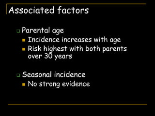
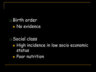
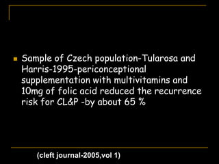
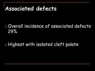
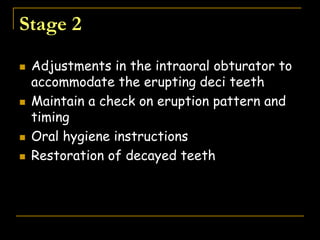
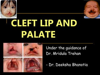
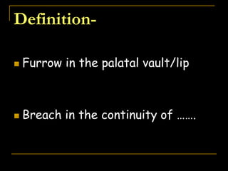
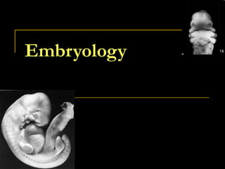
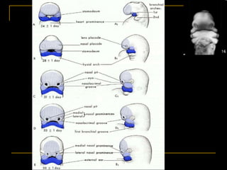
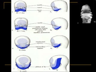
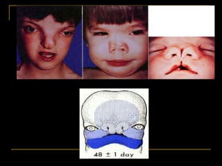
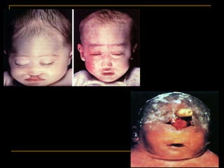
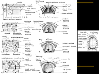
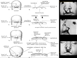
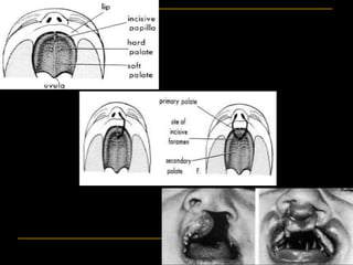
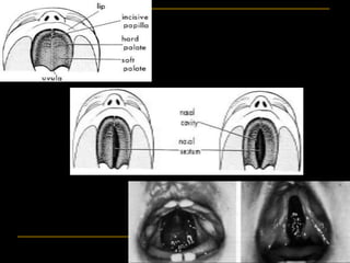
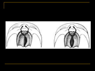
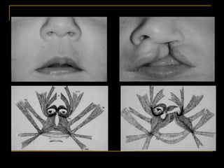
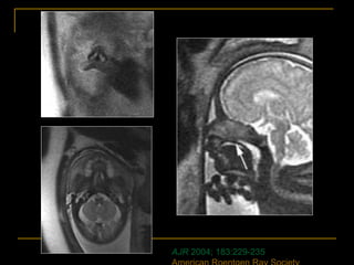
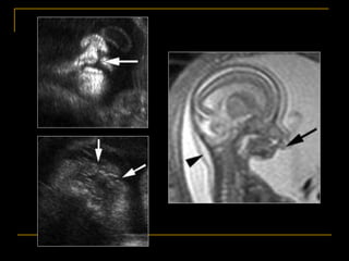
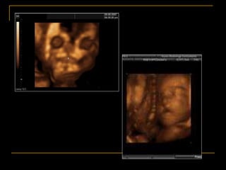
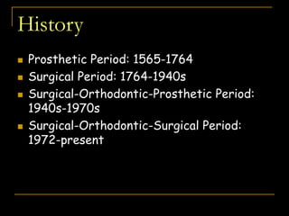
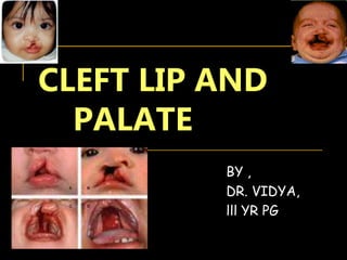
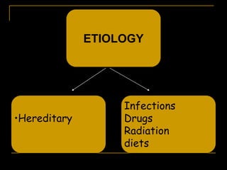
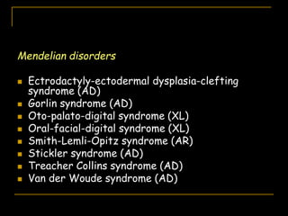
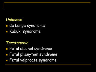
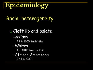
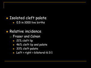
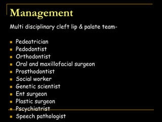
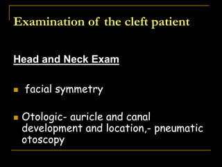
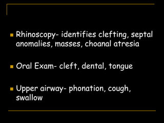
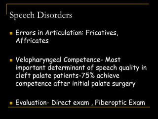
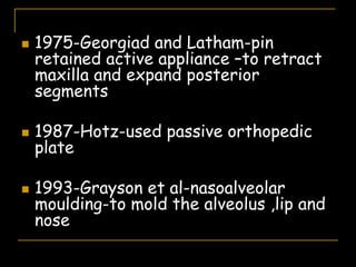
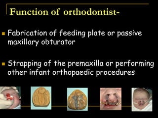
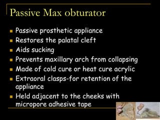
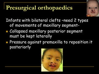
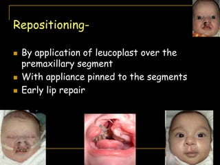
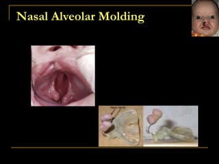
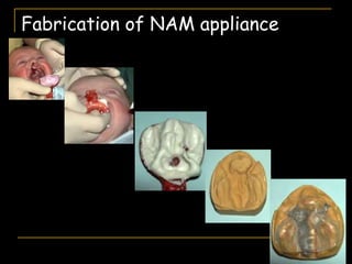
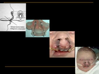
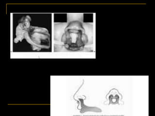
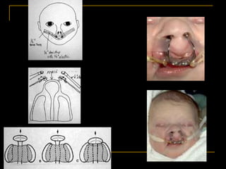
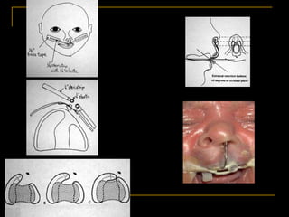
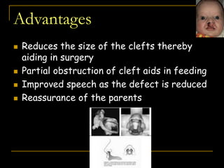
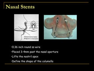
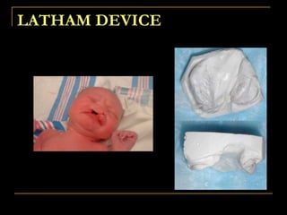
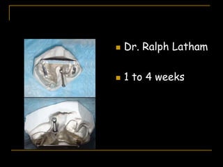
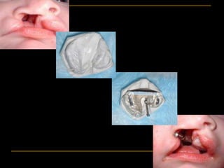
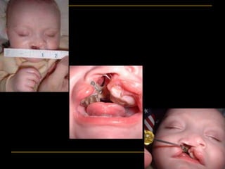
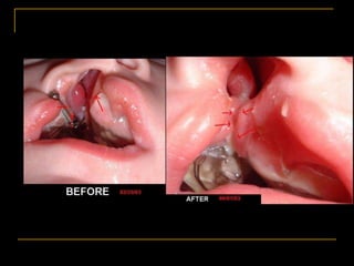
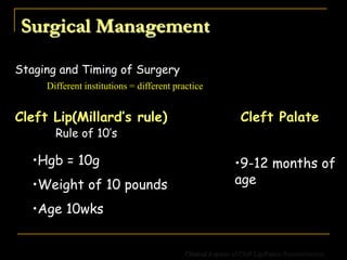
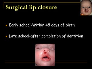
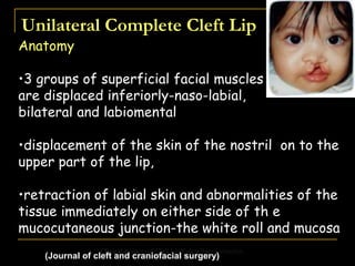
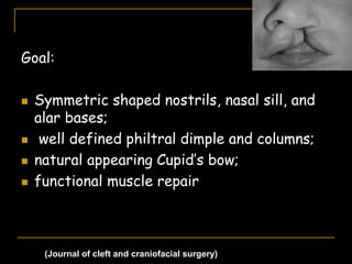
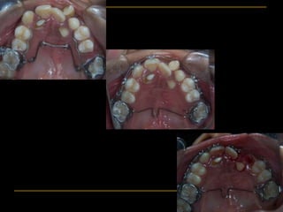
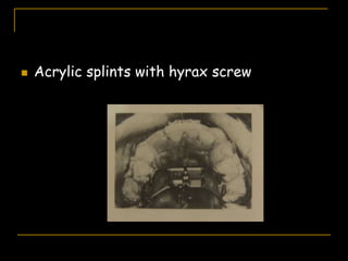
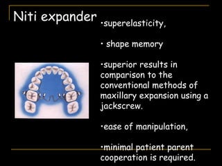
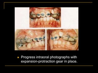
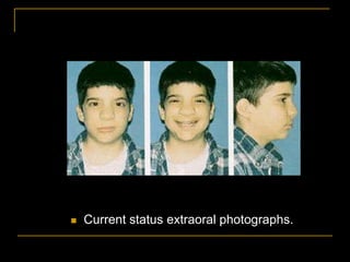
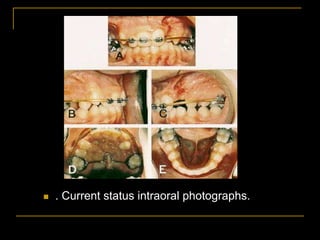
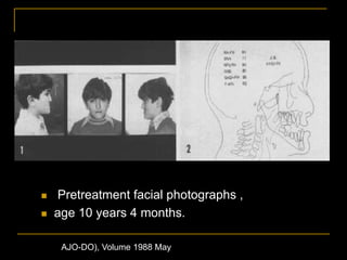
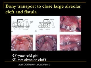
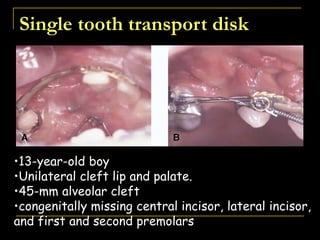
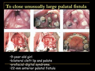
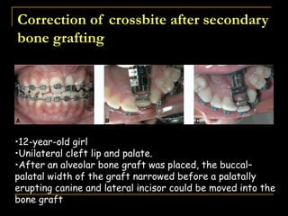
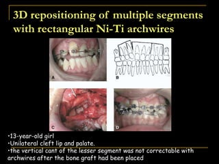
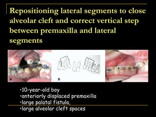
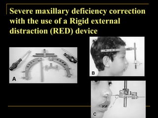
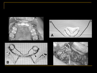
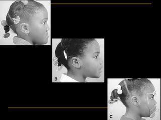
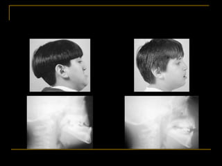
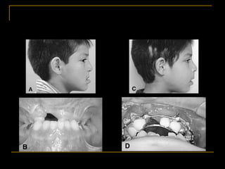
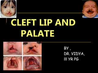
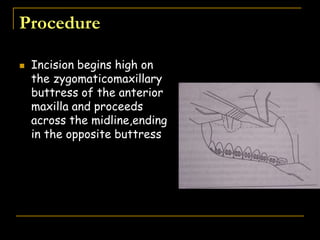
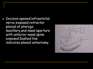
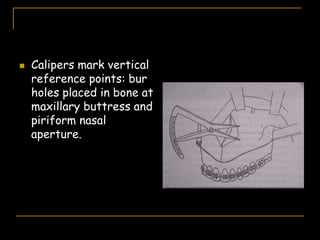
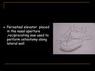
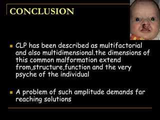
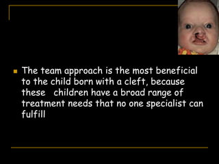
CLEFT LIP AND
PALATE
Under the guidance of
Dr. Mridula Trehan
- Dr. Deeksha Bhanotia
 “ BEING UNWANTED , UNLOVED AND
UNCARED, FORGOTTEN BY EVERY
BODY IS THE WORST KIND OF HUNGER,
MUCH GREATER POVERTY THAN A
PERSON WHO HAS NOTHING TO EAT”
-MOTHER THERESA
Contents
 Embryology
 Historical background
 Theories
 Classification
 Review of literature
 Etiology
 Treatment modalities
 conclusion
Definition-
 Furrow in the palatal vault/lip
 Breach in the continuity of …….
Embryology
AJR 2004; 183:229-235
History
 Prosthetic Period: 1565-1764
 Surgical Period: 1764-1940s
 Surgical-Orthodontic-Prosthetic Period:
1940s-1970s
 Surgical-Orthodontic-Surgical Period:
1972-present
 Prosthetic Period: 1565-1764. In this period,
prostheses were designed to obturate an
unrepaired cleft of the hard or soft palate.
 Developed by Ambroise Pare. Techniques for
cleft closure have been altered in search of the
ideal material:
 Sponges
 Wax
 Silver
 Gold
 Cold and hard-cooled acrylic
 Surgical Period: 1764-1940s
 Le Monnier proposed a 3-stage surgery for
closure of the congenital cleft palate:
 Introduce sutures
 Cauterize the cleft edges
 Approximate cleft edges with sutures
 Bone grafting of the maxilla did not
appear until the 1900s.
 1901 - Von Eiselsburg performed the
first bone graft on an adult with cleft lip
and palate.
 1908 - Lexer performed the first bone
graft on a growing patient with a cleft
palate.
 1914 - Dracther performed the first
bone graft on a patient with an
alveolar cleft.
 1931 - Veau was the first surgeon to
correct a cleft defect with
autogenous tibial chips.
 1952 - Auxhausen defined the challenge of
cleft palate correction:
 Lack of subsequent bone healing between the
premaxilla and the lateral segments after soft
tissue lip and palate repair

 Induced healing (by some means) would yield
preservation of well-formed incisors
 Surgical-Orthodontic-Prosthetic
Period: 1940s-1970s.
 New technology points to conservative
approaches.
 Quantitative facial growth assessment is
made possible by cephalometric facial
analysis and knowledge of craniofacial
growth patterns.
 Orthodontists role:
 Coordinate and manage care of patients
with cleft lip and palate
 Correct deviant growth patterns and
surgical failures when feasible
 Conservative surgery gains popularity.
 Soft tissue lip and palate repair in
infancy or childhood
 Later orthodontic expansion of collapsed
segments
 Straightening of teeth
 Prosthodontist then treats patient
 Bridging of dental gaps
 Obturating residual fistulas
 Insertion of a speech bulb to correct
hypernasal speech
 Surgical-Orthodontic-Surgical Period:
1972-present
 Surgical correction of the cleft alveolus,
maxilla and base of nose, soft tissue
fistulas and nasal scarring.
Timing of bone grafting
 Primary
 secondary
 delayed
(AJO, Vol 1987 Sep)
 ''Primary" bone grafting - before
eruption of the primary dentition or
before 1 year of age.
 ''Secondary" bone grafting -Bone grafting
performed after development of the permanent
dentition .
1. "early secondary'‘- taking place between 5 and 6
years
2. ''secondary'' -taking place between 9 and 11
years or before permanent canine eruption
3. "late secondary" or "delayed" grafting- taking
place after eruption of the permanent canine.
Advantages of primary bone grafting include :
 prevention of maxillary collapse,
 improved bony support that enhances soft-
tissue repair,
 support for the alar base,
 improved ability to eat, and
 enhanced potential to develop normal
dentition.
(AJO, Vol 1987 Sep)
Disadvantages of primary bone grafting :
 the graft does not keep up with vertical
development of the alveolar process and
 inhibits lateral and anterior growth of the
maxilla.
 long-term result is a more unfavorable
facial growth pattern and development of
the dentition
Advantages of secondary grafting :
 little disruption to facial growth because a larger
percentage of the adult size has been achieved
before grafting.
 the canines are expected to migrate and erupt
through the grafted area resulting in improved
development of the dentition,
 improved bony environment to facilitate orthodontic
and prosthodontic treatment,
 improved stability and health of the periodontium.
(AJO, Vol 1987 Sep)
Disadvantages of secondary grafting :
 bone does not show apposition on the graft
surface, which results in the graft's
inability to keep pace with vertical alveolar
development and subsequent compromised
support for the adjacent teeth.
 postponement of grafting jeopardizes
teeth adjacent to the cleft because of
lack of sufficient bone support.
 Recently, delayed grafting has been
reported as a possible method to achieve a
firm anatomic base to aid orthodontic or
prosthodontic management while avoiding
interference with facial growth.
 "Primary" bone grafting is abandoned
by most surgeons.
 A high frequency of resulting maxillary
growth restriction .
 "Early secondary" alveolar bone grafting:
Bone grafting before eruption of permanent
canine, after palate repair, and in the mixed
dentition
 Avoids frequency of maxillary retrusion
seen following "primary" grafting
 Achieves positive alveolar effects of
grafting, especially eruption and migration
of canine teeth
 Occlusal and periodontal benefits
 Bergland's and colleagues' study of 300
patients convinced cleft care specialists
to adopt "early secondary" alveolar bone
grafting.
 Noted success in elimination of
perialveolar oro-nasal fistula, graft take,
eruption of the permanent canine tooth,
and orthodontic closure of cleft-dental
gap
 Turvey's and colleagues' study
successfully placed an autogenous
cancellous (hip) graft before eruption
of the canine tooth, showing the
periodontal benefits of this technique.
 Hall and Posnick noted a 98% success
rate of autogenous cancellous iliac
grafts and closures of oro-nasal
fistulas in 106 patients.
Theories
Two Theories About Clefting:
 Mesoderm & Ectoderm Layers “Migrate &
Flow”
 Multiplication of cells Which makes facial
structures “grow & merge”
Classification
Davis and Ritchie's classification-1922
 Based on location of the cleft relative to
the alveolar process
 3 groups-
1. Pre alveolar
2. Post alveolar
3. Alveolar
 Fog anderson-1942
 Schuchardt and Pfeiffer's symbolic
classification
 Kernahan and stark -1958
Kernahan’s stripped ‘y’ classification-
1971
 Incisive foramen as
focal point
 1,4-lip
 2,5-alveolus
 3,6-Hp ant to IF
 7,8-Hp post to If
 9-soft palate
Millard’s modification of the
Kernahan’s stripped ‘y’
classification
Iowa Classification
Group I
Clefts of lip only
Group II
Clefts of palate only
Group III
Clefts of lip,
alveolus, palate
Group IV
Clefts of lip and
alveolus (primary
cleft palate and
lip)
Group V
Miscellaneous
Clinical Aspects of Cleft Lip/Palate Reconstruction
 Elsahy’s modification of the
kernahan’s stripped ‘y’ classification
 International confederation for
plastic and reconstructive surgery
classification-1968
 American cleft palate association’s
classification-1962
1.clefts of pre palate
2.clefts of palate
3. cleft of hard and soft palates
4.clefts of pre palate and palate
Lahshal classification-Okreins-1987
LAH-R
Mechanism of formation of
isolated cleft
 agenesis or hypoplasia of the tissues
involved
 Palatal tissues may be obstructed from
moving dorsal to tongue by lack of intrinsic
or extrinsic motive force or by physical
obstruction
 Poor adherence of medial edge epithelium
of the palatal process to each other or a
delay in transposition until fusion capacity
is lost
 Persistence of midline seam due to failure
of cellular degeneration might result in
post-fusion breakdown in the midline
 Lack of mesenchymal growth in the midline
region may result in submucous cleft
formation
To be continued-
•Etiology
•Epidemiology
•Management of cleft lip and palate
References
 CONTEMPORARY ORTHODONTICS-PROFFITT
 HUMAN EMBRYOLOGY-INDERBIR SINGH,PAL
 TEXT BOOK OF ORTHODONTICS-BISHARA
 CLINICAL ASPECTS OF CLEFT LIP AND PALATE-JC-
1975
 ORTHODONTICS:CURRENT PRINCIPLES AND
TECHNIQUES-GRABER, VANARSDALL,VIG
• AJO, Vol 1987 Sep
• AJO, Vol 1995 Feb : Bone grafting in patients with cleft
lip and cleft palate Andlin-Sobocki, Eliasson, and Paulin
 AJO, Vol 1986 Jul : Tooth formation in children with
cleft lip/palate – Ranta
 AJO, Vol 1985 Jun : Dentofacial relationships in
persons with unoperated clefts - Bishara, de Arrendondo,
Vales, and Jakobsen
CLEFT LIP AND
PALATE
BY ,
DR. VIDYA,
lll YR PG
Contents
 Etiology
 Epidemiology
 Complications
 Management
ETIOLOGY
•Hereditary
Infections
Drugs
Radiation
diets
 Gene IRF 65-normal development of lips,
palate,skin,and genetelia
 Genes TGFA,IRF 6,TGFB-2,TGFB-3 & MSX
16- contributes for CL&P in different
ethnic populations
Syndromes associated with cleft lip
and palate
Chromosomal
 Trisomy 13
 Trisomy 18
 Velocardiofacial syndrome (22q11 deletion)
Non-Mendelian
 Pierre Robin sequence
 CHARGE association
 Goldenhar syndrome
(cleft journal-2005,vol 1)
Mendelian disorders
 Ectrodactyly-ectodermal dysplasia-clefting
syndrome (AD)
 Gorlin syndrome (AD)
 Oto-palato-digital syndrome (XL)
 Oral-facial-digital syndrome (XL)
 Smith-Lemli-Opitz syndrome (AR)
 Stickler syndrome (AD)
 Treacher Collins syndrome (AD)
 Van der Woude syndrome (AD)
Unknown
 de Lange syndrome
 Kabuki syndrome
Teratogenic
 Fetal alcohol syndrome
 Fetal phenytoin syndrome
 Fetal valproate syndrome
Epidemiology
Racial heterogeneity
 Cleft lip and palate
-Asians
2.1 in 1000 live births
-Whites
1 in 1000 live births
-African Americans
0.41 in 1000
 Isolated cleft palate
 0.5 in 1000 live births
 Relative incidence
 Fraser and Calnan
 21% cleft lip
 46% cleft lip and palate
 33% cleft palate
 Left > right > bilateral-6:3:1
Associated factors
 Parental age
 Incidence increases with age
 Risk highest with both parents
over 30 years
 Seasonal incidence
 No strong evidence
 Birth order
 No evidence
 Social class
 High incidence in low socio economic
status
 Poor nutrition
 Sample of Czech population-Tularosa and
Harris-1995-periconceptional
supplementation with multivitamins and
10mg of folic acid reduced the recurrence
risk for CL&P -by about 65 %
(cleft journal-2005,vol 1)
Associated defects
 Overall incidence of associated defects
29%
 Highest with isolated cleft palate
Cranio facial abnormalities-32%
 Ocular coloboma
 Prominent occiput
 Low set ears
 Hypertelorism
 Slanting palpebral fissures
 Depressed nasal bridge
 micrognathia
Musculoskeletal system-12 %
 Club foot
 Club hand
 Polydactyly
 Limb deficiency
 syndactyly
Ear defects-6%
 Loss of hearing
Eye defects-10 %
 Coloboma
 Micropthalmia
 Anopthalmia
 Cataract
Skin manifestations-32 %
 Scalp agenesia
Cvs-4 %
 Truncus arteriosus
 Transposition
 Tetralogy of fallot
 Double outlet right ventricle
 Pulmonary stenosis
 aortic stenosis
 Ventricular septal defect
 Atrioventricular septal defect
Miscellaneous-
 Ankyloglossia
 Lingual thyroid
 Diabetes
 Hepatitis
 Nasal polyp
 hernia
Complications
General
•Psychological
•Esthetic
•Speech and
hearing
Dental
REVIEW OF THE LITERATURE -
Tooth formation
 Lateral incisor in the cleft area- Fukuhara
and Saito, Woolf, and Broadbent -LI occurs more
often on the distal than the mesial side of the
cleft.
 rotation and crowding of the maxillary frontal
teeth
 congenitally missing lateral and central incisors
AJO, Vol 1986 Jul (11 - 18) Tooth formation in children with cleft lip/palate
 Formation and eruption of the permanent
teeth - formation of permanent teeth in children
with cleft lip or palate, or both, was delayed
approximately 6 months.
 premolars and canines on the cleft side of the
upper jaw erupt later than the corresponding
teeth on the noncleft side of the upper jaw.
 the crown-to-root formation - asymmetric
 Shape and size of the permanent teeth -Hellquist
and associates, Kraus, Jordan and Pruzansky,
Schroeder and Green
 extremely high incidence of enamel hypoplasia in
the incisors of both dentitions
 Jordan, Kraus, and Neptune - high degree of
tooth malformation in the entire dentition of
children and also of fetuses affected with cleft
lip and palate
 Foster and Lavelle- The teeth of the cleft
group were significantly smaller than those
of the control group.
 The normal sex differences in tooth
dimensions were to some extent reversed in
the cleft palate subjects and several of the
dimensions were significantly larger for the
females than for the males.
 Adams and Niswander -familial cleft lip and/or
palate showed a propensity for greater
asymmetry than in the controls.
 According to Sofaer, this generalized
developmental instability may, to some extent,
be under genetic control, since cases with
positive family histories showed some signs of
greater asymmetry than those with negative
family histories.
 The mandible in the cleft groups displayed
shorter mandibular ramus and body length,
 BCLP results in an anteriorly progressive
constriction of the upper dental arch in
both genders.
 The BCLP group has a significantly longer
maxillary dental arch, which is attributed
to the premaxillary anterior projection.
AJO-DO), Volume 1998 Aug- Influence of cleft type on mandibular growth,
Multi disciplinary cleft lip & palate team-
 Pedeatrician
 Pedodontist
 Orthodontist
 Oral and maxillofacial surgeon
 Prosthodontist
 Social worker
 Genetic scientist
 Ent surgeon
 Plastic surgeon
 Pscychiatrist
 Speech pathologist
Management
Examination of the cleft patient
Head and Neck Exam
 facial symmetry
 Otologic- auricle and canal
development and location,- pneumatic
otoscopy
 Rhinoscopy- identifies clefting, septal
anomalies, masses, choanal atresia
 Oral Exam- cleft, dental, tongue
 Upper airway- phonation, cough,
swallow
Speech Disorders
 Errors in Articulation: Fricatives,
Affricates
 Velopharyngeal Competence- Most
important determinant of speech quality in
cleft palate patients-75% achieve
competence after initial palate surgery
 Evaluation- Direct exam , Fiberoptic Exam
Stages
1. Birth -18 months
2. 18 months -5th year-primary dentition
stage
3. 6-11 yr-mixed dentition stage
4. 12 – 18 yrs-permanent dentition stage
STAGE 1
 Fabrication of passive obturator
 Presurgical orthopaedics
 Surgical management of cleft lip
 Surgical management of cleft
palate
History of presurgical
orthopedics
 1689-Hoffmann-facial binding
 1790-Dessault-similar technique
to retract max
 1844-Hullihen-adhesive tape
 1927-Brophy-passed silver wire
through both the ends of cleft
alveolus and tightened
 1950-Mc Neil-used series of plates to
mold alveolar segments
 1975-Georgiad and Latham-pin
retained active appliance –to retract
maxilla and expand posterior
segments
 1987-Hotz-used passive orthopedic
plate
 1993-Grayson et al-nasoalveolar
moulding-to mold the alveolus ,lip and
nose
 Fabrication of feeding plate or passive
maxillary obturator
 Strapping of the premaxilla or performing
other infant orthopaedic procedures
Function of orthodontist-
appliance.
appliance.
Passive Max obturator
 Passive prosthetic appliance
 Restores the palatal cleft
 Aids sucking
 Prevents maxillary arch from collapsing
 Made of cold cure or heat cure acrylic
 Extraoral clasps-for retention of the
appliance
 Held adjacent to the cheeks with
micropore adhesive tape
Infants with bilateral clefts –need 2 types
of movements of maxillary segment-
 Collapsed maxillary posterior segment
must be kept laterally
 Pressure against premaxilla to reposition it
posteriorly
Presurgical orthopaedics
Repositioning-
 By application of leucoplast over the
premaxillary segment
 With appliance pinned to the segments
 Early lip repair
Nasal Alveolar Molding
Fabrication of NAM appliance
 Reduces the size of the clefts thereby
aiding in surgery
 Partial obstruction of cleft aids in feeding
 Improved speech as the defect is reduced
 Reassurance of the parents
Advantages
Nasal Stents
•0.36 inch round ss wire
•Placed 3-4mm past the nasal aperture
•Lifts the nostril apex
•Define the shape of the columella
LATHAM DEVICE
 Dr. Ralph Latham
 1 to 4 weeks
Contents
 Surgical management
 Arch expansion
 Sec bone grafting
Surgical Management
Staging and Timing of Surgery
Different institutions = different practice
Rule of 10’s
•Hgb = 10g
•Weight of 10 pounds
•Age 10wks
Cleft Lip(Millard’s rule) Cleft Palate
•9-12 months of
age
Clinical Aspects of Cleft Lip/Palate Reconstruction
Surgical lip closure
 Early school-Within 45 days of birth
 Late school-after completion of dentition
Unilateral Complete Cleft Lip
Anatomy
•3 groups of superficial facial muscles
are displaced inferiorly-naso-labial,
bilateral and labiomental
•displacement of the skin of the nostril on to the
upper part of the lip,
•retraction of labial skin and abnormalities of the
tissue immediately on either side of th e
mucocutaneous junction-the white roll and mucosa
Clinical Aspects of Cleft Lip/Palate Reconstruction
(Journal of cleft and craniofacial surgery)
Flap designs:
1) Triangular flap(Tennison-Randall)-1952
2) Quadrangular flap (Le Mesurier)-1955
3) Arciform flaps (Millard, Mohler)-1960
4) Undulating flap (Pfeifer)-1966
Goal:
 Symmetric shaped nostrils, nasal sill, and
alar bases;
 well defined philtral dimple and columns;
 natural appearing Cupid’s bow;
 functional muscle repair
(Journal of cleft and craniofacial surgery)
Millard Technique
•“Cut as you go” technique
• Preserves’ cupid’s bow and philtral dimple
•Scar placed in more anatomically correct
position along philtral column
•Tension of closure under the alar base;
reduces flair and promotes better molding of
the underlying alveolar processes
Clinical Aspects of Cleft Lip/Palate Reconstruction
1) Medial flap rotates downward to achieve
necessary lengthening
2) Lateral flap advances into the defect
produced by downward displacement of
medial flap
Clinical Aspects of Cleft Lip/Palate Reconstruction
3) Small pennant-shaped medial flap can be
used to restore nostril sill or lengthen the
columella
Bilateral cleft lip
Anatomy
 Skin of the prolabium is retracted and
raised by the underlying cellular tissue
 True vermillion is replaced by excess
mucosa
 Transverse development of the premaxilla
is reduced –due to the absense of
development of median interincisive suture
Surgical technique
 An incision made at the junction between
the skin of nasal origin and the skin of
labial origin,commencing at the lateral part
of the alar base-(pt A)
 This extends to the mucocutaneous
junction which it meets at pt B and to
which it is perpendicular
 Second incision is made along the
mucocutaneous junction which it meets at
the point where the white roll just begins
to disappear (pt C) extending to the
superior part of the lip mucosa
 Incision made from points 2 to 3 (convex)
following the mucocutaneous junction
 From point 3 on either side ,the curve of
incision is reversed until they both meet in
the mid point in line with the labial frenum
Post-op Management
Inform the parents of:
•Scar contracture
•Erythema
•Firmness
Avoid placing in direct sunlight until the scar
fully matures
Clinical Aspects of Cleft Lip/Palate Reconstruction
Surgical palate closure
 Between 9-12 mon of age
 Facilitates normal speech , hearing and
improves swallowing
 Bone transplants taken from rib,iliac
bone,mand symphysis,tibial bone,or outer
table of parietal bone
Goal: Production of a competent
velopharyngeal sphincter
Most common repairs:
1) V-Y (Veau-Wardill-Kilner) technique
2) von Langenbeck
3)Widmaier technique-1964
4)Malek and Psaume technique-1983
Clinical Aspects of Cleft Lip/Palate Reconstruction
Anatomy
 Median part of the palatal vault is absent
 Width of the cleft is due to atrophy of
the palatal shelves
 lateral displacement of the maxillary
segments due to a nonfunctioning soft
palate
 Palatal fibromucosa is reduced
 vomer is reduced
Wardill-Kilner technique
1)Incisions made along free margins of cleft
and extend anteriorly to apex
2)Dissection continued posteriorly along oral
side of alveolar ridge to retromolar trigone
Clinical Aspects of Cleft Lip/Palate Reconstruction
3) Mucoperiosteal flaps are
elevated from nasal/oral
surfaces of bony palate
4) Dissection of the greater
palatine vessels from the
foramen lengthens the pedicle
5) Tensor veli palatini muscle is
elevated off the hamulus to aid in
relaxing the midline closure
Clinical Aspects of Cleft Lip/Palate Reconstruction
6) Nasal mucosa freed from
bony palate and closed to
either side, or if necessary
closed by using vomer flaps
7) Muscle and oral mucosa
closed in a second single layer
in a horizontal fashion
Clinical Aspects of Cleft Lip/Palate Reconstruction
8) Anteriorly, the oral
mucoperiosteal flaps are
attached to the mucosa
overlying the primary palate
9) Posteriorly, the palate is
closed in 3 layers
Nasal mucosa
Levator muscle
Oral mucosa
Clinical Aspects of Cleft Lip/Palate Reconstruction
Post-op Management
Complications
 Airway obstruction
 Intraoperative bleeding
 Palatal fistula
 Midface abnormalities (early interventions)
Clinical Aspects of Cleft Lip/Palate Reconstruction
Stage 2
 Adjustments in the intraoral obturator to
accommodate the erupting deci teeth
 Maintain a check on eruption pattern and
timing
 Oral hygiene instructions
 Restoration of decayed teeth
Stage 3
 Arch expansion- Buccal segment cross
bites-quad helix / expansion screws
 Correction of anterior cross bites-
removable or fixed appliances
 Sec bone grafting
 Maxillary protraction-reverse pull hg
 Alignment with fixed appli
PRESURGICAL ORTHODONTIC MANAGEMENT
AND SECONDARY
BONE GRAFT IN THE CLEFT PATIENTS
*ABIDA IJAZ, sayeeED ASHRAF CHEEMA,
Upper Occlusal View
Post-Expansion Retention (Nance with
Extended Arm)
Occlusal View at 11/2 months
after Secondary Graft
ARCH EXPANSION
ESTIMATE OF EXPANSION
NEEDED
1.width b/n the MB cusp tips of the max 1st
molars-width b/n the buccal grooves of
mand 1st molars
2.to this add 2/3mm for over correction as
protection against relapse
Quad-helix appliance
Advantages -
 excellent anchorage and retention,
 minimum effect on speech,
 continuous force delivery over time,
 and no adjustment responsibility for the patient.
 cleft palate patients- continuous force delivery
characteristics and the ability to use more force
anteriorly than posteriorly as needed.
AJO-DO), Volume 1988 Oct
 fabricated from stainless steel or Elgiloy blue
arch wires with diameters of 0.038 inch .
 In vitro measurements by Chaconas and Caputo
have shown that activation of 8 mm produces a
force of approximately 400 g .
 An activation of 8 mm is frequently the initial
expansion added to the appliance before
cementation and corresponds to the average
buccal-lingual width of deciduous maxillary
second molars.
RAKSHA AFSARI
15/F
2727
PRE-TREATMENT
MOHAMMED AHMED
13/M
26404
PRE-TREATMENT
 Acrylic splints with hyrax screw
Niti expander •superelasticity,
• shape memory
•superior results in
comparison to the
conventional methods of
maxillary expansion using a
jackscrew.
•ease of manipulation,
•minimal patient parent
cooperation is required.
Secondary Bone Grafting
-Boyne and Sands in the 1970s.
 The technique involves autogenous transplantation of
highly cellular cancellous bone from the iliac crest to
the alveolar ridge, completely filling in the residual
alveolar cleft.
 usually performed at age 9 to 12, immediately prior to
the eruption of the permanent canine.
 The ideal timing, regardless of chronological age, is
when the roots of the canines show one-fourth to
three-fourths of their development.
(JCO, Vol 1998 Jul)
Secondary bone grafting offers
the following benefits:
• Stabilization of the maxillary segments,
mainly the premaxilla, in bilateral cleft
cases.
• Bone support for the alar cartilage.
• Continuity of the alveolar ridge from the
creation of new alveolar bone in the cleft
area.
 Continuity of the dental arch, with the
possibility of moving teeth through the
grafted area.
 Opportunity for spontaneous or induced
eruption of the permanent canine as the
periodontium develops.
 Easier orthodontic closure of the cleft
space and more favorable height of
interdental bone septum result where bone
has been grafted prior to eruption of the
permanent canine.
Autogenous bone grafts
Donor sites include-
 the ilium,
 mandibular symphysis,
 rib,
 cranial vault.
(Angle Orthodontist, 1996 No. 1)
Disadvantage in using autogenous
tissues-
 The necessity of a second surgical site for
harvesting the donor bone. This harvesting
procedure carries some morbidity.
 An average of 355 ml of blood is lost
following iliac crest and alveolar grafting
procedures.
 Harvesting of donor bone from either the
rib or the cranial vault is not readily
acceptable among some ethnic groups,
especially the Chinese, because of cultural
beliefs.
 Cranial bone has already been reported to
be unsatisfactory, as ingrowth of fibrous
tissue and periodontal defects can form
around the adjacent teeth in the grafted
area.
 Delayed ambulation ranges from 8 to 10
days.
 Other complications include pain, wound
dehiscence, hematoma, seroma,
paraesthesia, potential disturbance of
ilium development in young children and
potential risk to apices of anterior teeth in
harvesting mandibular symphyseal bone
Allogenic bone grafts(1950) -
 frozen or freeze-dried (lyophilized)
cortical and cancellous explants
 in the maxillary alveolar clefts
 reduce morbidity by rendering the
harvesting of autogenous bone
unnecessary.
 bone became available with the
establishment of allogenic bone banks
Disadvantages
 its antigenic constituents will evoke a localized
cellular immunodefensive reaction
 retard revascularization and osteoinduction.
 increased risk of dehiscence and sequestration.
 availability of bank bone in some communities has
always been very limited, because donations are
generally nontraditional–even unacceptable–among
some ethnic groups.
Bioceramic hydroxyapatite
 has greater compressive and transverse
strength than enamel.
 shows no evidence of adverse
inflammatory or cytotoxic local or
systemic foreign-body response
 excellent histocompatibility and some
osteoconduction,
 do not possess any osteoinductive
activity.
 the long-term effects on the growing
maxilla and eruption of teeth in humans
are unknown.Thus, hydroxylapatite is
not used to repair maxillary alveolar
clefts, especially in young children
between 9 and 11.
HCL-demineralized bone chips from
Ox tibiae
 were implanted into canine cranial defects.
 Although the healing was incomplete, bony
ingrowth was observed at the margins of the
defects.
 the decalcification process had provided a
more antiseptic environment for bone
growth.
 Demineralized long bone fragments
stimulate osteogenesis in subcutaneous
sites.
 The cellular events of osteoinduction involve
transformation of undifferentiated
mesenchymal cells to chondroblasts and
osteoblasts.
Bone from the third molar regions
 Third molars are impacted in around
20% of individuals.
 Early removal of third molars - reduces
postoperative morbidity, better healing,
quicker recovery, and better periodontal
reattachment.
(Angle Orthodontist, 1996 No. 1)
 impacted teeth should be considered for
removal as soon as they are diagnosed,i.e.,
when it first becomes apparent that the third
molars do not have enough room for occlusion.
 ideal time for removal of impacted third
molars, -as early as the late teenage years,
between 16 and 18 years, when the roots are
more than one-third but less than two-thirds
formed.
Harvesting bone from the third
molar regions-
 allows not only the removal of impacted third
molars during the same surgical procedure,
 but also eliminates the morbidity associated
with additional surgical sites such as the ilium
or mandibular symphysis.
 excellent histocompatibility and
osteoinduction,
Contents
 PROTRACTION HG
 LEFORT 1 OSTEOTOMY
 DISTRACTION OSTEOGENESIS
 RETENTION
 CONCLUSION
PROTRACTION HG
 Facial growth in children with cleft lip and palate is
characterized by underdevelopment of the maxilla,
mainly due to surgical repair of palatal clefts. This
causes skeletal discrepancies between the upper and
lower jaws, which frequently result in anterior and/or
posterior crossbite (Pruzansky and Aduss, 1967; Ross
and Johnston, 1972; Nordén et al., 1973; Bergland and
Sidhu, 1974; Sakuda, 1978; Dahl and Hanusardottir,
1979).
 Midfacial growth disturbance also contributes to the
characteristic unattractiveness of soft tissue facial
profiles in cleft patients (Ross, 1987; Chen and So,
1997).
(The Cleft Palate-Craniofacial Journal: Vol. 37)
 Patient age has been considered important
among the factors that influence the effects
of the maxillary protraction of the cleft lip
and palate (Irie et al., 1973; Friede and
Lennartsson, 1981; Rygh and Tindlund, 1982;
Ranta, 1988).
 Several clinical studies have shown
considerable forward displacement of the
maxilla when the treatment begins at an early
age (Irie and Nakamura, 1975; Friede and
Lennartsson, 1981; Rygh and Tindlund, 1982;
Ranta, 1988; Ishikawa et al., 1996).
 Several clinical studies have shown favorable
effects on the maxillary forward growth when
the treatment was started at the deciduous
or early mixed dentition stages (Irie and
Nakamura, 1975; Friede and Lennartsson,
1981; Rygh and Tindlund, 1982; Ranta, 1988;
Ishikawa et al., 1996).
 Because circummaxillary sutural growth is
active during the early juvenile period (Scott,
1956; Björk, 1966), it seems reasonable and
advantageous to facilitate the forward
growth of the maxilla at these stages
 The mean changes in the cephalometric
variables during the first year of treatment
showed forward displacement, with a slight
counterclockwise rotation of the maxilla and
backward rotation of the mandible.
 The magnitude of force exerted on both jaws
does not strongly influence the treatment
effects if the force is greater than a certain
value. Delaire (1997) suggested that 200 g on
each side was enough for protraction
Factors to be considered for
facemask therapy
 There should be evidence of only mild
max hypoplasia rather than true max
ankylosis
 It is only suitable for mild skeletal
discrepancies where it is considered
desirable to obtain a class 1 incisor
relationship
 Well motivated and cooperative patient
 Occasionally true mandibular prognathism
is responsible for the creation of a class lll
malocclusion rather than max
development.Careful clinical and
cephalometric analysis is essential .
Results obtained with face mask:
(1) After maxillary protraction, the maxilla
displaces anteriorly, whereas the mandible
rotates posteriorly
(2) the maxillary incisors move in the
anterior direction, whereas the mandibular
incisors move posteriorly
(3) the mandibular plane angle and anterior
lower and total face heights increase
(4) these changes were reflected in the
profile, whereby the skeletal profile
convexity increases
(5) the Class III concave profile becomes
more balanced, with the upper lip area
becoming more marked
 11-year, old boy
 complete unilateral cleft lip and palate on the left side
AJO-DO), Volume 1994 Jun
 Initial intraoral photographs.
 Progress intraoral photographs with
expansion-protraction gear in place.
 Current status extraoral photographs.
 . Current status intraoral photographs.
 Pretreatment facial photographs ,
 age 10 years 4 months.
AJO-DO), Volume 1988 May
 Pretreatment study models.
 The patient wearing an orthopedic face mask
appliance.
 Posttreatment photographs
 Postretention photographs
Distraction osteogenesis
 also termed bony transport,
uses-
 severe cleft lip and palate deformities who
have inadequate maxillary arch forms,
 large cleft spaces,
 Vertically canted segments,
 restricted skeletal development
 Distraction osteogenesis (DO) was
first used for correction of the
craniofacial skeleton in the early
1990s.
 McCarthy et al reported using
distraction to lengthen the mandible in
patients with hemifacial microsomia.
History
 Figuero and Polley reported success
with no significant complications when
DO was used to advance the maxilla in
children with cleft lip and palate.
 Recently, DO was used to advance the
maxilla in patients with cleft palate
Treatment of large cleft spaces and segment
positions by the gradual movement of a
transported segment has several benefits.
1.the repositioning of a distracted segment
is under the control of the orthodontist
and can be changed during treatment.
2.there are no immediate segmental
movements that can compromise blood
supply to the osteotomized segment.
3.the morbidity associated with grafting large
alveolar clefts can be reduced by making the
cleft spaces smaller and more manageable.
4.the osteotomy or corticotomy used in these
procedures is well tolerated by patients and
heals quickly.
5.new bone is generated behind the
transported segment or disk of bone by
distraction osteogenesis.
Bony transport to close large alveolar
cleft and fistula
AJO-DOVolume 127, Number 2
•17-year-old girl
•21 mm alveolar cleft.
Single tooth transport disk
•13-year-old boy
•Unilateral cleft lip and palate.
•45-mm alveolar cleft
•congenitally missing central incisor, lateral incisor,
and first and second premolars
To close unusually large palatal fistula
•9-year-old girl
•bilateral cleft lip and palate
•orofacial-digital syndrome.
•22-mm anterior palatal fistula
Correction of crossbite after secondary
bone grafting
•12-year-old girl
•Unilateral cleft lip and palate.
•After an alveolar bone graft was placed, the buccal–
palatal width of the graft narrowed before a palatally
erupting canine and lateral incisor could be moved into the
bone graft
3D repositioning of multiple segments
with rectangular Ni-Ti archwires
•13-year-old girl
•Unilateral cleft lip and palate.
•the vertical cant of the lesser segment was not correctable with
archwires after the bone graft had been placed
Repositioning lateral segments to close
alveolar cleft and correct vertical step
between premaxilla and lateral
segments
•10-year-old boy
•anteriorly displaced premaxilla
•large palatal fistula,
•large alveolar cleft spaces
Severe maxillary deficiency correction
with the use of a Rigid external
distraction (RED) device
CLEFT LIP AND
PALATE
BY ,
DR. VIDYA,
lll YR PG
 Cheever was the first to do downfracture
of the maxilla as early as 1864
 In 1960 - Obwegeser started to perform
maxillary surgery and described a large
series of LeFort I osteotomies in 1969
LeFort I maxillary advancement
osteotomy
 Surgical technique must be based on
additional considerations of blood supply
 The lesser segment has an
unpredictable blood supply,premaxillary
segment has a labial blood supply
AJO-DO), Volume 1983 Aug
 The major concerns had been intraoperative
bleeding, revascularization and healing of the
maxilla.
 After studies (Bell et al. 1975, Turvey & Fonseca
1980).of vascular perfusion and the anatomy and
relevance of the maxillary artery, it was found
that the most important thing would be to
preserve a wide, intact palatal and maxillary soft
tissue pedicle attached to the ostetomized
segments.
 This allows good healing and minimizes the risk of
tissue necrosis.
Procedure
 Incision begins high on
the zygomaticomaxillary
buttress of the anterior
maxilla and proceeds
across the midline,ending
in the opposite buttress
 Incision opened:infraorbital
nerve exposed,retractor
placed at pterygo
maxillary,and nasal aparture
with anterior nasal spine
exposed.Dashed line
indicates planed osteotomy.
 Calipers mark vertical
reference points: bur
holes placed in bone at
maxillary buttress and
piriform nasal
aperture.
 Periosteal elevator placed
in the nasal aperture
,reciprocating saw used to
perform osteotomy along
lateral wall
 Caliper used to
measure amount of
bone to be removed at
piriform region after
initial osteotomy
 Fixation is achieved by using 2 miniplates
for each segment and without
intermaxillary fixation or any other type
of fixation
 Postoperative elastic guidance may be used
to assist in maintenance of the achieved
occlusion or even to improve the dental
interdigitation before postsurgical
orthodontics can be started
Relapse-
 20-25 %-horizontal plane
 Upto 33 %-transverse plane
 22 % -vertical plane
AJO-DO), Volume 1983 Aug
Velopharyngeal Incompetence
 Difficulty closing off the nose from the
mouth while speaking, allowing too much air to
escape through the nose, causing a speech
defect.
 occurs because the repaired soft palate is
too short or does not move adequately.
 special diagnostic procedures such as
nasoendoscopy and videofluoroscopy of
speech may be required to directly
visualize the soft palate during speech
 Techniques-
 Superior based pharyngeal flap-Hogan’s
tech
 Sphincter pharyngoplasty-Jackson’s tech
 Posterior tonsillar
pillars,containing the
palatopharyngeus muscles
are raised ,transposed
horizontally and sutured to
each other in the midline
thus creating sphincter
action in the midline
Sphincter Pharyngoplasty
Procedure
 Each posterior pillar is
outlined on the
pharyngeal wall ,leaving
the posterior wall intact
 A horizontal incision joins
the 2 medial limbs of the
pillar flaps across the
posterior pharyngeal wall
 Each lateral flap
,composed of mucosa
and palatopharyngeus
muscle is raised and
transposed
horizontally
 Muscle and mucosa are
sutured in the midline
 The medial edge of
each flap is sutured to
the upper border of the
horizontal incision and
the lateral edge to the
lower border of the
horizontal incision
Retention
 long term or perm retention
 incisor rotations -bonding a multistranded
wire to the palatal aspects of the teeth
CONCLUSION
 CLP has been described as multifactorial
and also multidimensional.the dimensions of
this common malformation extend
from,structure,function and the very
psyche of the individual
 A problem of such amplitude demands far
reaching solutions
 Cleft lip, with or without palate involvement, is a
common congenital deformity. Although it is not
fatal unless associated with other serious
congenital diseases, it is an important public
health problem worldwide because it carries a
great deal of social, functional, and psychological
morbidity
 The association of defects ,malformations and
syndromes have been studied to a vast extent
and is still being studied till date,
 The team approach is the most beneficial
to the child born with a cleft, because
these children have a broad range of
treatment needs that no one specialist can
fulfill
THANQ
References
 CONTEMPORARY ORTHODONTICS-PROFFITT
 TEXT BOOK OF ORTHODONTICS-BISHARA
 CLINICAL ASPECTS OF CLEFT LIP AND PALATE-JC-
1975
 ORTHODONTICS:CURRENT PRINCIPLES AND
TECHNIQUES-GRABER, VANARSDALL,VIG
 PRESURGICAL ORTHODONTIC MANAGEMENT AND
SECONDARY
BONE GRAFT IN THE CLEFT PATIENTS
*ABIDA IJAZ,ASHRAF CHEEMA
 CLINICAL ASPECTS OF CLEFT AND PALATE
 JOURNAL OF CLEFT AND CRANIOFACIAL SURGERY
 AJO-DO), VOLUME 1983 AUG : LATE DEFINITIVE CORRECTION
OF THE OROFACIAL CLEFT - KINNEBREW AND KENT
 THE CLEFT PALATE-CRANIOFACIAL JOURNAL: VOL. 37, -
EFFECTS OF MAXILLARY PROTRACTION COMBINED WITH
CHIN-CAP THERAPY IN UNILATERAL CLEFT LIP AND PALATE
PATIENTS,HIROYUKI ISHIKAWA, HINICHI KITAZAWA, KIROSHI
IWASAKI, SHINJI NAKAMURA,
 AJO-DO VOLUME 1994 JUN - CASE REPORT - BHATIA AND
NANDA
 AJO-DO, VOLUME 1988 MAY - USE OF FACE MASK IN
TREATMENT OF MAXILLARY SKELETAL RETRUSION -
ROBERTS AND SUBTELNY ,
 THE ANGLE ORTHODONTIST: VOL. 73, USE OF DISTRACTION
OSTEOGENESIS IN CLEFT PALATE PATIENTS-KI CHUL TAE,
DDS, MSD
• AJO-DO 1998 VOL, 2-YEN ET AL
MANAGEMENT OF SEVERE CLEFT MAXILLARY
DEFICIENCY WITH DISTRACTION OSTEOGENESIS:
PROCEDURE AND RESULTS
 AJO-DO, VOLUME 1983 AUG - LATE DEFINITIVE
CORRECTION OF THE OROFACIAL CLEFT - KINNEBREW
AND KENT
References
 CONTEMPORARY ORTHODONTICS-PROFFITT
 HUMAN EMBRYOLOGY-INDERBIR SINGH,PAL
 TEXT BOOK OF ORTHODONTICS-BISHARA
 CLINICAL ASPECTS OF CLEFT LIP AND PALATE-JC-
1975
 ORTHODONTICS:CURRENT PRINCIPLES AND
TECHNIQUES-GRABER, VANARSDALL,VIG
 AJO-DO), Volume 1986 Jul (11 - 18): Tooth
formation in children with cleft lip/palate –
Ranta
 AJO-DO), Volume 1998 Aug (154 - 161):
Upper dental arch morphology of adult
unoperated complete bilateral cleft lip and
palate Omar Gabriel da Silva Filho

CLEFT LIP AND PALATE

  • 1.
    CLEFT LIP AND PALATE Underthe guidance of Dr. Mridula Trehan - Dr. Deeksha Bhanotia
  • 2.
     “ BEINGUNWANTED , UNLOVED AND UNCARED, FORGOTTEN BY EVERY BODY IS THE WORST KIND OF HUNGER, MUCH GREATER POVERTY THAN A PERSON WHO HAS NOTHING TO EAT” -MOTHER THERESA
  • 3.
    Contents  Embryology  Historicalbackground  Theories  Classification  Review of literature  Etiology  Treatment modalities  conclusion
  • 4.
    Definition-  Furrow inthe palatal vault/lip  Breach in the continuity of …….
  • 5.
  • 16.
  • 19.
    History  Prosthetic Period:1565-1764  Surgical Period: 1764-1940s  Surgical-Orthodontic-Prosthetic Period: 1940s-1970s  Surgical-Orthodontic-Surgical Period: 1972-present
  • 20.
     Prosthetic Period:1565-1764. In this period, prostheses were designed to obturate an unrepaired cleft of the hard or soft palate.  Developed by Ambroise Pare. Techniques for cleft closure have been altered in search of the ideal material:  Sponges  Wax  Silver  Gold  Cold and hard-cooled acrylic
  • 21.
     Surgical Period:1764-1940s  Le Monnier proposed a 3-stage surgery for closure of the congenital cleft palate:  Introduce sutures  Cauterize the cleft edges  Approximate cleft edges with sutures
  • 22.
     Bone graftingof the maxilla did not appear until the 1900s.  1901 - Von Eiselsburg performed the first bone graft on an adult with cleft lip and palate.  1908 - Lexer performed the first bone graft on a growing patient with a cleft palate.
  • 23.
     1914 -Dracther performed the first bone graft on a patient with an alveolar cleft.  1931 - Veau was the first surgeon to correct a cleft defect with autogenous tibial chips.
  • 24.
     1952 -Auxhausen defined the challenge of cleft palate correction:  Lack of subsequent bone healing between the premaxilla and the lateral segments after soft tissue lip and palate repair   Induced healing (by some means) would yield preservation of well-formed incisors
  • 25.
     Surgical-Orthodontic-Prosthetic Period: 1940s-1970s. New technology points to conservative approaches.  Quantitative facial growth assessment is made possible by cephalometric facial analysis and knowledge of craniofacial growth patterns.
  • 26.
     Orthodontists role: Coordinate and manage care of patients with cleft lip and palate  Correct deviant growth patterns and surgical failures when feasible
  • 27.
     Conservative surgerygains popularity.  Soft tissue lip and palate repair in infancy or childhood  Later orthodontic expansion of collapsed segments  Straightening of teeth
  • 28.
     Prosthodontist thentreats patient  Bridging of dental gaps  Obturating residual fistulas  Insertion of a speech bulb to correct hypernasal speech
  • 29.
     Surgical-Orthodontic-Surgical Period: 1972-present Surgical correction of the cleft alveolus, maxilla and base of nose, soft tissue fistulas and nasal scarring.
  • 30.
    Timing of bonegrafting  Primary  secondary  delayed (AJO, Vol 1987 Sep)
  • 31.
     ''Primary" bonegrafting - before eruption of the primary dentition or before 1 year of age.
  • 32.
     ''Secondary" bonegrafting -Bone grafting performed after development of the permanent dentition . 1. "early secondary'‘- taking place between 5 and 6 years 2. ''secondary'' -taking place between 9 and 11 years or before permanent canine eruption 3. "late secondary" or "delayed" grafting- taking place after eruption of the permanent canine.
  • 33.
    Advantages of primarybone grafting include :  prevention of maxillary collapse,  improved bony support that enhances soft- tissue repair,  support for the alar base,  improved ability to eat, and  enhanced potential to develop normal dentition. (AJO, Vol 1987 Sep)
  • 34.
    Disadvantages of primarybone grafting :  the graft does not keep up with vertical development of the alveolar process and  inhibits lateral and anterior growth of the maxilla.  long-term result is a more unfavorable facial growth pattern and development of the dentition
  • 35.
    Advantages of secondarygrafting :  little disruption to facial growth because a larger percentage of the adult size has been achieved before grafting.  the canines are expected to migrate and erupt through the grafted area resulting in improved development of the dentition,  improved bony environment to facilitate orthodontic and prosthodontic treatment,  improved stability and health of the periodontium. (AJO, Vol 1987 Sep)
  • 36.
    Disadvantages of secondarygrafting :  bone does not show apposition on the graft surface, which results in the graft's inability to keep pace with vertical alveolar development and subsequent compromised support for the adjacent teeth.  postponement of grafting jeopardizes teeth adjacent to the cleft because of lack of sufficient bone support.
  • 37.
     Recently, delayedgrafting has been reported as a possible method to achieve a firm anatomic base to aid orthodontic or prosthodontic management while avoiding interference with facial growth.
  • 38.
     "Primary" bonegrafting is abandoned by most surgeons.  A high frequency of resulting maxillary growth restriction .
  • 39.
     "Early secondary"alveolar bone grafting: Bone grafting before eruption of permanent canine, after palate repair, and in the mixed dentition  Avoids frequency of maxillary retrusion seen following "primary" grafting  Achieves positive alveolar effects of grafting, especially eruption and migration of canine teeth
  • 40.
     Occlusal andperiodontal benefits  Bergland's and colleagues' study of 300 patients convinced cleft care specialists to adopt "early secondary" alveolar bone grafting.  Noted success in elimination of perialveolar oro-nasal fistula, graft take, eruption of the permanent canine tooth, and orthodontic closure of cleft-dental gap
  • 41.
     Turvey's andcolleagues' study successfully placed an autogenous cancellous (hip) graft before eruption of the canine tooth, showing the periodontal benefits of this technique.  Hall and Posnick noted a 98% success rate of autogenous cancellous iliac grafts and closures of oro-nasal fistulas in 106 patients.
  • 42.
    Theories Two Theories AboutClefting:  Mesoderm & Ectoderm Layers “Migrate & Flow”  Multiplication of cells Which makes facial structures “grow & merge”
  • 43.
    Classification Davis and Ritchie'sclassification-1922  Based on location of the cleft relative to the alveolar process  3 groups- 1. Pre alveolar 2. Post alveolar 3. Alveolar
  • 44.
     Fog anderson-1942 Schuchardt and Pfeiffer's symbolic classification  Kernahan and stark -1958
  • 45.
    Kernahan’s stripped ‘y’classification- 1971  Incisive foramen as focal point  1,4-lip  2,5-alveolus  3,6-Hp ant to IF  7,8-Hp post to If  9-soft palate
  • 46.
    Millard’s modification ofthe Kernahan’s stripped ‘y’ classification
  • 47.
    Iowa Classification Group I Cleftsof lip only Group II Clefts of palate only Group III Clefts of lip, alveolus, palate Group IV Clefts of lip and alveolus (primary cleft palate and lip) Group V Miscellaneous Clinical Aspects of Cleft Lip/Palate Reconstruction
  • 48.
     Elsahy’s modificationof the kernahan’s stripped ‘y’ classification  International confederation for plastic and reconstructive surgery classification-1968
  • 49.
     American cleftpalate association’s classification-1962 1.clefts of pre palate 2.clefts of palate 3. cleft of hard and soft palates 4.clefts of pre palate and palate
  • 50.
  • 51.
    Mechanism of formationof isolated cleft  agenesis or hypoplasia of the tissues involved  Palatal tissues may be obstructed from moving dorsal to tongue by lack of intrinsic or extrinsic motive force or by physical obstruction  Poor adherence of medial edge epithelium of the palatal process to each other or a delay in transposition until fusion capacity is lost
  • 52.
     Persistence ofmidline seam due to failure of cellular degeneration might result in post-fusion breakdown in the midline  Lack of mesenchymal growth in the midline region may result in submucous cleft formation
  • 53.
  • 54.
    References  CONTEMPORARY ORTHODONTICS-PROFFITT HUMAN EMBRYOLOGY-INDERBIR SINGH,PAL  TEXT BOOK OF ORTHODONTICS-BISHARA  CLINICAL ASPECTS OF CLEFT LIP AND PALATE-JC- 1975  ORTHODONTICS:CURRENT PRINCIPLES AND TECHNIQUES-GRABER, VANARSDALL,VIG
  • 55.
    • AJO, Vol1987 Sep • AJO, Vol 1995 Feb : Bone grafting in patients with cleft lip and cleft palate Andlin-Sobocki, Eliasson, and Paulin  AJO, Vol 1986 Jul : Tooth formation in children with cleft lip/palate – Ranta  AJO, Vol 1985 Jun : Dentofacial relationships in persons with unoperated clefts - Bishara, de Arrendondo, Vales, and Jakobsen
  • 56.
    CLEFT LIP AND PALATE BY, DR. VIDYA, lll YR PG
  • 57.
    Contents  Etiology  Epidemiology Complications  Management
  • 58.
  • 59.
     Gene IRF65-normal development of lips, palate,skin,and genetelia  Genes TGFA,IRF 6,TGFB-2,TGFB-3 & MSX 16- contributes for CL&P in different ethnic populations
  • 60.
    Syndromes associated withcleft lip and palate Chromosomal  Trisomy 13  Trisomy 18  Velocardiofacial syndrome (22q11 deletion) Non-Mendelian  Pierre Robin sequence  CHARGE association  Goldenhar syndrome (cleft journal-2005,vol 1)
  • 61.
    Mendelian disorders  Ectrodactyly-ectodermaldysplasia-clefting syndrome (AD)  Gorlin syndrome (AD)  Oto-palato-digital syndrome (XL)  Oral-facial-digital syndrome (XL)  Smith-Lemli-Opitz syndrome (AR)  Stickler syndrome (AD)  Treacher Collins syndrome (AD)  Van der Woude syndrome (AD)
  • 62.
    Unknown  de Langesyndrome  Kabuki syndrome Teratogenic  Fetal alcohol syndrome  Fetal phenytoin syndrome  Fetal valproate syndrome
  • 63.
    Epidemiology Racial heterogeneity  Cleftlip and palate -Asians 2.1 in 1000 live births -Whites 1 in 1000 live births -African Americans 0.41 in 1000
  • 64.
     Isolated cleftpalate  0.5 in 1000 live births  Relative incidence  Fraser and Calnan  21% cleft lip  46% cleft lip and palate  33% cleft palate  Left > right > bilateral-6:3:1
  • 65.
    Associated factors  Parentalage  Incidence increases with age  Risk highest with both parents over 30 years  Seasonal incidence  No strong evidence
  • 66.
     Birth order No evidence  Social class  High incidence in low socio economic status  Poor nutrition
  • 67.
     Sample ofCzech population-Tularosa and Harris-1995-periconceptional supplementation with multivitamins and 10mg of folic acid reduced the recurrence risk for CL&P -by about 65 % (cleft journal-2005,vol 1)
  • 68.
    Associated defects  Overallincidence of associated defects 29%  Highest with isolated cleft palate
  • 69.
    Cranio facial abnormalities-32% Ocular coloboma  Prominent occiput  Low set ears  Hypertelorism  Slanting palpebral fissures  Depressed nasal bridge  micrognathia
  • 70.
    Musculoskeletal system-12 % Club foot  Club hand  Polydactyly  Limb deficiency  syndactyly
  • 71.
    Ear defects-6%  Lossof hearing Eye defects-10 %  Coloboma  Micropthalmia  Anopthalmia  Cataract Skin manifestations-32 %  Scalp agenesia
  • 72.
    Cvs-4 %  Truncusarteriosus  Transposition  Tetralogy of fallot  Double outlet right ventricle  Pulmonary stenosis  aortic stenosis  Ventricular septal defect  Atrioventricular septal defect
  • 73.
    Miscellaneous-  Ankyloglossia  Lingualthyroid  Diabetes  Hepatitis  Nasal polyp  hernia
  • 74.
  • 75.
    REVIEW OF THELITERATURE - Tooth formation  Lateral incisor in the cleft area- Fukuhara and Saito, Woolf, and Broadbent -LI occurs more often on the distal than the mesial side of the cleft.  rotation and crowding of the maxillary frontal teeth  congenitally missing lateral and central incisors AJO, Vol 1986 Jul (11 - 18) Tooth formation in children with cleft lip/palate
  • 76.
     Formation anderuption of the permanent teeth - formation of permanent teeth in children with cleft lip or palate, or both, was delayed approximately 6 months.  premolars and canines on the cleft side of the upper jaw erupt later than the corresponding teeth on the noncleft side of the upper jaw.  the crown-to-root formation - asymmetric
  • 77.
     Shape andsize of the permanent teeth -Hellquist and associates, Kraus, Jordan and Pruzansky, Schroeder and Green  extremely high incidence of enamel hypoplasia in the incisors of both dentitions  Jordan, Kraus, and Neptune - high degree of tooth malformation in the entire dentition of children and also of fetuses affected with cleft lip and palate
  • 78.
     Foster andLavelle- The teeth of the cleft group were significantly smaller than those of the control group.  The normal sex differences in tooth dimensions were to some extent reversed in the cleft palate subjects and several of the dimensions were significantly larger for the females than for the males.
  • 79.
     Adams andNiswander -familial cleft lip and/or palate showed a propensity for greater asymmetry than in the controls.  According to Sofaer, this generalized developmental instability may, to some extent, be under genetic control, since cases with positive family histories showed some signs of greater asymmetry than those with negative family histories.
  • 80.
     The mandiblein the cleft groups displayed shorter mandibular ramus and body length,  BCLP results in an anteriorly progressive constriction of the upper dental arch in both genders.  The BCLP group has a significantly longer maxillary dental arch, which is attributed to the premaxillary anterior projection. AJO-DO), Volume 1998 Aug- Influence of cleft type on mandibular growth,
  • 81.
    Multi disciplinary cleftlip & palate team-  Pedeatrician  Pedodontist  Orthodontist  Oral and maxillofacial surgeon  Prosthodontist  Social worker  Genetic scientist  Ent surgeon  Plastic surgeon  Pscychiatrist  Speech pathologist Management
  • 82.
    Examination of thecleft patient Head and Neck Exam  facial symmetry  Otologic- auricle and canal development and location,- pneumatic otoscopy
  • 83.
     Rhinoscopy- identifiesclefting, septal anomalies, masses, choanal atresia  Oral Exam- cleft, dental, tongue  Upper airway- phonation, cough, swallow
  • 84.
    Speech Disorders  Errorsin Articulation: Fricatives, Affricates  Velopharyngeal Competence- Most important determinant of speech quality in cleft palate patients-75% achieve competence after initial palate surgery  Evaluation- Direct exam , Fiberoptic Exam
  • 85.
    Stages 1. Birth -18months 2. 18 months -5th year-primary dentition stage 3. 6-11 yr-mixed dentition stage 4. 12 – 18 yrs-permanent dentition stage
  • 86.
    STAGE 1  Fabricationof passive obturator  Presurgical orthopaedics  Surgical management of cleft lip  Surgical management of cleft palate
  • 87.
    History of presurgical orthopedics 1689-Hoffmann-facial binding  1790-Dessault-similar technique to retract max  1844-Hullihen-adhesive tape
  • 88.
     1927-Brophy-passed silverwire through both the ends of cleft alveolus and tightened  1950-Mc Neil-used series of plates to mold alveolar segments
  • 89.
     1975-Georgiad andLatham-pin retained active appliance –to retract maxilla and expand posterior segments  1987-Hotz-used passive orthopedic plate  1993-Grayson et al-nasoalveolar moulding-to mold the alveolus ,lip and nose
  • 90.
     Fabrication offeeding plate or passive maxillary obturator  Strapping of the premaxilla or performing other infant orthopaedic procedures Function of orthodontist- appliance. appliance.
  • 91.
    Passive Max obturator Passive prosthetic appliance  Restores the palatal cleft  Aids sucking  Prevents maxillary arch from collapsing  Made of cold cure or heat cure acrylic  Extraoral clasps-for retention of the appliance  Held adjacent to the cheeks with micropore adhesive tape
  • 92.
    Infants with bilateralclefts –need 2 types of movements of maxillary segment-  Collapsed maxillary posterior segment must be kept laterally  Pressure against premaxilla to reposition it posteriorly Presurgical orthopaedics
  • 93.
    Repositioning-  By applicationof leucoplast over the premaxillary segment  With appliance pinned to the segments  Early lip repair
  • 94.
  • 95.
  • 100.
     Reduces thesize of the clefts thereby aiding in surgery  Partial obstruction of cleft aids in feeding  Improved speech as the defect is reduced  Reassurance of the parents Advantages
  • 101.
    Nasal Stents •0.36 inchround ss wire •Placed 3-4mm past the nasal aperture •Lifts the nostril apex •Define the shape of the columella
  • 102.
  • 103.
     Dr. RalphLatham  1 to 4 weeks
  • 107.
    Contents  Surgical management Arch expansion  Sec bone grafting
  • 108.
    Surgical Management Staging andTiming of Surgery Different institutions = different practice Rule of 10’s •Hgb = 10g •Weight of 10 pounds •Age 10wks Cleft Lip(Millard’s rule) Cleft Palate •9-12 months of age Clinical Aspects of Cleft Lip/Palate Reconstruction
  • 109.
    Surgical lip closure Early school-Within 45 days of birth  Late school-after completion of dentition
  • 110.
    Unilateral Complete CleftLip Anatomy •3 groups of superficial facial muscles are displaced inferiorly-naso-labial, bilateral and labiomental •displacement of the skin of the nostril on to the upper part of the lip, •retraction of labial skin and abnormalities of the tissue immediately on either side of th e mucocutaneous junction-the white roll and mucosa Clinical Aspects of Cleft Lip/Palate Reconstruction (Journal of cleft and craniofacial surgery)
  • 111.
    Flap designs: 1) Triangularflap(Tennison-Randall)-1952 2) Quadrangular flap (Le Mesurier)-1955 3) Arciform flaps (Millard, Mohler)-1960 4) Undulating flap (Pfeifer)-1966
  • 112.
    Goal:  Symmetric shapednostrils, nasal sill, and alar bases;  well defined philtral dimple and columns;  natural appearing Cupid’s bow;  functional muscle repair (Journal of cleft and craniofacial surgery)
  • 113.
    Millard Technique •“Cut asyou go” technique • Preserves’ cupid’s bow and philtral dimple •Scar placed in more anatomically correct position along philtral column •Tension of closure under the alar base; reduces flair and promotes better molding of the underlying alveolar processes Clinical Aspects of Cleft Lip/Palate Reconstruction
  • 114.
    1) Medial flaprotates downward to achieve necessary lengthening 2) Lateral flap advances into the defect produced by downward displacement of medial flap
  • 115.
    Clinical Aspects ofCleft Lip/Palate Reconstruction 3) Small pennant-shaped medial flap can be used to restore nostril sill or lengthen the columella
  • 116.
    Bilateral cleft lip Anatomy Skin of the prolabium is retracted and raised by the underlying cellular tissue  True vermillion is replaced by excess mucosa  Transverse development of the premaxilla is reduced –due to the absense of development of median interincisive suture
  • 117.
    Surgical technique  Anincision made at the junction between the skin of nasal origin and the skin of labial origin,commencing at the lateral part of the alar base-(pt A)  This extends to the mucocutaneous junction which it meets at pt B and to which it is perpendicular
  • 118.
     Second incisionis made along the mucocutaneous junction which it meets at the point where the white roll just begins to disappear (pt C) extending to the superior part of the lip mucosa
  • 119.
     Incision madefrom points 2 to 3 (convex) following the mucocutaneous junction  From point 3 on either side ,the curve of incision is reversed until they both meet in the mid point in line with the labial frenum
  • 120.
    Post-op Management Inform theparents of: •Scar contracture •Erythema •Firmness Avoid placing in direct sunlight until the scar fully matures Clinical Aspects of Cleft Lip/Palate Reconstruction
  • 121.
    Surgical palate closure Between 9-12 mon of age  Facilitates normal speech , hearing and improves swallowing  Bone transplants taken from rib,iliac bone,mand symphysis,tibial bone,or outer table of parietal bone
  • 122.
    Goal: Production ofa competent velopharyngeal sphincter Most common repairs: 1) V-Y (Veau-Wardill-Kilner) technique 2) von Langenbeck 3)Widmaier technique-1964 4)Malek and Psaume technique-1983 Clinical Aspects of Cleft Lip/Palate Reconstruction
  • 123.
    Anatomy  Median partof the palatal vault is absent  Width of the cleft is due to atrophy of the palatal shelves  lateral displacement of the maxillary segments due to a nonfunctioning soft palate  Palatal fibromucosa is reduced  vomer is reduced
  • 124.
    Wardill-Kilner technique 1)Incisions madealong free margins of cleft and extend anteriorly to apex 2)Dissection continued posteriorly along oral side of alveolar ridge to retromolar trigone Clinical Aspects of Cleft Lip/Palate Reconstruction
  • 125.
    3) Mucoperiosteal flapsare elevated from nasal/oral surfaces of bony palate 4) Dissection of the greater palatine vessels from the foramen lengthens the pedicle 5) Tensor veli palatini muscle is elevated off the hamulus to aid in relaxing the midline closure Clinical Aspects of Cleft Lip/Palate Reconstruction
  • 126.
    6) Nasal mucosafreed from bony palate and closed to either side, or if necessary closed by using vomer flaps 7) Muscle and oral mucosa closed in a second single layer in a horizontal fashion Clinical Aspects of Cleft Lip/Palate Reconstruction
  • 127.
    8) Anteriorly, theoral mucoperiosteal flaps are attached to the mucosa overlying the primary palate 9) Posteriorly, the palate is closed in 3 layers Nasal mucosa Levator muscle Oral mucosa Clinical Aspects of Cleft Lip/Palate Reconstruction
  • 128.
    Post-op Management Complications  Airwayobstruction  Intraoperative bleeding  Palatal fistula  Midface abnormalities (early interventions) Clinical Aspects of Cleft Lip/Palate Reconstruction
  • 129.
    Stage 2  Adjustmentsin the intraoral obturator to accommodate the erupting deci teeth  Maintain a check on eruption pattern and timing  Oral hygiene instructions  Restoration of decayed teeth
  • 130.
    Stage 3  Archexpansion- Buccal segment cross bites-quad helix / expansion screws  Correction of anterior cross bites- removable or fixed appliances  Sec bone grafting  Maxillary protraction-reverse pull hg  Alignment with fixed appli
  • 131.
    PRESURGICAL ORTHODONTIC MANAGEMENT ANDSECONDARY BONE GRAFT IN THE CLEFT PATIENTS *ABIDA IJAZ, sayeeED ASHRAF CHEEMA, Upper Occlusal View Post-Expansion Retention (Nance with Extended Arm) Occlusal View at 11/2 months after Secondary Graft ARCH EXPANSION
  • 132.
    ESTIMATE OF EXPANSION NEEDED 1.widthb/n the MB cusp tips of the max 1st molars-width b/n the buccal grooves of mand 1st molars 2.to this add 2/3mm for over correction as protection against relapse
  • 133.
    Quad-helix appliance Advantages - excellent anchorage and retention,  minimum effect on speech,  continuous force delivery over time,  and no adjustment responsibility for the patient.  cleft palate patients- continuous force delivery characteristics and the ability to use more force anteriorly than posteriorly as needed. AJO-DO), Volume 1988 Oct
  • 134.
     fabricated fromstainless steel or Elgiloy blue arch wires with diameters of 0.038 inch .  In vitro measurements by Chaconas and Caputo have shown that activation of 8 mm produces a force of approximately 400 g .  An activation of 8 mm is frequently the initial expansion added to the appliance before cementation and corresponds to the average buccal-lingual width of deciduous maxillary second molars.
  • 135.
  • 137.
  • 139.
     Acrylic splintswith hyrax screw
  • 140.
    Niti expander •superelasticity, •shape memory •superior results in comparison to the conventional methods of maxillary expansion using a jackscrew. •ease of manipulation, •minimal patient parent cooperation is required.
  • 141.
    Secondary Bone Grafting -Boyneand Sands in the 1970s.  The technique involves autogenous transplantation of highly cellular cancellous bone from the iliac crest to the alveolar ridge, completely filling in the residual alveolar cleft.  usually performed at age 9 to 12, immediately prior to the eruption of the permanent canine.  The ideal timing, regardless of chronological age, is when the roots of the canines show one-fourth to three-fourths of their development. (JCO, Vol 1998 Jul)
  • 142.
    Secondary bone graftingoffers the following benefits: • Stabilization of the maxillary segments, mainly the premaxilla, in bilateral cleft cases. • Bone support for the alar cartilage. • Continuity of the alveolar ridge from the creation of new alveolar bone in the cleft area.
  • 143.
     Continuity ofthe dental arch, with the possibility of moving teeth through the grafted area.  Opportunity for spontaneous or induced eruption of the permanent canine as the periodontium develops.  Easier orthodontic closure of the cleft space and more favorable height of interdental bone septum result where bone has been grafted prior to eruption of the permanent canine.
  • 144.
    Autogenous bone grafts Donorsites include-  the ilium,  mandibular symphysis,  rib,  cranial vault. (Angle Orthodontist, 1996 No. 1)
  • 145.
    Disadvantage in usingautogenous tissues-  The necessity of a second surgical site for harvesting the donor bone. This harvesting procedure carries some morbidity.  An average of 355 ml of blood is lost following iliac crest and alveolar grafting procedures.
  • 146.
     Harvesting ofdonor bone from either the rib or the cranial vault is not readily acceptable among some ethnic groups, especially the Chinese, because of cultural beliefs.  Cranial bone has already been reported to be unsatisfactory, as ingrowth of fibrous tissue and periodontal defects can form around the adjacent teeth in the grafted area.
  • 147.
     Delayed ambulationranges from 8 to 10 days.  Other complications include pain, wound dehiscence, hematoma, seroma, paraesthesia, potential disturbance of ilium development in young children and potential risk to apices of anterior teeth in harvesting mandibular symphyseal bone
  • 148.
    Allogenic bone grafts(1950)-  frozen or freeze-dried (lyophilized) cortical and cancellous explants  in the maxillary alveolar clefts  reduce morbidity by rendering the harvesting of autogenous bone unnecessary.  bone became available with the establishment of allogenic bone banks
  • 149.
    Disadvantages  its antigenicconstituents will evoke a localized cellular immunodefensive reaction  retard revascularization and osteoinduction.  increased risk of dehiscence and sequestration.  availability of bank bone in some communities has always been very limited, because donations are generally nontraditional–even unacceptable–among some ethnic groups.
  • 150.
    Bioceramic hydroxyapatite  hasgreater compressive and transverse strength than enamel.  shows no evidence of adverse inflammatory or cytotoxic local or systemic foreign-body response  excellent histocompatibility and some osteoconduction,
  • 151.
     do notpossess any osteoinductive activity.  the long-term effects on the growing maxilla and eruption of teeth in humans are unknown.Thus, hydroxylapatite is not used to repair maxillary alveolar clefts, especially in young children between 9 and 11.
  • 152.
    HCL-demineralized bone chipsfrom Ox tibiae  were implanted into canine cranial defects.  Although the healing was incomplete, bony ingrowth was observed at the margins of the defects.  the decalcification process had provided a more antiseptic environment for bone growth.
  • 153.
     Demineralized longbone fragments stimulate osteogenesis in subcutaneous sites.  The cellular events of osteoinduction involve transformation of undifferentiated mesenchymal cells to chondroblasts and osteoblasts.
  • 154.
    Bone from thethird molar regions  Third molars are impacted in around 20% of individuals.  Early removal of third molars - reduces postoperative morbidity, better healing, quicker recovery, and better periodontal reattachment. (Angle Orthodontist, 1996 No. 1)
  • 155.
     impacted teethshould be considered for removal as soon as they are diagnosed,i.e., when it first becomes apparent that the third molars do not have enough room for occlusion.  ideal time for removal of impacted third molars, -as early as the late teenage years, between 16 and 18 years, when the roots are more than one-third but less than two-thirds formed.
  • 156.
    Harvesting bone fromthe third molar regions-  allows not only the removal of impacted third molars during the same surgical procedure,  but also eliminates the morbidity associated with additional surgical sites such as the ilium or mandibular symphysis.  excellent histocompatibility and osteoinduction,
  • 157.
    Contents  PROTRACTION HG LEFORT 1 OSTEOTOMY  DISTRACTION OSTEOGENESIS  RETENTION  CONCLUSION
  • 158.
    PROTRACTION HG  Facialgrowth in children with cleft lip and palate is characterized by underdevelopment of the maxilla, mainly due to surgical repair of palatal clefts. This causes skeletal discrepancies between the upper and lower jaws, which frequently result in anterior and/or posterior crossbite (Pruzansky and Aduss, 1967; Ross and Johnston, 1972; Nordén et al., 1973; Bergland and Sidhu, 1974; Sakuda, 1978; Dahl and Hanusardottir, 1979).  Midfacial growth disturbance also contributes to the characteristic unattractiveness of soft tissue facial profiles in cleft patients (Ross, 1987; Chen and So, 1997). (The Cleft Palate-Craniofacial Journal: Vol. 37)
  • 159.
     Patient agehas been considered important among the factors that influence the effects of the maxillary protraction of the cleft lip and palate (Irie et al., 1973; Friede and Lennartsson, 1981; Rygh and Tindlund, 1982; Ranta, 1988).  Several clinical studies have shown considerable forward displacement of the maxilla when the treatment begins at an early age (Irie and Nakamura, 1975; Friede and Lennartsson, 1981; Rygh and Tindlund, 1982; Ranta, 1988; Ishikawa et al., 1996).
  • 160.
     Several clinicalstudies have shown favorable effects on the maxillary forward growth when the treatment was started at the deciduous or early mixed dentition stages (Irie and Nakamura, 1975; Friede and Lennartsson, 1981; Rygh and Tindlund, 1982; Ranta, 1988; Ishikawa et al., 1996).  Because circummaxillary sutural growth is active during the early juvenile period (Scott, 1956; Björk, 1966), it seems reasonable and advantageous to facilitate the forward growth of the maxilla at these stages
  • 161.
     The meanchanges in the cephalometric variables during the first year of treatment showed forward displacement, with a slight counterclockwise rotation of the maxilla and backward rotation of the mandible.  The magnitude of force exerted on both jaws does not strongly influence the treatment effects if the force is greater than a certain value. Delaire (1997) suggested that 200 g on each side was enough for protraction
  • 162.
    Factors to beconsidered for facemask therapy  There should be evidence of only mild max hypoplasia rather than true max ankylosis  It is only suitable for mild skeletal discrepancies where it is considered desirable to obtain a class 1 incisor relationship
  • 163.
     Well motivatedand cooperative patient  Occasionally true mandibular prognathism is responsible for the creation of a class lll malocclusion rather than max development.Careful clinical and cephalometric analysis is essential .
  • 164.
    Results obtained withface mask: (1) After maxillary protraction, the maxilla displaces anteriorly, whereas the mandible rotates posteriorly (2) the maxillary incisors move in the anterior direction, whereas the mandibular incisors move posteriorly
  • 165.
    (3) the mandibularplane angle and anterior lower and total face heights increase (4) these changes were reflected in the profile, whereby the skeletal profile convexity increases (5) the Class III concave profile becomes more balanced, with the upper lip area becoming more marked
  • 166.
     11-year, oldboy  complete unilateral cleft lip and palate on the left side AJO-DO), Volume 1994 Jun
  • 167.
  • 168.
     Progress intraoralphotographs with expansion-protraction gear in place.
  • 169.
     Current statusextraoral photographs.
  • 170.
     . Currentstatus intraoral photographs.
  • 171.
     Pretreatment facialphotographs ,  age 10 years 4 months. AJO-DO), Volume 1988 May
  • 172.
     Pretreatment studymodels.  The patient wearing an orthopedic face mask appliance.
  • 173.
  • 174.
  • 175.
    Distraction osteogenesis  alsotermed bony transport, uses-  severe cleft lip and palate deformities who have inadequate maxillary arch forms,  large cleft spaces,  Vertically canted segments,  restricted skeletal development
  • 176.
     Distraction osteogenesis(DO) was first used for correction of the craniofacial skeleton in the early 1990s.  McCarthy et al reported using distraction to lengthen the mandible in patients with hemifacial microsomia. History
  • 177.
     Figuero andPolley reported success with no significant complications when DO was used to advance the maxilla in children with cleft lip and palate.  Recently, DO was used to advance the maxilla in patients with cleft palate
  • 178.
    Treatment of largecleft spaces and segment positions by the gradual movement of a transported segment has several benefits. 1.the repositioning of a distracted segment is under the control of the orthodontist and can be changed during treatment. 2.there are no immediate segmental movements that can compromise blood supply to the osteotomized segment.
  • 179.
    3.the morbidity associatedwith grafting large alveolar clefts can be reduced by making the cleft spaces smaller and more manageable. 4.the osteotomy or corticotomy used in these procedures is well tolerated by patients and heals quickly. 5.new bone is generated behind the transported segment or disk of bone by distraction osteogenesis.
  • 180.
    Bony transport toclose large alveolar cleft and fistula AJO-DOVolume 127, Number 2 •17-year-old girl •21 mm alveolar cleft.
  • 181.
    Single tooth transportdisk •13-year-old boy •Unilateral cleft lip and palate. •45-mm alveolar cleft •congenitally missing central incisor, lateral incisor, and first and second premolars
  • 182.
    To close unusuallylarge palatal fistula •9-year-old girl •bilateral cleft lip and palate •orofacial-digital syndrome. •22-mm anterior palatal fistula
  • 183.
    Correction of crossbiteafter secondary bone grafting •12-year-old girl •Unilateral cleft lip and palate. •After an alveolar bone graft was placed, the buccal– palatal width of the graft narrowed before a palatally erupting canine and lateral incisor could be moved into the bone graft
  • 184.
    3D repositioning ofmultiple segments with rectangular Ni-Ti archwires •13-year-old girl •Unilateral cleft lip and palate. •the vertical cant of the lesser segment was not correctable with archwires after the bone graft had been placed
  • 185.
    Repositioning lateral segmentsto close alveolar cleft and correct vertical step between premaxilla and lateral segments •10-year-old boy •anteriorly displaced premaxilla •large palatal fistula, •large alveolar cleft spaces
  • 186.
    Severe maxillary deficiencycorrection with the use of a Rigid external distraction (RED) device
  • 192.
    CLEFT LIP AND PALATE BY, DR. VIDYA, lll YR PG
  • 193.
     Cheever wasthe first to do downfracture of the maxilla as early as 1864  In 1960 - Obwegeser started to perform maxillary surgery and described a large series of LeFort I osteotomies in 1969 LeFort I maxillary advancement osteotomy
  • 194.
     Surgical techniquemust be based on additional considerations of blood supply  The lesser segment has an unpredictable blood supply,premaxillary segment has a labial blood supply AJO-DO), Volume 1983 Aug
  • 195.
     The majorconcerns had been intraoperative bleeding, revascularization and healing of the maxilla.  After studies (Bell et al. 1975, Turvey & Fonseca 1980).of vascular perfusion and the anatomy and relevance of the maxillary artery, it was found that the most important thing would be to preserve a wide, intact palatal and maxillary soft tissue pedicle attached to the ostetomized segments.  This allows good healing and minimizes the risk of tissue necrosis.
  • 196.
    Procedure  Incision beginshigh on the zygomaticomaxillary buttress of the anterior maxilla and proceeds across the midline,ending in the opposite buttress
  • 197.
     Incision opened:infraorbital nerveexposed,retractor placed at pterygo maxillary,and nasal aparture with anterior nasal spine exposed.Dashed line indicates planed osteotomy.
  • 198.
     Calipers markvertical reference points: bur holes placed in bone at maxillary buttress and piriform nasal aperture.
  • 199.
     Periosteal elevatorplaced in the nasal aperture ,reciprocating saw used to perform osteotomy along lateral wall
  • 200.
     Caliper usedto measure amount of bone to be removed at piriform region after initial osteotomy
  • 201.
     Fixation isachieved by using 2 miniplates for each segment and without intermaxillary fixation or any other type of fixation  Postoperative elastic guidance may be used to assist in maintenance of the achieved occlusion or even to improve the dental interdigitation before postsurgical orthodontics can be started
  • 202.
    Relapse-  20-25 %-horizontalplane  Upto 33 %-transverse plane  22 % -vertical plane
  • 203.
  • 205.
    Velopharyngeal Incompetence  Difficultyclosing off the nose from the mouth while speaking, allowing too much air to escape through the nose, causing a speech defect.  occurs because the repaired soft palate is too short or does not move adequately.
  • 206.
     special diagnosticprocedures such as nasoendoscopy and videofluoroscopy of speech may be required to directly visualize the soft palate during speech  Techniques-  Superior based pharyngeal flap-Hogan’s tech  Sphincter pharyngoplasty-Jackson’s tech
  • 207.
     Posterior tonsillar pillars,containingthe palatopharyngeus muscles are raised ,transposed horizontally and sutured to each other in the midline thus creating sphincter action in the midline Sphincter Pharyngoplasty
  • 208.
    Procedure  Each posteriorpillar is outlined on the pharyngeal wall ,leaving the posterior wall intact  A horizontal incision joins the 2 medial limbs of the pillar flaps across the posterior pharyngeal wall
  • 209.
     Each lateralflap ,composed of mucosa and palatopharyngeus muscle is raised and transposed horizontally
  • 210.
     Muscle andmucosa are sutured in the midline  The medial edge of each flap is sutured to the upper border of the horizontal incision and the lateral edge to the lower border of the horizontal incision
  • 211.
    Retention  long termor perm retention  incisor rotations -bonding a multistranded wire to the palatal aspects of the teeth
  • 212.
    CONCLUSION  CLP hasbeen described as multifactorial and also multidimensional.the dimensions of this common malformation extend from,structure,function and the very psyche of the individual  A problem of such amplitude demands far reaching solutions
  • 213.
     Cleft lip,with or without palate involvement, is a common congenital deformity. Although it is not fatal unless associated with other serious congenital diseases, it is an important public health problem worldwide because it carries a great deal of social, functional, and psychological morbidity  The association of defects ,malformations and syndromes have been studied to a vast extent and is still being studied till date,
  • 214.
     The teamapproach is the most beneficial to the child born with a cleft, because these children have a broad range of treatment needs that no one specialist can fulfill
  • 215.
  • 216.
    References  CONTEMPORARY ORTHODONTICS-PROFFITT TEXT BOOK OF ORTHODONTICS-BISHARA  CLINICAL ASPECTS OF CLEFT LIP AND PALATE-JC- 1975  ORTHODONTICS:CURRENT PRINCIPLES AND TECHNIQUES-GRABER, VANARSDALL,VIG
  • 217.
     PRESURGICAL ORTHODONTICMANAGEMENT AND SECONDARY BONE GRAFT IN THE CLEFT PATIENTS *ABIDA IJAZ,ASHRAF CHEEMA  CLINICAL ASPECTS OF CLEFT AND PALATE  JOURNAL OF CLEFT AND CRANIOFACIAL SURGERY  AJO-DO), VOLUME 1983 AUG : LATE DEFINITIVE CORRECTION OF THE OROFACIAL CLEFT - KINNEBREW AND KENT
  • 218.
     THE CLEFTPALATE-CRANIOFACIAL JOURNAL: VOL. 37, - EFFECTS OF MAXILLARY PROTRACTION COMBINED WITH CHIN-CAP THERAPY IN UNILATERAL CLEFT LIP AND PALATE PATIENTS,HIROYUKI ISHIKAWA, HINICHI KITAZAWA, KIROSHI IWASAKI, SHINJI NAKAMURA,  AJO-DO VOLUME 1994 JUN - CASE REPORT - BHATIA AND NANDA  AJO-DO, VOLUME 1988 MAY - USE OF FACE MASK IN TREATMENT OF MAXILLARY SKELETAL RETRUSION - ROBERTS AND SUBTELNY ,  THE ANGLE ORTHODONTIST: VOL. 73, USE OF DISTRACTION OSTEOGENESIS IN CLEFT PALATE PATIENTS-KI CHUL TAE, DDS, MSD
  • 219.
    • AJO-DO 1998VOL, 2-YEN ET AL MANAGEMENT OF SEVERE CLEFT MAXILLARY DEFICIENCY WITH DISTRACTION OSTEOGENESIS: PROCEDURE AND RESULTS  AJO-DO, VOLUME 1983 AUG - LATE DEFINITIVE CORRECTION OF THE OROFACIAL CLEFT - KINNEBREW AND KENT
  • 220.
    References  CONTEMPORARY ORTHODONTICS-PROFFITT HUMAN EMBRYOLOGY-INDERBIR SINGH,PAL  TEXT BOOK OF ORTHODONTICS-BISHARA  CLINICAL ASPECTS OF CLEFT LIP AND PALATE-JC- 1975  ORTHODONTICS:CURRENT PRINCIPLES AND TECHNIQUES-GRABER, VANARSDALL,VIG
  • 221.
     AJO-DO), Volume1986 Jul (11 - 18): Tooth formation in children with cleft lip/palate – Ranta  AJO-DO), Volume 1998 Aug (154 - 161): Upper dental arch morphology of adult unoperated complete bilateral cleft lip and palate Omar Gabriel da Silva Filho