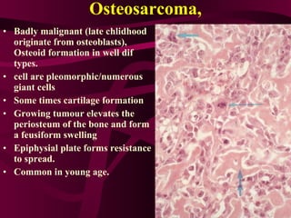
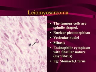
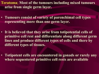
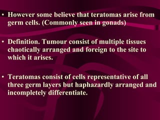
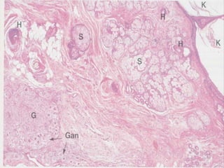
Neoplasia Malignant connective tissue tumours, Teratomas,Hamartomas
Malignant tumours of connective tissue origin (Sarcomata) Not common as carcinomas, But can get at any age prognosis worse compared to carcinomas. Spread through blood.  They form sheaths made up from merged neoplastic cells. They are inseparably merged in to stroma .
Eg: Fibrosarcomas  Osteosarcomas Rhabdomyosaecomas Leiomyosarcomas Liposarcomas.
Fibrosarcoma;   Counter part of the benign tumour Elongate large vesicular  nucleus  Atypical mitosis Giant cell commonly seen /collagen formation is scanty nuclear pleomorphism hypercromatism Haemorrege and necrosis is common infiltration and metastasis
Osteosarcoma, Badly malignant (late chlidhood originate from osteoblasts),  Osteoid formation in well dif types. cell are pleomorphic/numerous giant cells Some times cartilage formation Growing tumour elevates the periosteum of the bone and form a feusiform swelling Epiphysial plate forms resistance to spread. Common in young age.
Leiomyosarcoma The tumour cells are spindle shaped. Nuclear pleomorphism Vesicular nuclei Mitosis Eosinophilic cytoplasm with fibrillar nature (myofibrils) Eg: Stomach,Uterus
Teratoma ;   Most of the tumours including mixed tumours arise from single germ layer. Tumours consist of variety of parenchimal cell types representing more than one germ layer.  It is believed that they arise from totipotential cells of primitive cell rest and differentiate along different germ lines and produce different types of cells and there by different types of tissues.  Totipotent cells are encountered in gonads or rarely any where sequestered primitive cell rests are available
Teratomas are common in ovaries and testes  Other sites: mediastinum, retroperitoneal tissues Endless verity of tissues: Cartilage, Bone, Teeth, Hair, Epithelium Specialised types of tissues: like Hepatic,    Renal, Nervous, Occular etc--- Teratomas may Malignant  Benign
However some believe that teratomas arise from germ cells. (Commonly seen in gonads) Definition. Tumour consist of multiple tissues chaotically arranged and foreign to the site to which it arises.   Teratomas consist of cells representative of all three germ layers but haphazardly arranged and incompletely differentiate.
 
Ovarian teratomas Well differentiated, benign, (young or middle age women) Histologically; Thin-walled cystic mass filled with sebaceous keratinous debris and matted hair. Wall is usually stratified squamous epithelium .  In the eminence of the wall- Teeth, tongue like structure, even bone and cartilage,  Including other types of epithelia tissues  respiratory, intestinal also can be seen in eminence.
Rarely entire teratoma has a one type of tissue  eg Struma ovarii (Thyroid tissues)  Malignant changes are uncommon ,  Malignant teratomas metastasis rapidly via blood and lymph. Secondary lesion may be  A mixture of tissues  One type or  type which has not seen in primary
 
Testicular teratoma Young middle aged males/ some in children  Majority highly malignant (undifferentiated) Benign; Well differentiated and behave in a benign  manner  Malignant intermediate;  Both oraganoid formation  and undifferentiated cells. Malignant undifferentiated type; Anaplastic cells Malignant trophoblastic type; Trophoblastic cells highly malignant.
Less malignant types  are solid but often with cystic spaces. Histologically  - irregular gland like spaces lined with atypical cells which may resemble squamous respiratory and intestinal epithelium in different places.  Masses of cartilage bones and muscles etc inter-spread with epithelial elements and supported by connective tissue stroma. Highly malignant types-  consist of anaplastic sheets of cells sometimes with glandular patterns and papillary formation
Hamartomas;   Tumour like malformation. Tissues of the particular part of the body are haphazardly arranged with an excess of one or more of its components. Eg Hamartoma in lung tissue Hyaline cartilage is similar to found in the  bronchi There are clefts lined with respiratory  epithelium In the middle smooth muscle and connective  tissues.
Haemangioma:  Cavernous type  Capillary type
Tumours of nerve sheath origin Both schwannomas and neurofibromas are nerve sheeth tumours. Schwannomas Arise from Schwann cells. Neurofibromas arise from Perinural cells and fibroblasts. However, it is believed these lesions are two entities of the same spectrum.
Neurofibroma Cutaneous neurofibroma/ Solitary form in a periperal nerve. Plexiform neuroma
Neurofibroma: Slow growing, Benign, Well circumscribe  Not encapsulated. Lesions may be multiple & solitary  Histology:  Mixed population of  Schwann cells  Fibroblasts  Histology Spindle shaped cells with ill defined cell margins. Nuclei are ovoid or elongated but uniform. Cells dispersed in a connective tissue matrix which has collagen and fibroblasts. Myxomatous changes and nerve fibrous can be seen in the matrix.
 
Plexiform neuroma Large/multiple  Pendulous/Flabby Consist of thicken tortuous and small nerves. Schwann cells/fibroblasts Schwann cells produced myxoid connective tissue which expands each nerve.
Schwannoma(Neurilemmoma);   Cranial/spinal/peripheral nerves can de affected.  Encapsulated and appears to arise focally on the nerve trunk. So nerve stretched over the tumour. Microscopically tumour composed of elongated cells disposal in one of these two patterns.  Antoni type A - Arranged in an organised impact  manner. Palisading  of nuclei, and pink area between two palisadings (Varocay Body) Antoni type B-  cells are lossely arranged and scattered in the matrix.
 

Cancer

  • 1.
    Neoplasia Malignant connectivetissue tumours, Teratomas,Hamartomas
  • 2.
    Malignant tumours ofconnective tissue origin (Sarcomata) Not common as carcinomas, But can get at any age prognosis worse compared to carcinomas. Spread through blood. They form sheaths made up from merged neoplastic cells. They are inseparably merged in to stroma .
  • 3.
    Eg: Fibrosarcomas Osteosarcomas Rhabdomyosaecomas Leiomyosarcomas Liposarcomas.
  • 4.
    Fibrosarcoma; Counter part of the benign tumour Elongate large vesicular nucleus Atypical mitosis Giant cell commonly seen /collagen formation is scanty nuclear pleomorphism hypercromatism Haemorrege and necrosis is common infiltration and metastasis
  • 5.
    Osteosarcoma, Badly malignant(late chlidhood originate from osteoblasts), Osteoid formation in well dif types. cell are pleomorphic/numerous giant cells Some times cartilage formation Growing tumour elevates the periosteum of the bone and form a feusiform swelling Epiphysial plate forms resistance to spread. Common in young age.
  • 6.
    Leiomyosarcoma The tumourcells are spindle shaped. Nuclear pleomorphism Vesicular nuclei Mitosis Eosinophilic cytoplasm with fibrillar nature (myofibrils) Eg: Stomach,Uterus
  • 7.
    Teratoma ; Most of the tumours including mixed tumours arise from single germ layer. Tumours consist of variety of parenchimal cell types representing more than one germ layer. It is believed that they arise from totipotential cells of primitive cell rest and differentiate along different germ lines and produce different types of cells and there by different types of tissues. Totipotent cells are encountered in gonads or rarely any where sequestered primitive cell rests are available
  • 8.
    Teratomas are commonin ovaries and testes Other sites: mediastinum, retroperitoneal tissues Endless verity of tissues: Cartilage, Bone, Teeth, Hair, Epithelium Specialised types of tissues: like Hepatic, Renal, Nervous, Occular etc--- Teratomas may Malignant Benign
  • 9.
    However some believethat teratomas arise from germ cells. (Commonly seen in gonads) Definition. Tumour consist of multiple tissues chaotically arranged and foreign to the site to which it arises. Teratomas consist of cells representative of all three germ layers but haphazardly arranged and incompletely differentiate.
  • 10.
  • 11.
    Ovarian teratomas Welldifferentiated, benign, (young or middle age women) Histologically; Thin-walled cystic mass filled with sebaceous keratinous debris and matted hair. Wall is usually stratified squamous epithelium . In the eminence of the wall- Teeth, tongue like structure, even bone and cartilage, Including other types of epithelia tissues respiratory, intestinal also can be seen in eminence.
  • 12.
    Rarely entire teratomahas a one type of tissue eg Struma ovarii (Thyroid tissues) Malignant changes are uncommon , Malignant teratomas metastasis rapidly via blood and lymph. Secondary lesion may be A mixture of tissues One type or type which has not seen in primary
  • 13.
  • 14.
    Testicular teratoma Youngmiddle aged males/ some in children Majority highly malignant (undifferentiated) Benign; Well differentiated and behave in a benign manner Malignant intermediate; Both oraganoid formation and undifferentiated cells. Malignant undifferentiated type; Anaplastic cells Malignant trophoblastic type; Trophoblastic cells highly malignant.
  • 15.
    Less malignant types are solid but often with cystic spaces. Histologically - irregular gland like spaces lined with atypical cells which may resemble squamous respiratory and intestinal epithelium in different places. Masses of cartilage bones and muscles etc inter-spread with epithelial elements and supported by connective tissue stroma. Highly malignant types- consist of anaplastic sheets of cells sometimes with glandular patterns and papillary formation
  • 16.
    Hamartomas; Tumour like malformation. Tissues of the particular part of the body are haphazardly arranged with an excess of one or more of its components. Eg Hamartoma in lung tissue Hyaline cartilage is similar to found in the bronchi There are clefts lined with respiratory epithelium In the middle smooth muscle and connective tissues.
  • 17.
    Haemangioma: Cavernoustype Capillary type
  • 18.
    Tumours of nervesheath origin Both schwannomas and neurofibromas are nerve sheeth tumours. Schwannomas Arise from Schwann cells. Neurofibromas arise from Perinural cells and fibroblasts. However, it is believed these lesions are two entities of the same spectrum.
  • 19.
    Neurofibroma Cutaneous neurofibroma/Solitary form in a periperal nerve. Plexiform neuroma
  • 20.
    Neurofibroma: Slow growing,Benign, Well circumscribe Not encapsulated. Lesions may be multiple & solitary Histology: Mixed population of Schwann cells Fibroblasts Histology Spindle shaped cells with ill defined cell margins. Nuclei are ovoid or elongated but uniform. Cells dispersed in a connective tissue matrix which has collagen and fibroblasts. Myxomatous changes and nerve fibrous can be seen in the matrix.
  • 21.
  • 22.
    Plexiform neuroma Large/multiple Pendulous/Flabby Consist of thicken tortuous and small nerves. Schwann cells/fibroblasts Schwann cells produced myxoid connective tissue which expands each nerve.
  • 23.
    Schwannoma(Neurilemmoma); Cranial/spinal/peripheral nerves can de affected. Encapsulated and appears to arise focally on the nerve trunk. So nerve stretched over the tumour. Microscopically tumour composed of elongated cells disposal in one of these two patterns. Antoni type A - Arranged in an organised impact manner. Palisading of nuclei, and pink area between two palisadings (Varocay Body) Antoni type B- cells are lossely arranged and scattered in the matrix.
  • 24.