Downloaded 198 times

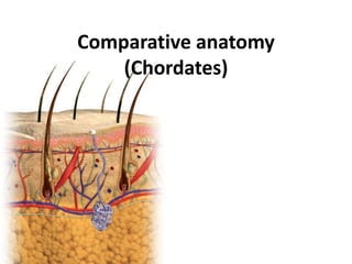

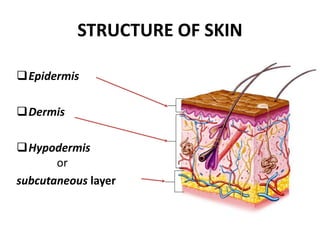
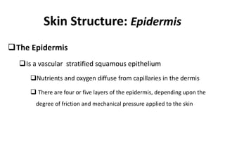
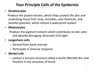
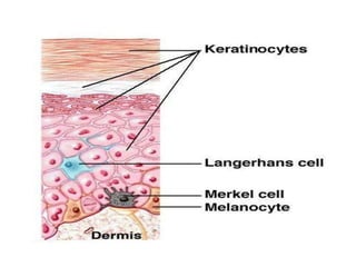
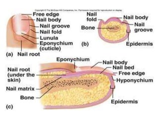
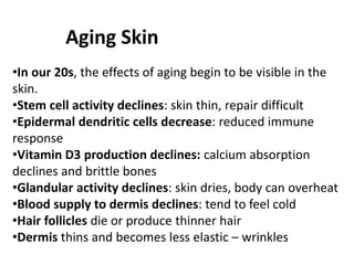

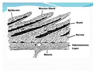
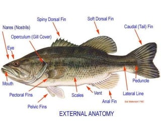
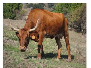

This document provides information on the integumentary system of several chordates including humans, fish, birds, cattle/horses, and ruminants. It describes the basic structure of skin which consists of an epidermis, dermis, and hypodermis layers across these species. Key differences are highlighted such as fish having scales instead of hair and birds having feathers. The structure and function of hair, nails, hooves and horns are also reviewed for different animals.


![[TRANS] HES 029 - Lecture 3 (The Integumentary System).pdf](https://cdn.slidesharecdn.com/ss_thumbnails/transhes029-lecture3theintegumentarysystem-221001083441-a0e9cb33-thumbnail.jpg?width=640&height=640&fit=bounds)


