Downloaded 303 times
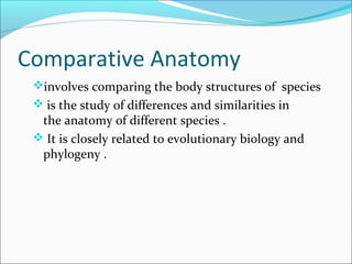


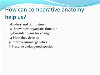


Comparative anatomy involves comparing the body structures of different species to understand evolutionary relationships and physiological similarities and differences. The document discusses key topics in comparative anatomy including homologous and analogous structures, directional terms, skeletal systems in humans and other vertebrates like fish, birds and cattle. Examples are provided of skeletal features like pneumatic and medullary bones that are specialized adaptations for respiration and calcium storage.