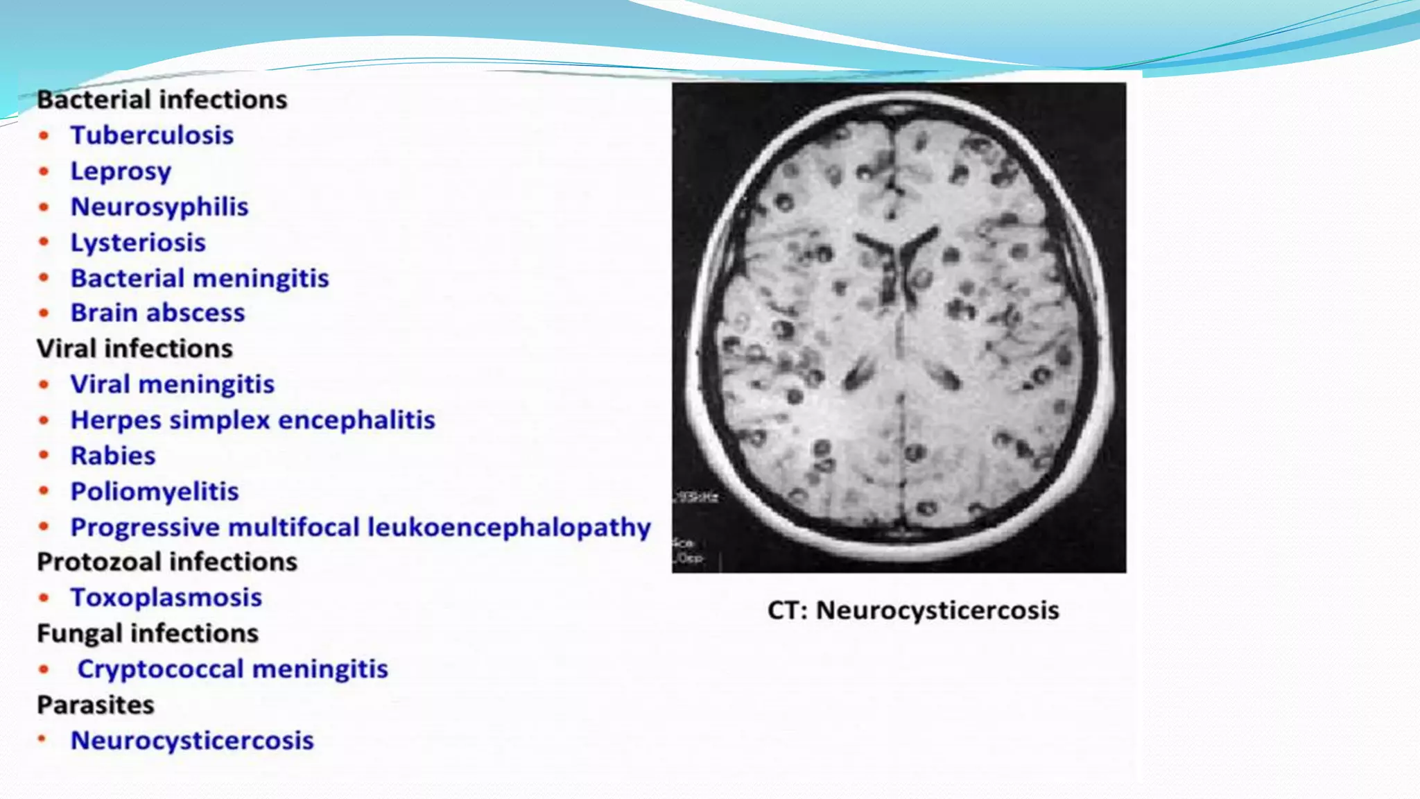
SlideShare a Scribd company logo
Dr Biplave Karki
Resident, Internal Medicine
Kathmandu University School of Medical Science
Overview
 Acute infections of the nervous system are among the most important
problems in medicine.
 Early recognition, efficient decision making, and rapid institution of
therapy can be lifesaving.
 Each may present with a nonspecific prodrome of fever and headache
(may initially be thought to be benign, until altered consciousness, focal
neurologic signs, or seizures appear.
 Key goals of early management are to emergently distinguish between
these conditions, identify the responsible pathogen, and initiate
appropriate antimicrobial therapy.
Lecture outline
 Acute bacterial meningitis
 Acute Viral meningitis
 Encephalitis
 TB meningitis
 Fungal infections
 Focal infections such as brain abscess
Acute Bacterial Meningitis
 Bacterial meningitis is an acute purulent infection within the
subarachnoid space.
 It is associated with a CNS inflammatory reaction that may result in
decreased consciousness, seizures, raised intracranial pressure (ICP), and
stroke.
 The meninges, subarachnoid space, and brain parenchyma are all
frequently involved in the inflammatory reaction (meningoencephalitis).
Etiology
 Streptococcus pneumoniae 50%
 Neisseria meningitidis 25%
 Group B Streptococci 15%
 Listeria monocytogenes 10%
 Haemophilus influenzae <10%
 Streptococcus pneumoniae
 Most common cause in adults >20 years.
 Predisposing conditions:
Pneumococcal pneumonia
Acute/chronic Pneumococcal
sinusitis/otitis media
Alcoholism
Diabetes
 Mortality ~20% despite antibiotic therapy.
•Splenectomy
•Hypogammaglobulinemia
•Complement deficiency
•Basilar skull fracture
 Neisseria meningitides
 Common serogroups: A, C, B, Y, W-135
 In some cases: fulminant, leading to death within a few hours.
 Petechial/purpuric skin lesions: can provide an important clue to diagnosis.
 The risk of invasive disease following nasopharyngeal colonization depends on both
bacterial virulence factors and host immune defense mechanisms, including the
host’s capacity
 to produce antimeningococcal antibodies and
 to lyse meningococci by both classic and alternative complement pathways.
 Individuals with deficiencies of any of the complement components, including
properdin, are highly susceptible to meningococcal infections.
 Gram negative bacilli
 Individuals with chronic debilitating conditions
 (diabetes, cirrhosis, alcoholism, chronic UTI)
 Neurosurgical procedures (craniotomy), head trauma with CSF rhinorrhoea or
otorrhoea.
 Listeria monocytogenes:
 important cause in neonates, pregnancy, elderly and immunocompromised individuals of all
ages.
 Haemophilus influenzae type b (Hib):
 unvaccinates children and older adults.
 Non-b H. influenzae: an emerging pathogen.
 Group B Streptococcus:
 previously mostly in neonates
 increasing in >50 years, particularly those with chronic underlying diseases.
 Staphylococcus aureus, CoNS:
 important causes of meningitis following invasive procedures
Otitis media, mastoiditis, sinusitis Streptococci
Gram negative anaerobes
S. aureus, Haemophilus sp.
Bacterial endocarditis Viridans Streptococci, S. bovis
S. aureus
HACEK, Enterococci
Pathophysiology
 Route of infection:
 Most common: hematogenous.
 May be from contiguous route
 (sinusitis, otitis media, mastoiditis, orbital cellulitis, cranial or vertebral osteomyelitis).
 May be from direct inoculation
 penetrating trauma, dermal sinus tract, meningomyelocele, surgery.
 Bloodborne bacteria can invade intraventricular choroid plexus
 directly infect choroid plexus epithelial cells and gain access to CSF.
 Can readily multiply within CSF
 absence of effective host immune defenses.
 A critical event in the pathogenesis:
 inflammatory reaction induced by the invading bacteria.
 Many of the neurologic manifestations and complications of bacterial
meningitis:
 immune response to the invading pathogen rather than from direct bacteria-
induced tissue injury.
 Neurologic injury can progress even after the CSF has been sterilized by
antibiotic therapy.
 Increase in CSF protein:
 due to increased vascular permeability with leakage of plasma proteins into
CSF.
 Hypoglucorrhachia:
 due to reduced glucose transport by cerebral tissue into CSF.
Pathology
 Leptomeninges
 infiltrated with inflammatory infiltrate.
 Cortex:
 edema, exudate, proliferation of microglia.
 Meningeal purulent exudates
 around cerebral veins, venous sinuses, convexities of brain, cerebellum, sulci,
sylvian fissure, basal cistern, spinal cord.
 Ventriculitis with bacterial and inflammatory cells in CSF.
 Subdural effusion, rarely empyema.
 Exudates may block foramen of Magendie, foramen of Lushka:
 internal hydrocephalus.
 Cerebral infarction
 (vascular occlusion due to phlebitis, vasospasm, thrombosis).
 Inflammation of
 spinal nerves (meningeal signs),
 cranial nerves (cranial neuropathies (II, VI, VII, VIII).
 Damage to cerebral cortex:
 Focal/diffuse effect of vascular occlusion.
 Hypoxia
 Bacterial invasion (cerebritis)
 Toxic encephalopathy (bacterial toxins)
 Raised ICP
 Ventriculitis
Clinical Presentation
 Can present as either an acute fulminant illness that progresses rapidly in
a few hours or as a subacute infection that progressively worsens over
several days.
 The classic clinical triad of meningitis
 fever, headache, and nuchal rigidity, but the classic triad may not be present.
 Nonspecific findings associated with systemic infection:
 Fever, headache
 Anorexia
 Myalgia, arthralgia
 URTI symptoms
 Tachycardia, hypotension
 Meningeal irritation:
 Nuchal rigidity
 Kernig sign
 Brudzinski sign
 Features suggesting raised ICP
 Headache
 Emesis
 CN III, VI palsy
 Papilloedema
 Hypertension, bradycardia, irregular respiration (Cushing reflex).
 >90% patients:
 CSF opening pressure >180 mmHg.
 20%:
 CSF opening pressure >400 mmHg
 Most disastrous complication:
 cerebral herniation (1-8%).
• Apnea or hyperventilation
• Decorticate/Decerebrate posturing
• Stupor/coma
 Seizures:
 May occur as part of initial presentation or during course of illness in 20-40%
 Focal seizures due to:
 Focal arterial ischemia or infarction
 Cortical venous thrombosis with hemorrhage
 Focal edema
 Generalized seizure due to:
 Hyponatremia
 Cerebral anoxia
 Toxic effects of antimicrobial agents
 Alteration of mental status
 Irritability, lethargy, stupor, obtundation, coma.
 Due to:
 Raised ICP
 Cerebritis
 Hypotension
 Atypical presentation may be observed in certain groups
 Elderly,
 especially with underlying comorbidities (eg, diabetes, renal and liver disease),
 may present with lethargy and an absence of meningeal symptoms.
 Patients with neutropenia
 may present with subtle symptoms of meningeal irritation.
 Other –
 immunocompromised hosts, including organ and tissue transplant recipients and
 patients with HIV and AIDS.
 Patients with aseptic meningitis syndrome usually appear clinically nontoxic with no vascular
instability.
 Special features of particular forms:
 Meningococcal meningitis:
 Petechiae/purpura/ecchymotic rashes over skin/mucosa.
 Petechiae over conjunctiva
 Rapid progression to shock, hypotension, coma, death within 24 hours.
 Pneumococcal meningitis:
 Preceeding pneumonia/otitis media/sinusitis/head injury.
 Subdural effusion: a common complication.
 The presence of a murmur suggests infective endocarditis with secondary
bacterial seeding of the meninges.
Diagnosis
 Clinically: history, examination.
 When suspected, cultures should be obtained immediately and empirical
antibiotics and adjunctive steroid therapy initiated without delay.
 Diagnosis by: CSF examination
 Investigations:
 Haematology, Biochemistry, Random blood sugar
 Viral markers
 Blood culture
 Culture from other possible sites of infection
 CSF examination
 Imaging (CT/MRI)
Lumbar puncture
 Crucial for:
 Establishing the diagnosis
 Identifying the causative organism
 Performing in-vitro susceptibility testing.
 Every patient suspected of meningitis should have CSF obtained unless lumbar puncture is
contraindicated.
 Send for:
 Cell count, differential
 Sugar, Protein, ADA
 Gram stain, AFB
 Culture and sensitivity
 Others: India Ink, viral serology, latex agglutination, PCR
 Indications of CT before LP (2004 IDSA guidelines)
 Immunocompromised state (HIV, immunosuppressive therapy, transplant
recipient)
 History of CNS disease (mass lesion, stroke or focal infection)
 New onset seizure (within one week of presentation)
 Papilloedema
 Abnormal level of consciousness
 Focal neurologic deficit (including dilated non-reactive pupils, abnormalities
of ocular motility, abnormal visual fields, gaze palsy, arm or leg drift)
 If LP is delayed or deferred
 Blood culture
 Empiric antibiotics before the imaging
 Followed by LP asap.
 Dexamethasone 0.15 mg/kg 20min before or at the same time as antibiotics,
then 6hrly for 4 days. No benefit after 6 hr of antibiotics.
 Prior administration of antibiotics tends to have minimal effects on the
chemistry and cytology findings but can reduce the yield of Gram stain and
culture.
 Relative contraindications
 Evidence of raised ICP
 Thrombocytopenia, other bleeding diathesis.
 Spinal epidural abscess
 Complications
 Cerebral herniation
 Postdural puncture headache
 Traumatic tap
 Spinal trauma
Neuroimaging
 MRI preferred over CT because of its superiority in demonstrationg areas
of cerebral edema and ischemia.
 Diffuse meningeal enhancement often seen after the administration of
Gadolinium.
 Meningeal enhancement is not diagnostic of meningitis but occurs in any
CNS disease with increased blood brain barrier.
Management
Treatment
 Empirical Antimicrobial
therapy
 Goal: begin antibiotic
therapy within 60 minutes.
 Acyclovir is commonly
added to the regime, as HSV
encephalitis is an important
differential.
 Specific antimicrobial therapy
BACTERIAL MENINGITIS (Duration of Antibiotic)
Pathogen Duration of Rx (d)
H. influenzae 7
N. meningitidis 7
S. pneumoniae 10-14
L. monocytogenes 14-21
Group B strep 14-21
GNRs 21
NEJM 1997;336:708
Adjunctive therapy: Dexamethasone
 Release of bacterial cell wall components induces production of
inflammatory cytokines IL-1B and TNFα.
 The results of clinical trials of dexamethasone therapy in meningitis due
to H. influenzae, S. pneumoniae, and N. meningitides have demonstrated
its efficacy in decreasing meningeal inflammation and neurologic
sequelae.
Adjunctive therapy: Dexamethasone
 IDSA Guidelines:
 Dexamethasone (0.15 mg/kg q6h for 2–4 days with the first dose administered 10–20
min before, or at least concomitant with, the first dose of antimicrobial therapy) in
adults with suspected or proven pneumococcal meningitis.
 Dexamethasone should only be continued if the CSF Gram stain reveals gram-
positive diplococci, or if blood or CSF cultures are positive for S. pneumoniae.
 Adjunctive dexamethasone should not be given to adult patients who have already
received antimicrobial therapy, because administration of dexamethasone in this
circumstance is unlikely to improve patient outcome.
 The data are inadequate to recommend adjunctive dexamethasone to adults with
meningitis caused by other bacterial pathogens, although some authorities would
initiate dexamethasone in all adults, because the etiology of meningitis is not always
ascertained at initial evaluation .
Raised ICP
Prognosis
 Mortality
 3-7% for meningitis due to H. influenzae, N. meningitidis, group B streptococci
 15% for that due to L. monocytogenes
 20% for S. pneumoniae
 Risk of death increases with:
 Decreased level of consciousness during admission.
 Onset of seizures within 24 hours of admission
 Signs of raised ICP.
 Infancy and >50 years.
 Comorbid conditions including shock and need for mechanical ventilation
 Delay in initiation of treatment.
 CSF glucose <40 and protein >300 predictive of increased mortality and poorer
outcomes.
 Moderate to severe sequelae in 25% of survivors.
Complications
 CNS:
 Acute:
Seizures Raised ICP
Cranial nerve palsies Stroke
Cerebral herniation Thrombosis of dural venous sinuses
 Subdural effusion/empyema
 Ventriculitis, Arachnoiditis
 Brain abscess
 Hydrocephalus
 Long-term neurologic deficits
Hemiplegia Aphasia
Hearing loss Ocular palsies
Hemianopia, Blindness Mental retardation
 Systemic: Shock, myocarditis, SIADH
Acute Viral Meningitis
Introduction
 Viral infection is the most common cause of meningitis, and usually results in a
benign and self-limiting illness requiring no specific therapy.
 It is a much less serious illness than bacterial meningitis unless there is
associated encephalitis.
Etiology
Clinical Manifestations
 Immunocompetent adults usually present with headache, fever and signs
of meningeal irritation coupled with inflammatory CSF profile.
 Headache:
 Almost always present.
 Often frontal or retroorbital
 Frequently associated with photophobia and pain on moving the eyes.
 Nuchal rigidity:
 May be mild and present only near the limit of neck anti-flexion.
 Constitutional features.
Diagnosis
 Investigations:
 Haematology, Renal and liver function tests, Random blood sugar
 ESR, CRP
 Viral markers
 Blood culture
 Culture from other possible sites of infection
 CSF examination
 Imaging (CT/MRI)
CSF findings
 Lymphocytic pleocytosis.
 Count typically 25-500/mm3.
 Rarely, may reach thousands.
 Rarely, PMNs may predominate in the first 48 hours
 Normal or slightly elevated protein (20-80 mg/dL)
 Normal glucose concentration.
 Normal or mildly elevated opening pressure (10-35 cmH2O).
 PCR amplification of viral nucleic acid:
 Single most important method for diagnosing CNS viral infections.
 Viral culture
 Poor sensitivity.
 In addition to CSF, specific viruses may also be isolated from throat swabs,
stool, blood and urine.
 Serologic studies
 For viruses with low sero-prevalence in population, diagnosis can be made
 by documenting sero-conversion between acute phase and convalescent sera (typically
obtained after 2-4 weeks) or
 by demonstrating presence of virus specific IgM antibodies.
 Serum antibody determination less useful for viruses with high
seroprevalence in general population such as HSV, VZV, CMV, EBV.
 Demonstration of virus specific antibody in CSF may be useful.
Neuroimaging
 Neuroimaging studies (MRI preferable to CT) are not absolutely necessary
in patients with uncomplicated viral meningitis.
 Should be performed in patients with altered consciousness, seizures,
focal neurologic signs or symptoms, atypical CSF profiles, or underlying
immunocompromising treatments or conditions.
Bacterial vs Viral – IDSA Guidelines
 Bacterial meningitis score,
 probability of bacterial meningitis is 0.3% or less in children with CSF
pleocytosis who have
1) a negative CSF Gram’s stain,
2) CSF neutrophil count <1000 cells/μL,
3) CSF protein <80 mg/dL,
4) peripheral absolute neutrophil count of <10,000 cells/μL, and
5) no prior history or current presence of seizures
Treatment
 Most cases: primarily symptomatic
 Analgesics, antipyretics, antiemetics.
 Optimization of fluid and electrolytes.
 Empirical antibiotics in whom bacterial etiology cannot be ruled out.
 Hospitalization:
 Immunocompromized patients
 Alteration in consciousness, seizures, focal signs
 Symptoms suggesting the possibility of encephalitis or parenchymal brain
involvement
 Atypical CSF profile.
Treatment
 Acyclovir
 May be of benefit in: HSV-1, 2, severe EBV or VZV.
 Seriously ill patients:
 IV Acyclovir 15-30 mg/kg per day in three divided doses.
Followed by oral drug
 Acyclovir 800 mg five times daily or
 Famciclovir 500 mg TDS or
 Valacyclovir 1000 mg TDS) for a total of 7-14 days.
 Patients who are less ill can be treated with oral drugs alone.
Treatment
 HIV meningitis:
 HAART
 No specific therapy of proven benefit for arbovirus.
Prognosis
 Adults: good
 Rare patients may complain of persisting headache, mild mental impairment,
incoordination, generalized asthenia for weeks to months.
 Infants and children: outcome less certain.
 May develop intellectual impairement, learning disabilities, hearing loss and
other lasting sequelae.
Prevention
 Vaccination
 Poliovirus
 Mumps
 Measles
 Rubella
 Varicella
 Can prevent development of meningitis and other neurologic complications.
Viral Encephalitis
Introduction
 In contrast to viral meningitis, where the infectious process and associated
inflammatory response are limited largely to the meninges, in encephalitis the
brain parenchyma is also involved.
 Many patients with encephalitis also have evidence of associated meningitis
(meningo-encephalitis) and, in some cases, involvement of the spinal cord or
nerve roots (encephalo-myelitis, encephalo-myelo-radiculitis).
Etiology
Clinical manifestations
 In addition to features of meningitis:
 Altered level of consciousness (confusion or behavioral abnormalities) or a
depressed level of consciousness (lethargy to coma).
 Evidence of either focal or diffuse neurologic signs and symptoms.
 Focal or generalized seizures occur in many patients.
 Involvement of the hypothalamic-pituitary axis may result in temperature
dysregulation, diabetes insipidus, or the development of the syndrome of
inappropriate secretion of antidiuretic hormone (SIADH).
Diagnosis
 Routine investigations, CSF study:
 similar as in viral meningitis.
 MRI/CT/EEG:
 Help to identify or exclude alternative diagnosis.
 Assist in differentiating focal or diffuse encephalitic process
[Focal findings: should always raise possibility of HSV encephalitis].
 Brain biopsy: reserved for
 CSF PCR studies fail to lead to a specific diagnosis,
 Focal abnormalities on MRI, and who continue to show progressive clinical
deterioration despite treatment with acyclovir and supportive therapy.
Treatment
 General measures:
 Vital functions including respiration and blood pressure should be
monitored and supported as required.
 Careful ICP monitoring, fluid restriction, avoidance of hypotonic IV
solutions, suppression of fever.
 Seizures: standard anticonvulsant regimen. Prophylactic therapy should
be considered in view of high frequency of seizures in severe cases.
 Prophylaxis/treatment of aspiration pneumonia, decubitus ulcers,
contractures, DVT, infections of indwelling lines and catheters.
Treatment
 Acyclovir
 Benefit in HSV, EBV or VZV. Should be started empirically in patients with
suspected viral encephalitis especially if focal features are present.
 Should be discontinued if found not to have HSV, EBV or VZV.
 10 mg/kg IV TDS for 14-21 days. (Neonates: 20 mg/kg TDS for a minimum of
21 days). Repeat CSF PCR at completion of this course and if positive, give
another course.
 Ganciclovir and Foscarnet alone or in combination often used in CMV
related encephalitis.
 Cidofovir may provide an alternative.
Tuberculous Meningitis
Introduction
 Most commonly occurs shortly after a primary infection in childhood or as
part of miliary tuberculosis.
 Common in developing countries.
 A critical disease in terms of fatal outcome and permanent sequelae,
requiring rapid diagnosis and treatment.
Pathophysiology
 Develops in two steps:
 A transient bacteremia during formation of primary complex can seed the bacilli to other
organs. Seeding to meninges or brain parenchyma (more likely if miliary tuberculosis
develops) leads to formation of subpial or subependymal foci of metastatic caseous lesions
termed Rich focus.
 Rich focus increases in size until it ruptures into subarachnoid space, causing meningitis.
 Those deeper in the brain or spinal cord parenchyma cause tuberculoma or abscess.
Clinical Manifestation
 Presents subtly as headache and slight mental changes after a prodrome of
weeks of low-grade fever, malaise, anorexia, and irritability.
 Typically evolves over 1-2 weeks. May evolve acutely with severe headache,
confusion, lethargy, altered sensorium and neck rigidity.
 Paresis of cranial nerves (oculomotor nerves in particular) is a frequent
finding because meningeal involvement is common at the base.
 Visual symptoms include visual impairement or blindness and occasionally,
abrupt onset of painful ophthalmoplegia.
 Sudden onset of focal neurologic deficits including monoplegia, hemiplegia,
aphasia and tetraparesis have been reported.
 Tremors and less commonly, abnormal movements like choreoathetosis and
hemiballismus may ocuur (more in children)
 The ultimate evolution is towards coma, with hydrocephalus and intracranial
hypertension.
Staging
 Stage I (Early):
 non-specific symptoms and signs
 apathy, irritability, headache, malaise, fever, anorexia, nausea, vomiting, without any
alteration in consciousness.
 Stage II (Intermediate):
 Altered consciousness without coma or delirium but with minor focal neurological
signs;
 Symptoms and signs of meningism and meningitis are present, in addition
 Focal neurological deficits, isolated CN palsies, and abnormal involuntary
movements.
 Stage III (Advanced):
 Stupor or coma, dense neurological deficits, seizures, posturing, and/or abnormal
movements.
Diagnosis
 Lumbar puncture: cornerstone of diagnosis.
 CSF clear. When allowed to stand, ‘spider web’ may form.
 Total count up to 1000/mm3.
Predominanlty lymphocytes. May be neutrophils in early stages.
 Protein: 100-800 mg/dL
 Low glucose (<40 mg/dL)
 ADA: >10 U/L – 92-94% sensitivity, 90-97% specificity
 AFBs: infrequently seen.
 Culture: diagnostic in up to 80% of cases.
 Gene Xpert: 80% sensitivity.
 Imaging studies may show
 Hydrocephalus
 Abnormal enhancement of basal cisterns or ependyma.
 Infarct, edema
 Tuberculoma
Treatment
 Should be started as soon as the diagnosis is made or strongly suspected.
 Conflicting views in use of ethambutol or streptomycin and in duration of
therapy.
 Role of steroids
 Improves CSF abnormalities and elevated CSF pressure.
 Improves chances of survival but does not reduce frequency of neurological
sequelae.
 Dexamethasone:
 0.4 mg/kg/day IV, tapering by 0.1 mg/kg per week until the 4th week followed by 4
mg/day orally tapering by 1 mg per week until the 4th week.
Fungal Infections of CNS
CNS fungal infections in order of frequency
Meningitis Abscess/Granuloma Thrombus/Hemorrhage
Cryptococcus neoformans Aspergillus Aspergilus
Coccidiodes immitis Cryptococcus neoformans Zygomycetes
Histoplasma capsulatum Coccidioides immitis
Nocardia, Histoplasma
Mucor, Absidia, Rhizopus
Candida Cladophiaphora
Aspergillus Pseudoallescheria boydii
Blastomyces dermatidis Fusarium
Sporotrax schenckis
Cryptococcosis
 Cryptococcus neoformans is the most common fungal infection
affecting the nervous system.
 Cryptococcus neoformans
 Encapsulated form of yeast
 Capsule production varies with the strain and environmental conditions.
 Found throughout world, especially in soil contaminated with bird droppings
 Causes a chronic form of meningitis
 Third most common opportunistic infection in AIDS patients.
 In immuno-competent hosts, it mostly occurs in those who are
typically exposed to large amounts of the yeast from being in
contact with bird feces.
 The organism is inhaled and then spreads haematogenously.
 Cryptococcus typically causes chronic meningitis, which begins
insidiously with malaise, fever, photophobia, and meningism.
Clinical Manifestations
 Chronic headache and alteration of mental status including
confusion, hallucinations, and other psychotic symptoms.
 Meningitis can be complicated by encephalitis or formation of
granulomas that cause focal symptoms and signs referable to the
brain areas involved.
 In some cases, communicating hydrocephalus with vomiting,
blurred or double vision, obtundation, and other signs of
increased ICP.
Diagnosis
 CSF findings consistent with meningitis but routine stains and
bacterial antigens are negative.
 Special fungal stains such as Gomori silver methenamine and
India Ink are useful in demonstrating hyphae or yeast capsules.
 Culture, Serology.
CSF Analysis
 The characteristic CSF abnormalities in fungal meningitis are
 mononuclear or lymphocytic pleocytosis
 increased protein concentration
 decreased glucose concentration
 There may be eosinophils in the CSF in C. immitis meningitis.
 The cryptococcal polysaccharide antigen test is a highly sensitive
and specific test for cryptococcal meningitis
Treatment
 Meningitis due to C. neoformans in non-HIV, nontransplant patients
 Induction therapy with amphotericin B (AmB) (0.7 mg/kg IV per day) plus flucytosine
(100 mg/kg per day in four divided doses) for at least 4 weeks if CSF culture results are
negative after 2 weeks of treatment
 Therapy should be extended for a total of 6 weeks in the patient with neurologic
complications
 Induction therapy is followed by consolidation therapy with fluconazole (400 mg/day)
for 8 weeks.
 Organ transplant recipients
 liposomal AmB (3–4 mg/kg per day) or AmB lipid complex (ABLC) 5 mg/kg per day plus
flucytosine (100 mg/kg per day in four divided doses) for at least 2 weeks or until CSF
culture is sterile, followed by an 8- to 10-week course of fluconazole (400–800 mg/d).
If the CSF culture is sterile after 10 weeks of acute therapy, the dose of fluconazole is decreased to 200 mg/d for 6
months to a year.
 Patients with HIV infection
 AmB or a lipid formulation plus flucytosine for at least 2 weeks,
followed by fluconazole for a minimum of 8 weeks.
 Meningitis due to H. capsulatum
 AmB (0.7–1.0 mg/kg /day) for 4–12 wks,(total dose of 30 mg/kg)
 Therapy with AmB is not discontinued until fungal cultures are
sterile
 After completing a course of AmB, maintenance therapy with
itraconazole 200 mg bd is initiated and continued for at least 6
months to a year.
Complications
 The most common complication of fungal meningitis is
hydrocephalus.
 Patients who develop hydrocephalus should receive a CSF
diversion device.
 A ventriculostomy can be used until CSF fungal cultures are
sterile, at which time the ventriculostomy is replaced by a
ventriculo-peritoneal shunt.
Brain Abscess
Introduction
 Focal suppurative infection within the brain parenchyma, typically surrounded
by a vascularized capsule.
 The term Cerebritis is often employed to describe a non-encapsulated brain
abscess.
Epidemiology
 Incidence: 0.3-1.3 per 100,000 persons per year
 Predisposing conditions:
 Otitis media
 Mastoiditis
 Sinusitis
 Pyogenic infection in the chest or other body sites
 Penetrating head trauma
 Neurosurgical procedures
 Dental infection.
Etiology
 Immunocompetent individuals:
 Streptococcus spp (40%)
 Enterobacteriaceae (25%): Proteus, E. coli, Klebsiella
 Anaerobes (30%): Bacteroides, Fusobacterium
 Staphylococci (10%)
 Immunocompromised hosts
 Nocardia
 Toxoplasma
 Aspergillus
 Candida
 Cryptococcus
 Tuberculosis
 A brain abscess may develop
 By direct spread from a contiguous cranial site of infection
 Following head trauma or a neurosurgical procedure
 As a result of hematogenous spread from a remote site of infection.
 In about 25% of cases, no obvious primary source of infection is apparent
(Cryptogenic brain abscess).
Clinical Manifestation
 Typically presents as an expanding intracranial mass lesion rather than an
infectious process.
 Most present to hospital 11-12 days following onset of symptoms.
 Classic clinical triad:
 Headache (>75%)
 Fever (50%)
 Focal neurologic deficit (>60%)
 Features based on location of abscess
 Hemiparesis is the most common localizing sign of a frontal lobe abscess.
 Temporal lobe abscess: Dysphasia, upper homonymous quadrantopia.
 Cerebellar abscess : Nystagmus, ataxia .
 Meningismus is not present unless the abscess has ruptured into the ventricle or
the infection has spread to subarachnoid space.
Diagnosis
 Neuroimaging
 MRI better than CT for demonstrating abscess in the early stages (cerebritis)
and is superior to CT for identifying abscesses in posterior fossa.
 Microbiologic diagnosis of etiologic agent is most accurately determined
by Gram stain and culture of abscess material obtained by CT-guided
stereotactic needle aspiration.
 LP should not be done in known or suspected focal intracranial infection.
Contributes nothing to diagnosis and increases risk of herniation.
Treatment
 Combination of high dose parenteral antibiotic therapy and neurosurgical
drainage.
 Empiric therapy of community acquired brain abscess in a
immunocompetent patient:
 Third/fourth generation cephalosporin plus Metronidazole
 Penetrating head trauma or recent neurosurgical procedures
 Ceftazidime plus Vancomycin or
 Meropenem plus Vancomycin
 Duration: at least 6-8 weeks of parenteral antibiotics
 Medical management alone:
 Neurosurgically inaccessible
 Small (<2-3 cm) or non-encapsulated abscess (cerebritis)
 Unfit for surgery.
 Aspiration and drainage of the abscess under stereotactic guidance.
 Complete excision via craniotomy or craniectomy
 Multiloculated abscess
 Failed stereotactic aspiration.
 Prophylactic anticonvulsants
 High risk (35%) of focal or generalized seizures.
 Continued for at least 3 months following resolution of abscess. After that, decision to
withdraw is based on EEG.
 Steroids should not be given routinely.
 IV Dexamethasone 10 mg q6h reserved to those with substantial periabscess edema and
associated mass effect and raised ICP.
 Should be tapered as rapidly as possible to avoid delaying natural process of encapsulation of
the abscess.
 Serial CT/MRI every 1-2 month to document resolution of the abscess.
Prognosis
 Mortality rate <15%
 Sequelae: >20% of survivors.
 Seizures
 Persisting weakness
 Aphasia
 Mental impairement
THANK YOU

More Related Content

What's hot

Approach to a patient with vasculitis
Approach to a patient with vasculitisApproach to a patient with vasculitis
Approach to a patient with vasculitisaminanurnova
 
Cns infections
Cns infectionsCns infections
Cns infectionsraj kumar
 
Acute Viral encephalitis Dr. Shatdal Chaudhary
Acute Viral encephalitis Dr. Shatdal ChaudharyAcute Viral encephalitis Dr. Shatdal Chaudhary
Acute Viral encephalitis Dr. Shatdal ChaudharyShatdal Chaudhary
 
Immune reconstitution inflammatory syndrome-Hamisi Mkindi
Immune reconstitution inflammatory syndrome-Hamisi MkindiImmune reconstitution inflammatory syndrome-Hamisi Mkindi
Immune reconstitution inflammatory syndrome-Hamisi MkindiMkindi Mkindi
 
Ch. 24 Infections of Central Nervous System
Ch. 24 Infections of Central Nervous SystemCh. 24 Infections of Central Nervous System
Ch. 24 Infections of Central Nervous Systemkevperrino
 
Pediatric vasculitis dr inayat ullah
Pediatric vasculitis dr inayat ullahPediatric vasculitis dr inayat ullah
Pediatric vasculitis dr inayat ullahDr Inayat Ullah
 
PATHOLOGY OF UPPER RESPIRARTORY
PATHOLOGY OF UPPER RESPIRARTORY PATHOLOGY OF UPPER RESPIRARTORY
PATHOLOGY OF UPPER RESPIRARTORY دكتور مريض
 
Pseudotuberculosis
PseudotuberculosisPseudotuberculosis
PseudotuberculosisLadi Anudeep
 
Progressive multifocal leukoencephalopathy (PML)
Progressive multifocal leukoencephalopathy (PML)Progressive multifocal leukoencephalopathy (PML)
Progressive multifocal leukoencephalopathy (PML)Amr Hassan
 
CLINICAL FEATURES AND COMPLICATIONS OF MALARIA
CLINICAL FEATURES AND COMPLICATIONS OF MALARIACLINICAL FEATURES AND COMPLICATIONS OF MALARIA
CLINICAL FEATURES AND COMPLICATIONS OF MALARIAAbino David
 
Cryptococcal Meningitis SEMINAR
Cryptococcal Meningitis SEMINARCryptococcal Meningitis SEMINAR
Cryptococcal Meningitis SEMINARfareedresidency
 
Brain abscess
Brain abscessBrain abscess
Brain abscessjoemdas
 

What's hot (20)

Acute meningoencephalitis
Acute meningoencephalitisAcute meningoencephalitis
Acute meningoencephalitis
 
Encephalitis
EncephalitisEncephalitis
Encephalitis
 
Approach to a patient with vasculitis
Approach to a patient with vasculitisApproach to a patient with vasculitis
Approach to a patient with vasculitis
 
Viral meningitis
Viral meningitisViral meningitis
Viral meningitis
 
Cns infections
Cns infectionsCns infections
Cns infections
 
Acute Viral encephalitis Dr. Shatdal Chaudhary
Acute Viral encephalitis Dr. Shatdal ChaudharyAcute Viral encephalitis Dr. Shatdal Chaudhary
Acute Viral encephalitis Dr. Shatdal Chaudhary
 
Immune reconstitution inflammatory syndrome-Hamisi Mkindi
Immune reconstitution inflammatory syndrome-Hamisi MkindiImmune reconstitution inflammatory syndrome-Hamisi Mkindi
Immune reconstitution inflammatory syndrome-Hamisi Mkindi
 
Ch. 24 Infections of Central Nervous System
Ch. 24 Infections of Central Nervous SystemCh. 24 Infections of Central Nervous System
Ch. 24 Infections of Central Nervous System
 
meningitis
meningitismeningitis
meningitis
 
Pediatric vasculitis dr inayat ullah
Pediatric vasculitis dr inayat ullahPediatric vasculitis dr inayat ullah
Pediatric vasculitis dr inayat ullah
 
Meningitis
MeningitisMeningitis
Meningitis
 
PATHOLOGY OF UPPER RESPIRARTORY
PATHOLOGY OF UPPER RESPIRARTORY PATHOLOGY OF UPPER RESPIRARTORY
PATHOLOGY OF UPPER RESPIRARTORY
 
Pseudotuberculosis
PseudotuberculosisPseudotuberculosis
Pseudotuberculosis
 
Fungal infection of cns
Fungal infection of cnsFungal infection of cns
Fungal infection of cns
 
Viral encephalitis
Viral encephalitisViral encephalitis
Viral encephalitis
 
Pneumocystis jirovecii infection
Pneumocystis jirovecii infectionPneumocystis jirovecii infection
Pneumocystis jirovecii infection
 
Progressive multifocal leukoencephalopathy (PML)
Progressive multifocal leukoencephalopathy (PML)Progressive multifocal leukoencephalopathy (PML)
Progressive multifocal leukoencephalopathy (PML)
 
CLINICAL FEATURES AND COMPLICATIONS OF MALARIA
CLINICAL FEATURES AND COMPLICATIONS OF MALARIACLINICAL FEATURES AND COMPLICATIONS OF MALARIA
CLINICAL FEATURES AND COMPLICATIONS OF MALARIA
 
Cryptococcal Meningitis SEMINAR
Cryptococcal Meningitis SEMINARCryptococcal Meningitis SEMINAR
Cryptococcal Meningitis SEMINAR
 
Brain abscess
Brain abscessBrain abscess
Brain abscess
 

Similar to Cns infections biplave nams

04 Neurologic
04 Neurologic04 Neurologic
04 NeurologicDeep Deep
 
04 Neurologic
04 Neurologic04 Neurologic
04 NeurologicDeep Deep
 
22 Purulent Meningitis
22 Purulent Meningitis22 Purulent Meningitis
22 Purulent Meningitisghalan
 
Inflammatory conditions of brain ppt.pptx
Inflammatory conditions of brain ppt.pptxInflammatory conditions of brain ppt.pptx
Inflammatory conditions of brain ppt.pptxAshik Dhakal
 
Inflammatory conditions of brain ppt.pptx
Inflammatory conditions of brain ppt.pptxInflammatory conditions of brain ppt.pptx
Inflammatory conditions of brain ppt.pptxAshik Dhakal
 
CNS INFECTIONS.pdf
CNS INFECTIONS.pdfCNS INFECTIONS.pdf
CNS INFECTIONS.pdfNimonaAAyele
 
MENINGITIS & ENCEPHALITIS - Ayushi.pptx pdf
MENINGITIS & ENCEPHALITIS - Ayushi.pptx pdfMENINGITIS & ENCEPHALITIS - Ayushi.pptx pdf
MENINGITIS & ENCEPHALITIS - Ayushi.pptx pdfAditiShah380128
 
Cns infections
Cns infectionsCns infections
Cns infectionsLetaJarso
 
Short presentation version cns infections Lecture
Short presentation version cns infections LectureShort presentation version cns infections Lecture
Short presentation version cns infections Lecturetest
 
Cns Infxs
Cns InfxsCns Infxs
Cns Infxsmycomic
 
Brain Infxn 71609
Brain Infxn 71609Brain Infxn 71609
Brain Infxn 71609mycomic
 

Similar to Cns infections biplave nams (20)

04 Neurologic
04 Neurologic04 Neurologic
04 Neurologic
 
04 Neurologic
04 Neurologic04 Neurologic
04 Neurologic
 
MENINGITIS.ppt
MENINGITIS.pptMENINGITIS.ppt
MENINGITIS.ppt
 
22 Purulent Meningitis
22 Purulent Meningitis22 Purulent Meningitis
22 Purulent Meningitis
 
Inflammatory conditions of brain ppt.pptx
Inflammatory conditions of brain ppt.pptxInflammatory conditions of brain ppt.pptx
Inflammatory conditions of brain ppt.pptx
 
Inflammatory conditions of brain ppt.pptx
Inflammatory conditions of brain ppt.pptxInflammatory conditions of brain ppt.pptx
Inflammatory conditions of brain ppt.pptx
 
CNS INFECTIONS.pdf
CNS INFECTIONS.pdfCNS INFECTIONS.pdf
CNS INFECTIONS.pdf
 
MENINGITIS & ENCEPHALITIS - Ayushi.pptx pdf
MENINGITIS & ENCEPHALITIS - Ayushi.pptx pdfMENINGITIS & ENCEPHALITIS - Ayushi.pptx pdf
MENINGITIS & ENCEPHALITIS - Ayushi.pptx pdf
 
Meningitis
MeningitisMeningitis
Meningitis
 
Infectious diseases
Infectious diseasesInfectious diseases
Infectious diseases
 
Cns infections
Cns infectionsCns infections
Cns infections
 
Meningitis
MeningitisMeningitis
Meningitis
 
Short presentation version cns infections Lecture
Short presentation version cns infections LectureShort presentation version cns infections Lecture
Short presentation version cns infections Lecture
 
Meningitis
Meningitis Meningitis
Meningitis
 
13 meningitis.pptx
13 meningitis.pptx13 meningitis.pptx
13 meningitis.pptx
 
Cns Infxs
Cns InfxsCns Infxs
Cns Infxs
 
Brain Infxn 71609
Brain Infxn 71609Brain Infxn 71609
Brain Infxn 71609
 
Meningitis
MeningitisMeningitis
Meningitis
 
MENINGITIS - by DR K DELE
MENINGITIS - by DR K DELEMENINGITIS - by DR K DELE
MENINGITIS - by DR K DELE
 
CNS Infections
CNS InfectionsCNS Infections
CNS Infections
 

More from biplave karki

emphasis_HF_trial.ppt
emphasis_HF_trial.pptemphasis_HF_trial.ppt
emphasis_HF_trial.pptbiplave karki
 
Organophosphorus_Poisoning_final.pptx
Organophosphorus_Poisoning_final.pptxOrganophosphorus_Poisoning_final.pptx
Organophosphorus_Poisoning_final.pptxbiplave karki
 
Explorer HCM biplave.pptx
Explorer HCM biplave.pptxExplorer HCM biplave.pptx
Explorer HCM biplave.pptxbiplave karki
 
Myocardial Viability Biplave.pptx
Myocardial Viability Biplave.pptxMyocardial Viability Biplave.pptx
Myocardial Viability Biplave.pptxbiplave karki
 
Hepatic encephalopathy final
Hepatic encephalopathy finalHepatic encephalopathy final
Hepatic encephalopathy finalbiplave karki
 
Ankylosing spondilitis
Ankylosing spondilitisAnkylosing spondilitis
Ankylosing spondilitisbiplave karki
 
Impacts of diet on serum lipid profile
Impacts of diet on serum lipid profileImpacts of diet on serum lipid profile
Impacts of diet on serum lipid profilebiplave karki
 
Paraparesis biplave nams
Paraparesis biplave namsParaparesis biplave nams
Paraparesis biplave namsbiplave karki
 
Multiple sclerosis biplave
Multiple sclerosis biplaveMultiple sclerosis biplave
Multiple sclerosis biplavebiplave karki
 
Hemolytic anemia case
Hemolytic anemia caseHemolytic anemia case
Hemolytic anemia casebiplave karki
 
Diagnostic modalities in tuberculosis
Diagnostic modalities in tuberculosisDiagnostic modalities in tuberculosis
Diagnostic modalities in tuberculosisbiplave karki
 

More from biplave karki (15)

emphasis_HF_trial.ppt
emphasis_HF_trial.pptemphasis_HF_trial.ppt
emphasis_HF_trial.ppt
 
Organophosphorus_Poisoning_final.pptx
Organophosphorus_Poisoning_final.pptxOrganophosphorus_Poisoning_final.pptx
Organophosphorus_Poisoning_final.pptx
 
ABPM biplave.pptx
ABPM biplave.pptxABPM biplave.pptx
ABPM biplave.pptx
 
Explorer HCM biplave.pptx
Explorer HCM biplave.pptxExplorer HCM biplave.pptx
Explorer HCM biplave.pptx
 
Myocardial Viability Biplave.pptx
Myocardial Viability Biplave.pptxMyocardial Viability Biplave.pptx
Myocardial Viability Biplave.pptx
 
Approach to seizure
Approach to seizureApproach to seizure
Approach to seizure
 
Ulcerative collitis
Ulcerative collitisUlcerative collitis
Ulcerative collitis
 
Hepatic encephalopathy final
Hepatic encephalopathy finalHepatic encephalopathy final
Hepatic encephalopathy final
 
Uti
UtiUti
Uti
 
Ankylosing spondilitis
Ankylosing spondilitisAnkylosing spondilitis
Ankylosing spondilitis
 
Impacts of diet on serum lipid profile
Impacts of diet on serum lipid profileImpacts of diet on serum lipid profile
Impacts of diet on serum lipid profile
 
Paraparesis biplave nams
Paraparesis biplave namsParaparesis biplave nams
Paraparesis biplave nams
 
Multiple sclerosis biplave
Multiple sclerosis biplaveMultiple sclerosis biplave
Multiple sclerosis biplave
 
Hemolytic anemia case
Hemolytic anemia caseHemolytic anemia case
Hemolytic anemia case
 
Diagnostic modalities in tuberculosis
Diagnostic modalities in tuberculosisDiagnostic modalities in tuberculosis
Diagnostic modalities in tuberculosis
 

Recently uploaded

Non-Invasive assessment of arterial stiffness in advanced heart failure patie...
Non-Invasive assessment of arterial stiffness in advanced heart failure patie...Non-Invasive assessment of arterial stiffness in advanced heart failure patie...
Non-Invasive assessment of arterial stiffness in advanced heart failure patie...Catherine Liao
 
Anuman- An inference for helpful in diagnosis and treatment
Anuman- An inference for helpful in diagnosis and treatmentAnuman- An inference for helpful in diagnosis and treatment
Anuman- An inference for helpful in diagnosis and treatmentabdeli bhadarva
 
"Central Hypertension"‚ in China: Towards the nation-wide use of SphygmoCor t...
"Central Hypertension"‚ in China: Towards the nation-wide use of SphygmoCor t..."Central Hypertension"‚ in China: Towards the nation-wide use of SphygmoCor t...
"Central Hypertension"‚ in China: Towards the nation-wide use of SphygmoCor t...Catherine Liao
 
THORACOTOMY . SURGICAL PERSPECTIVES VOL 1
THORACOTOMY . SURGICAL PERSPECTIVES VOL 1THORACOTOMY . SURGICAL PERSPECTIVES VOL 1
THORACOTOMY . SURGICAL PERSPECTIVES VOL 1DR SETH JOTHAM
 
Factors Affecting child behavior in Pediatric Dentistry
Factors Affecting child behavior in Pediatric DentistryFactors Affecting child behavior in Pediatric Dentistry
Factors Affecting child behavior in Pediatric DentistryDr Simran Deepak Vangani
 
Lung Cancer: Artificial Intelligence, Synergetics, Complex System Analysis, S...
Lung Cancer: Artificial Intelligence, Synergetics, Complex System Analysis, S...Lung Cancer: Artificial Intelligence, Synergetics, Complex System Analysis, S...
Lung Cancer: Artificial Intelligence, Synergetics, Complex System Analysis, S...Oleg Kshivets
 
Fundamental of Radiobiology -SABBU.pptx
Fundamental of Radiobiology  -SABBU.pptxFundamental of Radiobiology  -SABBU.pptx
Fundamental of Radiobiology -SABBU.pptxSabbu Khatoon
 
Antiulcer drugs Advance Pharmacology .pptx
Antiulcer drugs Advance Pharmacology .pptxAntiulcer drugs Advance Pharmacology .pptx
Antiulcer drugs Advance Pharmacology .pptxRohit chaurpagar
 
Relationship between vascular system disfunction, neurofluid flow and Alzheim...
Relationship between vascular system disfunction, neurofluid flow and Alzheim...Relationship between vascular system disfunction, neurofluid flow and Alzheim...
Relationship between vascular system disfunction, neurofluid flow and Alzheim...Catherine Liao
 
US E-cigarette Summit: Taming the nicotine industrial complex
US E-cigarette Summit: Taming the nicotine industrial complexUS E-cigarette Summit: Taming the nicotine industrial complex
US E-cigarette Summit: Taming the nicotine industrial complexClive Bates
 
Temporal, Infratemporal & Pterygopalatine BY Dr.RIG.pptx
Temporal, Infratemporal & Pterygopalatine BY Dr.RIG.pptxTemporal, Infratemporal & Pterygopalatine BY Dr.RIG.pptx
Temporal, Infratemporal & Pterygopalatine BY Dr.RIG.pptxDr. Rabia Inam Gandapore
 
Book Trailer: PGMEE in a Nutshell (CEE MD/MS PG Entrance Examination)
Book Trailer: PGMEE in a Nutshell (CEE MD/MS PG Entrance Examination)Book Trailer: PGMEE in a Nutshell (CEE MD/MS PG Entrance Examination)
Book Trailer: PGMEE in a Nutshell (CEE MD/MS PG Entrance Examination)Dr. Aryan (Anish Dhakal)
 
Young at heart: Cardiovascular health stations to empower healthy lifestyle b...
Young at heart: Cardiovascular health stations to empower healthy lifestyle b...Young at heart: Cardiovascular health stations to empower healthy lifestyle b...
Young at heart: Cardiovascular health stations to empower healthy lifestyle b...Catherine Liao
 
ANATOMY OF THE LOWER URINARY TRACT AND MALE [Autosaved] [Autosaved].pptx
ANATOMY OF THE LOWER URINARY TRACT AND MALE [Autosaved] [Autosaved].pptxANATOMY OF THE LOWER URINARY TRACT AND MALE [Autosaved] [Autosaved].pptx
ANATOMY OF THE LOWER URINARY TRACT AND MALE [Autosaved] [Autosaved].pptxBright Chipili
 
5cl adbb 5cladba cheap and fine Telegram: +85297504341
5cl adbb 5cladba cheap and fine Telegram: +852975043415cl adbb 5cladba cheap and fine Telegram: +85297504341
5cl adbb 5cladba cheap and fine Telegram: +85297504341Sherrylee83
 
PT MANAGEMENT OF URINARY INCONTINENCE.pptx
PT MANAGEMENT OF URINARY INCONTINENCE.pptxPT MANAGEMENT OF URINARY INCONTINENCE.pptx
PT MANAGEMENT OF URINARY INCONTINENCE.pptxdrtabassum4
 
Retinal consideration in cataract surgery
Retinal consideration in cataract surgeryRetinal consideration in cataract surgery
Retinal consideration in cataract surgeryKafrELShiekh University
 
Is preeclampsia and spontaneous preterm delivery associate with vascular and ...
Is preeclampsia and spontaneous preterm delivery associate with vascular and ...Is preeclampsia and spontaneous preterm delivery associate with vascular and ...
Is preeclampsia and spontaneous preterm delivery associate with vascular and ...Catherine Liao
 
Why invest into infodemic management in health emergencies
Why invest into infodemic management in health emergenciesWhy invest into infodemic management in health emergencies
Why invest into infodemic management in health emergenciesTina Purnat
 

Recently uploaded (20)

Non-Invasive assessment of arterial stiffness in advanced heart failure patie...
Non-Invasive assessment of arterial stiffness in advanced heart failure patie...Non-Invasive assessment of arterial stiffness in advanced heart failure patie...
Non-Invasive assessment of arterial stiffness in advanced heart failure patie...
 
Anuman- An inference for helpful in diagnosis and treatment
Anuman- An inference for helpful in diagnosis and treatmentAnuman- An inference for helpful in diagnosis and treatment
Anuman- An inference for helpful in diagnosis and treatment
 
"Central Hypertension"‚ in China: Towards the nation-wide use of SphygmoCor t...
"Central Hypertension"‚ in China: Towards the nation-wide use of SphygmoCor t..."Central Hypertension"‚ in China: Towards the nation-wide use of SphygmoCor t...
"Central Hypertension"‚ in China: Towards the nation-wide use of SphygmoCor t...
 
THORACOTOMY . SURGICAL PERSPECTIVES VOL 1
THORACOTOMY . SURGICAL PERSPECTIVES VOL 1THORACOTOMY . SURGICAL PERSPECTIVES VOL 1
THORACOTOMY . SURGICAL PERSPECTIVES VOL 1
 
Factors Affecting child behavior in Pediatric Dentistry
Factors Affecting child behavior in Pediatric DentistryFactors Affecting child behavior in Pediatric Dentistry
Factors Affecting child behavior in Pediatric Dentistry
 
Lung Cancer: Artificial Intelligence, Synergetics, Complex System Analysis, S...
Lung Cancer: Artificial Intelligence, Synergetics, Complex System Analysis, S...Lung Cancer: Artificial Intelligence, Synergetics, Complex System Analysis, S...
Lung Cancer: Artificial Intelligence, Synergetics, Complex System Analysis, S...
 
Fundamental of Radiobiology -SABBU.pptx
Fundamental of Radiobiology  -SABBU.pptxFundamental of Radiobiology  -SABBU.pptx
Fundamental of Radiobiology -SABBU.pptx
 
Antiulcer drugs Advance Pharmacology .pptx
Antiulcer drugs Advance Pharmacology .pptxAntiulcer drugs Advance Pharmacology .pptx
Antiulcer drugs Advance Pharmacology .pptx
 
Relationship between vascular system disfunction, neurofluid flow and Alzheim...
Relationship between vascular system disfunction, neurofluid flow and Alzheim...Relationship between vascular system disfunction, neurofluid flow and Alzheim...
Relationship between vascular system disfunction, neurofluid flow and Alzheim...
 
US E-cigarette Summit: Taming the nicotine industrial complex
US E-cigarette Summit: Taming the nicotine industrial complexUS E-cigarette Summit: Taming the nicotine industrial complex
US E-cigarette Summit: Taming the nicotine industrial complex
 
Contact dermaititis (irritant and allergic).pdf
Contact dermaititis (irritant and allergic).pdfContact dermaititis (irritant and allergic).pdf
Contact dermaititis (irritant and allergic).pdf
 
Temporal, Infratemporal & Pterygopalatine BY Dr.RIG.pptx
Temporal, Infratemporal & Pterygopalatine BY Dr.RIG.pptxTemporal, Infratemporal & Pterygopalatine BY Dr.RIG.pptx
Temporal, Infratemporal & Pterygopalatine BY Dr.RIG.pptx
 
Book Trailer: PGMEE in a Nutshell (CEE MD/MS PG Entrance Examination)
Book Trailer: PGMEE in a Nutshell (CEE MD/MS PG Entrance Examination)Book Trailer: PGMEE in a Nutshell (CEE MD/MS PG Entrance Examination)
Book Trailer: PGMEE in a Nutshell (CEE MD/MS PG Entrance Examination)
 
Young at heart: Cardiovascular health stations to empower healthy lifestyle b...
Young at heart: Cardiovascular health stations to empower healthy lifestyle b...Young at heart: Cardiovascular health stations to empower healthy lifestyle b...
Young at heart: Cardiovascular health stations to empower healthy lifestyle b...
 
ANATOMY OF THE LOWER URINARY TRACT AND MALE [Autosaved] [Autosaved].pptx
ANATOMY OF THE LOWER URINARY TRACT AND MALE [Autosaved] [Autosaved].pptxANATOMY OF THE LOWER URINARY TRACT AND MALE [Autosaved] [Autosaved].pptx
ANATOMY OF THE LOWER URINARY TRACT AND MALE [Autosaved] [Autosaved].pptx
 
5cl adbb 5cladba cheap and fine Telegram: +85297504341
5cl adbb 5cladba cheap and fine Telegram: +852975043415cl adbb 5cladba cheap and fine Telegram: +85297504341
5cl adbb 5cladba cheap and fine Telegram: +85297504341
 
PT MANAGEMENT OF URINARY INCONTINENCE.pptx
PT MANAGEMENT OF URINARY INCONTINENCE.pptxPT MANAGEMENT OF URINARY INCONTINENCE.pptx
PT MANAGEMENT OF URINARY INCONTINENCE.pptx
 
Retinal consideration in cataract surgery
Retinal consideration in cataract surgeryRetinal consideration in cataract surgery
Retinal consideration in cataract surgery
 
Is preeclampsia and spontaneous preterm delivery associate with vascular and ...
Is preeclampsia and spontaneous preterm delivery associate with vascular and ...Is preeclampsia and spontaneous preterm delivery associate with vascular and ...
Is preeclampsia and spontaneous preterm delivery associate with vascular and ...
 
Why invest into infodemic management in health emergencies
Why invest into infodemic management in health emergenciesWhy invest into infodemic management in health emergencies
Why invest into infodemic management in health emergencies
 

Cns infections biplave nams

  • 1. Dr Biplave Karki Resident, Internal Medicine Kathmandu University School of Medical Science
  • 2. Overview  Acute infections of the nervous system are among the most important problems in medicine.  Early recognition, efficient decision making, and rapid institution of therapy can be lifesaving.
  • 3.
  • 4.  Each may present with a nonspecific prodrome of fever and headache (may initially be thought to be benign, until altered consciousness, focal neurologic signs, or seizures appear.  Key goals of early management are to emergently distinguish between these conditions, identify the responsible pathogen, and initiate appropriate antimicrobial therapy.
  • 5. Lecture outline  Acute bacterial meningitis  Acute Viral meningitis  Encephalitis  TB meningitis  Fungal infections  Focal infections such as brain abscess
  • 7. Acute Bacterial Meningitis  Bacterial meningitis is an acute purulent infection within the subarachnoid space.  It is associated with a CNS inflammatory reaction that may result in decreased consciousness, seizures, raised intracranial pressure (ICP), and stroke.  The meninges, subarachnoid space, and brain parenchyma are all frequently involved in the inflammatory reaction (meningoencephalitis).
  • 8. Etiology  Streptococcus pneumoniae 50%  Neisseria meningitidis 25%  Group B Streptococci 15%  Listeria monocytogenes 10%  Haemophilus influenzae <10%
  • 9.
  • 10.  Streptococcus pneumoniae  Most common cause in adults >20 years.  Predisposing conditions: Pneumococcal pneumonia Acute/chronic Pneumococcal sinusitis/otitis media Alcoholism Diabetes  Mortality ~20% despite antibiotic therapy. •Splenectomy •Hypogammaglobulinemia •Complement deficiency •Basilar skull fracture
  • 11.  Neisseria meningitides  Common serogroups: A, C, B, Y, W-135  In some cases: fulminant, leading to death within a few hours.  Petechial/purpuric skin lesions: can provide an important clue to diagnosis.  The risk of invasive disease following nasopharyngeal colonization depends on both bacterial virulence factors and host immune defense mechanisms, including the host’s capacity  to produce antimeningococcal antibodies and  to lyse meningococci by both classic and alternative complement pathways.  Individuals with deficiencies of any of the complement components, including properdin, are highly susceptible to meningococcal infections.
  • 12.  Gram negative bacilli  Individuals with chronic debilitating conditions  (diabetes, cirrhosis, alcoholism, chronic UTI)  Neurosurgical procedures (craniotomy), head trauma with CSF rhinorrhoea or otorrhoea.  Listeria monocytogenes:  important cause in neonates, pregnancy, elderly and immunocompromised individuals of all ages.  Haemophilus influenzae type b (Hib):  unvaccinates children and older adults.  Non-b H. influenzae: an emerging pathogen.
  • 13.  Group B Streptococcus:  previously mostly in neonates  increasing in >50 years, particularly those with chronic underlying diseases.  Staphylococcus aureus, CoNS:  important causes of meningitis following invasive procedures
  • 14. Otitis media, mastoiditis, sinusitis Streptococci Gram negative anaerobes S. aureus, Haemophilus sp. Bacterial endocarditis Viridans Streptococci, S. bovis S. aureus HACEK, Enterococci
  • 15. Pathophysiology  Route of infection:  Most common: hematogenous.  May be from contiguous route  (sinusitis, otitis media, mastoiditis, orbital cellulitis, cranial or vertebral osteomyelitis).  May be from direct inoculation  penetrating trauma, dermal sinus tract, meningomyelocele, surgery.  Bloodborne bacteria can invade intraventricular choroid plexus  directly infect choroid plexus epithelial cells and gain access to CSF.  Can readily multiply within CSF  absence of effective host immune defenses.
  • 16.  A critical event in the pathogenesis:  inflammatory reaction induced by the invading bacteria.  Many of the neurologic manifestations and complications of bacterial meningitis:  immune response to the invading pathogen rather than from direct bacteria- induced tissue injury.  Neurologic injury can progress even after the CSF has been sterilized by antibiotic therapy.
  • 17.
  • 18.  Increase in CSF protein:  due to increased vascular permeability with leakage of plasma proteins into CSF.  Hypoglucorrhachia:  due to reduced glucose transport by cerebral tissue into CSF.
  • 19. Pathology  Leptomeninges  infiltrated with inflammatory infiltrate.  Cortex:  edema, exudate, proliferation of microglia.  Meningeal purulent exudates  around cerebral veins, venous sinuses, convexities of brain, cerebellum, sulci, sylvian fissure, basal cistern, spinal cord.  Ventriculitis with bacterial and inflammatory cells in CSF.  Subdural effusion, rarely empyema.  Exudates may block foramen of Magendie, foramen of Lushka:  internal hydrocephalus.
  • 20.  Cerebral infarction  (vascular occlusion due to phlebitis, vasospasm, thrombosis).  Inflammation of  spinal nerves (meningeal signs),  cranial nerves (cranial neuropathies (II, VI, VII, VIII).  Damage to cerebral cortex:  Focal/diffuse effect of vascular occlusion.  Hypoxia  Bacterial invasion (cerebritis)  Toxic encephalopathy (bacterial toxins)  Raised ICP  Ventriculitis
  • 21. Clinical Presentation  Can present as either an acute fulminant illness that progresses rapidly in a few hours or as a subacute infection that progressively worsens over several days.  The classic clinical triad of meningitis  fever, headache, and nuchal rigidity, but the classic triad may not be present.
  • 22.  Nonspecific findings associated with systemic infection:  Fever, headache  Anorexia  Myalgia, arthralgia  URTI symptoms  Tachycardia, hypotension  Meningeal irritation:  Nuchal rigidity  Kernig sign  Brudzinski sign
  • 23.  Features suggesting raised ICP  Headache  Emesis  CN III, VI palsy  Papilloedema  Hypertension, bradycardia, irregular respiration (Cushing reflex).  >90% patients:  CSF opening pressure >180 mmHg.  20%:  CSF opening pressure >400 mmHg  Most disastrous complication:  cerebral herniation (1-8%). • Apnea or hyperventilation • Decorticate/Decerebrate posturing • Stupor/coma
  • 24.  Seizures:  May occur as part of initial presentation or during course of illness in 20-40%  Focal seizures due to:  Focal arterial ischemia or infarction  Cortical venous thrombosis with hemorrhage  Focal edema  Generalized seizure due to:  Hyponatremia  Cerebral anoxia  Toxic effects of antimicrobial agents
  • 25.  Alteration of mental status  Irritability, lethargy, stupor, obtundation, coma.  Due to:  Raised ICP  Cerebritis  Hypotension
  • 26.  Atypical presentation may be observed in certain groups  Elderly,  especially with underlying comorbidities (eg, diabetes, renal and liver disease),  may present with lethargy and an absence of meningeal symptoms.  Patients with neutropenia  may present with subtle symptoms of meningeal irritation.  Other –  immunocompromised hosts, including organ and tissue transplant recipients and  patients with HIV and AIDS.  Patients with aseptic meningitis syndrome usually appear clinically nontoxic with no vascular instability.
  • 27.  Special features of particular forms:  Meningococcal meningitis:  Petechiae/purpura/ecchymotic rashes over skin/mucosa.  Petechiae over conjunctiva  Rapid progression to shock, hypotension, coma, death within 24 hours.  Pneumococcal meningitis:  Preceeding pneumonia/otitis media/sinusitis/head injury.  Subdural effusion: a common complication.  The presence of a murmur suggests infective endocarditis with secondary bacterial seeding of the meninges.
  • 28. Diagnosis  Clinically: history, examination.  When suspected, cultures should be obtained immediately and empirical antibiotics and adjunctive steroid therapy initiated without delay.  Diagnosis by: CSF examination  Investigations:  Haematology, Biochemistry, Random blood sugar  Viral markers  Blood culture  Culture from other possible sites of infection  CSF examination  Imaging (CT/MRI)
  • 29. Lumbar puncture  Crucial for:  Establishing the diagnosis  Identifying the causative organism  Performing in-vitro susceptibility testing.  Every patient suspected of meningitis should have CSF obtained unless lumbar puncture is contraindicated.  Send for:  Cell count, differential  Sugar, Protein, ADA  Gram stain, AFB  Culture and sensitivity  Others: India Ink, viral serology, latex agglutination, PCR
  • 30.  Indications of CT before LP (2004 IDSA guidelines)  Immunocompromised state (HIV, immunosuppressive therapy, transplant recipient)  History of CNS disease (mass lesion, stroke or focal infection)  New onset seizure (within one week of presentation)  Papilloedema  Abnormal level of consciousness  Focal neurologic deficit (including dilated non-reactive pupils, abnormalities of ocular motility, abnormal visual fields, gaze palsy, arm or leg drift)
  • 31.  If LP is delayed or deferred  Blood culture  Empiric antibiotics before the imaging  Followed by LP asap.  Dexamethasone 0.15 mg/kg 20min before or at the same time as antibiotics, then 6hrly for 4 days. No benefit after 6 hr of antibiotics.  Prior administration of antibiotics tends to have minimal effects on the chemistry and cytology findings but can reduce the yield of Gram stain and culture.
  • 32.  Relative contraindications  Evidence of raised ICP  Thrombocytopenia, other bleeding diathesis.  Spinal epidural abscess  Complications  Cerebral herniation  Postdural puncture headache  Traumatic tap  Spinal trauma
  • 33.
  • 34. Neuroimaging  MRI preferred over CT because of its superiority in demonstrationg areas of cerebral edema and ischemia.  Diffuse meningeal enhancement often seen after the administration of Gadolinium.  Meningeal enhancement is not diagnostic of meningitis but occurs in any CNS disease with increased blood brain barrier.
  • 36. Treatment  Empirical Antimicrobial therapy  Goal: begin antibiotic therapy within 60 minutes.  Acyclovir is commonly added to the regime, as HSV encephalitis is an important differential.
  • 38. BACTERIAL MENINGITIS (Duration of Antibiotic) Pathogen Duration of Rx (d) H. influenzae 7 N. meningitidis 7 S. pneumoniae 10-14 L. monocytogenes 14-21 Group B strep 14-21 GNRs 21 NEJM 1997;336:708
  • 39. Adjunctive therapy: Dexamethasone  Release of bacterial cell wall components induces production of inflammatory cytokines IL-1B and TNFα.  The results of clinical trials of dexamethasone therapy in meningitis due to H. influenzae, S. pneumoniae, and N. meningitides have demonstrated its efficacy in decreasing meningeal inflammation and neurologic sequelae.
  • 40. Adjunctive therapy: Dexamethasone  IDSA Guidelines:  Dexamethasone (0.15 mg/kg q6h for 2–4 days with the first dose administered 10–20 min before, or at least concomitant with, the first dose of antimicrobial therapy) in adults with suspected or proven pneumococcal meningitis.  Dexamethasone should only be continued if the CSF Gram stain reveals gram- positive diplococci, or if blood or CSF cultures are positive for S. pneumoniae.  Adjunctive dexamethasone should not be given to adult patients who have already received antimicrobial therapy, because administration of dexamethasone in this circumstance is unlikely to improve patient outcome.  The data are inadequate to recommend adjunctive dexamethasone to adults with meningitis caused by other bacterial pathogens, although some authorities would initiate dexamethasone in all adults, because the etiology of meningitis is not always ascertained at initial evaluation .
  • 42. Prognosis  Mortality  3-7% for meningitis due to H. influenzae, N. meningitidis, group B streptococci  15% for that due to L. monocytogenes  20% for S. pneumoniae  Risk of death increases with:  Decreased level of consciousness during admission.  Onset of seizures within 24 hours of admission  Signs of raised ICP.  Infancy and >50 years.  Comorbid conditions including shock and need for mechanical ventilation  Delay in initiation of treatment.  CSF glucose <40 and protein >300 predictive of increased mortality and poorer outcomes.  Moderate to severe sequelae in 25% of survivors.
  • 43. Complications  CNS:  Acute: Seizures Raised ICP Cranial nerve palsies Stroke Cerebral herniation Thrombosis of dural venous sinuses  Subdural effusion/empyema  Ventriculitis, Arachnoiditis  Brain abscess  Hydrocephalus  Long-term neurologic deficits Hemiplegia Aphasia Hearing loss Ocular palsies Hemianopia, Blindness Mental retardation  Systemic: Shock, myocarditis, SIADH
  • 45. Introduction  Viral infection is the most common cause of meningitis, and usually results in a benign and self-limiting illness requiring no specific therapy.  It is a much less serious illness than bacterial meningitis unless there is associated encephalitis.
  • 47. Clinical Manifestations  Immunocompetent adults usually present with headache, fever and signs of meningeal irritation coupled with inflammatory CSF profile.  Headache:  Almost always present.  Often frontal or retroorbital  Frequently associated with photophobia and pain on moving the eyes.  Nuchal rigidity:  May be mild and present only near the limit of neck anti-flexion.  Constitutional features.
  • 48. Diagnosis  Investigations:  Haematology, Renal and liver function tests, Random blood sugar  ESR, CRP  Viral markers  Blood culture  Culture from other possible sites of infection  CSF examination  Imaging (CT/MRI)
  • 49. CSF findings  Lymphocytic pleocytosis.  Count typically 25-500/mm3.  Rarely, may reach thousands.  Rarely, PMNs may predominate in the first 48 hours  Normal or slightly elevated protein (20-80 mg/dL)  Normal glucose concentration.  Normal or mildly elevated opening pressure (10-35 cmH2O).
  • 50.  PCR amplification of viral nucleic acid:  Single most important method for diagnosing CNS viral infections.  Viral culture  Poor sensitivity.  In addition to CSF, specific viruses may also be isolated from throat swabs, stool, blood and urine.
  • 51.  Serologic studies  For viruses with low sero-prevalence in population, diagnosis can be made  by documenting sero-conversion between acute phase and convalescent sera (typically obtained after 2-4 weeks) or  by demonstrating presence of virus specific IgM antibodies.  Serum antibody determination less useful for viruses with high seroprevalence in general population such as HSV, VZV, CMV, EBV.  Demonstration of virus specific antibody in CSF may be useful.
  • 52. Neuroimaging  Neuroimaging studies (MRI preferable to CT) are not absolutely necessary in patients with uncomplicated viral meningitis.  Should be performed in patients with altered consciousness, seizures, focal neurologic signs or symptoms, atypical CSF profiles, or underlying immunocompromising treatments or conditions.
  • 53. Bacterial vs Viral – IDSA Guidelines  Bacterial meningitis score,  probability of bacterial meningitis is 0.3% or less in children with CSF pleocytosis who have 1) a negative CSF Gram’s stain, 2) CSF neutrophil count <1000 cells/μL, 3) CSF protein <80 mg/dL, 4) peripheral absolute neutrophil count of <10,000 cells/μL, and 5) no prior history or current presence of seizures
  • 54. Treatment  Most cases: primarily symptomatic  Analgesics, antipyretics, antiemetics.  Optimization of fluid and electrolytes.  Empirical antibiotics in whom bacterial etiology cannot be ruled out.  Hospitalization:  Immunocompromized patients  Alteration in consciousness, seizures, focal signs  Symptoms suggesting the possibility of encephalitis or parenchymal brain involvement  Atypical CSF profile.
  • 55. Treatment  Acyclovir  May be of benefit in: HSV-1, 2, severe EBV or VZV.  Seriously ill patients:  IV Acyclovir 15-30 mg/kg per day in three divided doses. Followed by oral drug  Acyclovir 800 mg five times daily or  Famciclovir 500 mg TDS or  Valacyclovir 1000 mg TDS) for a total of 7-14 days.  Patients who are less ill can be treated with oral drugs alone.
  • 56. Treatment  HIV meningitis:  HAART  No specific therapy of proven benefit for arbovirus.
  • 57. Prognosis  Adults: good  Rare patients may complain of persisting headache, mild mental impairment, incoordination, generalized asthenia for weeks to months.  Infants and children: outcome less certain.  May develop intellectual impairement, learning disabilities, hearing loss and other lasting sequelae.
  • 58. Prevention  Vaccination  Poliovirus  Mumps  Measles  Rubella  Varicella  Can prevent development of meningitis and other neurologic complications.
  • 60. Introduction  In contrast to viral meningitis, where the infectious process and associated inflammatory response are limited largely to the meninges, in encephalitis the brain parenchyma is also involved.  Many patients with encephalitis also have evidence of associated meningitis (meningo-encephalitis) and, in some cases, involvement of the spinal cord or nerve roots (encephalo-myelitis, encephalo-myelo-radiculitis).
  • 62. Clinical manifestations  In addition to features of meningitis:  Altered level of consciousness (confusion or behavioral abnormalities) or a depressed level of consciousness (lethargy to coma).  Evidence of either focal or diffuse neurologic signs and symptoms.  Focal or generalized seizures occur in many patients.  Involvement of the hypothalamic-pituitary axis may result in temperature dysregulation, diabetes insipidus, or the development of the syndrome of inappropriate secretion of antidiuretic hormone (SIADH).
  • 63. Diagnosis  Routine investigations, CSF study:  similar as in viral meningitis.  MRI/CT/EEG:  Help to identify or exclude alternative diagnosis.  Assist in differentiating focal or diffuse encephalitic process [Focal findings: should always raise possibility of HSV encephalitis].  Brain biopsy: reserved for  CSF PCR studies fail to lead to a specific diagnosis,  Focal abnormalities on MRI, and who continue to show progressive clinical deterioration despite treatment with acyclovir and supportive therapy.
  • 64. Treatment  General measures:  Vital functions including respiration and blood pressure should be monitored and supported as required.  Careful ICP monitoring, fluid restriction, avoidance of hypotonic IV solutions, suppression of fever.  Seizures: standard anticonvulsant regimen. Prophylactic therapy should be considered in view of high frequency of seizures in severe cases.  Prophylaxis/treatment of aspiration pneumonia, decubitus ulcers, contractures, DVT, infections of indwelling lines and catheters.
  • 65. Treatment  Acyclovir  Benefit in HSV, EBV or VZV. Should be started empirically in patients with suspected viral encephalitis especially if focal features are present.  Should be discontinued if found not to have HSV, EBV or VZV.  10 mg/kg IV TDS for 14-21 days. (Neonates: 20 mg/kg TDS for a minimum of 21 days). Repeat CSF PCR at completion of this course and if positive, give another course.  Ganciclovir and Foscarnet alone or in combination often used in CMV related encephalitis.  Cidofovir may provide an alternative.
  • 67. Introduction  Most commonly occurs shortly after a primary infection in childhood or as part of miliary tuberculosis.  Common in developing countries.  A critical disease in terms of fatal outcome and permanent sequelae, requiring rapid diagnosis and treatment.
  • 68. Pathophysiology  Develops in two steps:  A transient bacteremia during formation of primary complex can seed the bacilli to other organs. Seeding to meninges or brain parenchyma (more likely if miliary tuberculosis develops) leads to formation of subpial or subependymal foci of metastatic caseous lesions termed Rich focus.  Rich focus increases in size until it ruptures into subarachnoid space, causing meningitis.  Those deeper in the brain or spinal cord parenchyma cause tuberculoma or abscess.
  • 69. Clinical Manifestation  Presents subtly as headache and slight mental changes after a prodrome of weeks of low-grade fever, malaise, anorexia, and irritability.  Typically evolves over 1-2 weeks. May evolve acutely with severe headache, confusion, lethargy, altered sensorium and neck rigidity.  Paresis of cranial nerves (oculomotor nerves in particular) is a frequent finding because meningeal involvement is common at the base.
  • 70.  Visual symptoms include visual impairement or blindness and occasionally, abrupt onset of painful ophthalmoplegia.  Sudden onset of focal neurologic deficits including monoplegia, hemiplegia, aphasia and tetraparesis have been reported.  Tremors and less commonly, abnormal movements like choreoathetosis and hemiballismus may ocuur (more in children)  The ultimate evolution is towards coma, with hydrocephalus and intracranial hypertension.
  • 71. Staging  Stage I (Early):  non-specific symptoms and signs  apathy, irritability, headache, malaise, fever, anorexia, nausea, vomiting, without any alteration in consciousness.  Stage II (Intermediate):  Altered consciousness without coma or delirium but with minor focal neurological signs;  Symptoms and signs of meningism and meningitis are present, in addition  Focal neurological deficits, isolated CN palsies, and abnormal involuntary movements.  Stage III (Advanced):  Stupor or coma, dense neurological deficits, seizures, posturing, and/or abnormal movements.
  • 72. Diagnosis  Lumbar puncture: cornerstone of diagnosis.  CSF clear. When allowed to stand, ‘spider web’ may form.  Total count up to 1000/mm3. Predominanlty lymphocytes. May be neutrophils in early stages.  Protein: 100-800 mg/dL  Low glucose (<40 mg/dL)  ADA: >10 U/L – 92-94% sensitivity, 90-97% specificity  AFBs: infrequently seen.  Culture: diagnostic in up to 80% of cases.  Gene Xpert: 80% sensitivity.
  • 73.  Imaging studies may show  Hydrocephalus  Abnormal enhancement of basal cisterns or ependyma.  Infarct, edema  Tuberculoma
  • 74. Treatment  Should be started as soon as the diagnosis is made or strongly suspected.  Conflicting views in use of ethambutol or streptomycin and in duration of therapy.
  • 75.
  • 76.
  • 77.
  • 78.  Role of steroids  Improves CSF abnormalities and elevated CSF pressure.  Improves chances of survival but does not reduce frequency of neurological sequelae.  Dexamethasone:  0.4 mg/kg/day IV, tapering by 0.1 mg/kg per week until the 4th week followed by 4 mg/day orally tapering by 1 mg per week until the 4th week.
  • 80. CNS fungal infections in order of frequency Meningitis Abscess/Granuloma Thrombus/Hemorrhage Cryptococcus neoformans Aspergillus Aspergilus Coccidiodes immitis Cryptococcus neoformans Zygomycetes Histoplasma capsulatum Coccidioides immitis Nocardia, Histoplasma Mucor, Absidia, Rhizopus Candida Cladophiaphora Aspergillus Pseudoallescheria boydii Blastomyces dermatidis Fusarium Sporotrax schenckis
  • 81. Cryptococcosis  Cryptococcus neoformans is the most common fungal infection affecting the nervous system.  Cryptococcus neoformans  Encapsulated form of yeast  Capsule production varies with the strain and environmental conditions.  Found throughout world, especially in soil contaminated with bird droppings  Causes a chronic form of meningitis
  • 82.  Third most common opportunistic infection in AIDS patients.  In immuno-competent hosts, it mostly occurs in those who are typically exposed to large amounts of the yeast from being in contact with bird feces.  The organism is inhaled and then spreads haematogenously.  Cryptococcus typically causes chronic meningitis, which begins insidiously with malaise, fever, photophobia, and meningism.
  • 83. Clinical Manifestations  Chronic headache and alteration of mental status including confusion, hallucinations, and other psychotic symptoms.  Meningitis can be complicated by encephalitis or formation of granulomas that cause focal symptoms and signs referable to the brain areas involved.  In some cases, communicating hydrocephalus with vomiting, blurred or double vision, obtundation, and other signs of increased ICP.
  • 84. Diagnosis  CSF findings consistent with meningitis but routine stains and bacterial antigens are negative.  Special fungal stains such as Gomori silver methenamine and India Ink are useful in demonstrating hyphae or yeast capsules.  Culture, Serology.
  • 85. CSF Analysis  The characteristic CSF abnormalities in fungal meningitis are  mononuclear or lymphocytic pleocytosis  increased protein concentration  decreased glucose concentration  There may be eosinophils in the CSF in C. immitis meningitis.  The cryptococcal polysaccharide antigen test is a highly sensitive and specific test for cryptococcal meningitis
  • 86. Treatment  Meningitis due to C. neoformans in non-HIV, nontransplant patients  Induction therapy with amphotericin B (AmB) (0.7 mg/kg IV per day) plus flucytosine (100 mg/kg per day in four divided doses) for at least 4 weeks if CSF culture results are negative after 2 weeks of treatment  Therapy should be extended for a total of 6 weeks in the patient with neurologic complications  Induction therapy is followed by consolidation therapy with fluconazole (400 mg/day) for 8 weeks.  Organ transplant recipients  liposomal AmB (3–4 mg/kg per day) or AmB lipid complex (ABLC) 5 mg/kg per day plus flucytosine (100 mg/kg per day in four divided doses) for at least 2 weeks or until CSF culture is sterile, followed by an 8- to 10-week course of fluconazole (400–800 mg/d). If the CSF culture is sterile after 10 weeks of acute therapy, the dose of fluconazole is decreased to 200 mg/d for 6 months to a year.
  • 87.  Patients with HIV infection  AmB or a lipid formulation plus flucytosine for at least 2 weeks, followed by fluconazole for a minimum of 8 weeks.  Meningitis due to H. capsulatum  AmB (0.7–1.0 mg/kg /day) for 4–12 wks,(total dose of 30 mg/kg)  Therapy with AmB is not discontinued until fungal cultures are sterile  After completing a course of AmB, maintenance therapy with itraconazole 200 mg bd is initiated and continued for at least 6 months to a year.
  • 88. Complications  The most common complication of fungal meningitis is hydrocephalus.  Patients who develop hydrocephalus should receive a CSF diversion device.  A ventriculostomy can be used until CSF fungal cultures are sterile, at which time the ventriculostomy is replaced by a ventriculo-peritoneal shunt.
  • 90. Introduction  Focal suppurative infection within the brain parenchyma, typically surrounded by a vascularized capsule.  The term Cerebritis is often employed to describe a non-encapsulated brain abscess.
  • 91. Epidemiology  Incidence: 0.3-1.3 per 100,000 persons per year  Predisposing conditions:  Otitis media  Mastoiditis  Sinusitis  Pyogenic infection in the chest or other body sites  Penetrating head trauma  Neurosurgical procedures  Dental infection.
  • 92. Etiology  Immunocompetent individuals:  Streptococcus spp (40%)  Enterobacteriaceae (25%): Proteus, E. coli, Klebsiella  Anaerobes (30%): Bacteroides, Fusobacterium  Staphylococci (10%)  Immunocompromised hosts  Nocardia  Toxoplasma  Aspergillus  Candida  Cryptococcus  Tuberculosis
  • 93.  A brain abscess may develop  By direct spread from a contiguous cranial site of infection  Following head trauma or a neurosurgical procedure  As a result of hematogenous spread from a remote site of infection.  In about 25% of cases, no obvious primary source of infection is apparent (Cryptogenic brain abscess).
  • 94. Clinical Manifestation  Typically presents as an expanding intracranial mass lesion rather than an infectious process.  Most present to hospital 11-12 days following onset of symptoms.  Classic clinical triad:  Headache (>75%)  Fever (50%)  Focal neurologic deficit (>60%)
  • 95.  Features based on location of abscess  Hemiparesis is the most common localizing sign of a frontal lobe abscess.  Temporal lobe abscess: Dysphasia, upper homonymous quadrantopia.  Cerebellar abscess : Nystagmus, ataxia .  Meningismus is not present unless the abscess has ruptured into the ventricle or the infection has spread to subarachnoid space.
  • 96. Diagnosis  Neuroimaging  MRI better than CT for demonstrating abscess in the early stages (cerebritis) and is superior to CT for identifying abscesses in posterior fossa.
  • 97.  Microbiologic diagnosis of etiologic agent is most accurately determined by Gram stain and culture of abscess material obtained by CT-guided stereotactic needle aspiration.  LP should not be done in known or suspected focal intracranial infection. Contributes nothing to diagnosis and increases risk of herniation.
  • 98. Treatment  Combination of high dose parenteral antibiotic therapy and neurosurgical drainage.  Empiric therapy of community acquired brain abscess in a immunocompetent patient:  Third/fourth generation cephalosporin plus Metronidazole  Penetrating head trauma or recent neurosurgical procedures  Ceftazidime plus Vancomycin or  Meropenem plus Vancomycin  Duration: at least 6-8 weeks of parenteral antibiotics
  • 99.  Medical management alone:  Neurosurgically inaccessible  Small (<2-3 cm) or non-encapsulated abscess (cerebritis)  Unfit for surgery.  Aspiration and drainage of the abscess under stereotactic guidance.  Complete excision via craniotomy or craniectomy  Multiloculated abscess  Failed stereotactic aspiration.
  • 100.  Prophylactic anticonvulsants  High risk (35%) of focal or generalized seizures.  Continued for at least 3 months following resolution of abscess. After that, decision to withdraw is based on EEG.  Steroids should not be given routinely.  IV Dexamethasone 10 mg q6h reserved to those with substantial periabscess edema and associated mass effect and raised ICP.  Should be tapered as rapidly as possible to avoid delaying natural process of encapsulation of the abscess.  Serial CT/MRI every 1-2 month to document resolution of the abscess.
  • 101. Prognosis  Mortality rate <15%  Sequelae: >20% of survivors.  Seizures  Persisting weakness  Aphasia  Mental impairement

Editor's Notes

  1. Somatic Ag (O), flagellar Ag (H), capsular and fimbrial Ag (K)
  2. Properdin or Factor P is the only known positive regulator of complement activation that stabilizes the alternative pathway convertases. It is found in the blood serum of more complex animals
  3. A decreased level of consciousness occurs in >75% of patients and can vary from lethargy to coma. Fever and either headache, stiff neck, or an altered level of consciousness will be present in nearly every patient with bacterial meningitis. Nausea, vomiting, and photophobia are also common complaints.
  4. IDSA (Infectious Diseases Society of America)
  5. Limulus amebocyte lysate (LAL) is an aqueous extract of blood cells (amoebocytes) from the Atlantic horseshoe crab, Limulus polyphemus. LAL reacts with bacterial endotoxin lipopolysaccharide (LPS), which is a membrane component of gram negative bacteria
  6. HAART (highly active antiretroviral therapy)
  7. A thick gelatinous exudate infiltrates the cortical or meningeal blood vessels, producing inflammation, obstruction, or infarction. Basal meningitis accounts for frequent dysfunction of CN III, VI and VII, eventually leading to obstructive hydrocephalus from obstruction of basilar cisterns.