This document provides an overview of meningitis including:
- It is an inflammation of the meninges that can be caused by bacteria, viruses, fungi or tuberculosis. Common bacterial causes are Neisseria meningitidis, Streptococcus pneumoniae, and Haemophilus influenzae type b.
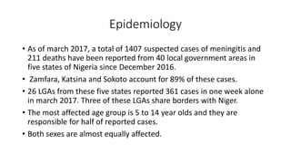
- An outbreak in Nigeria from 2016-2017 saw over 1,400 suspected cases mostly in children ages 5-14 in certain states. Risk factors include young age, close contacts, crowding and poverty.
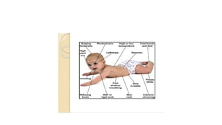
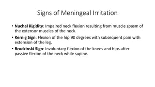
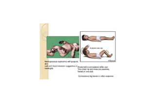
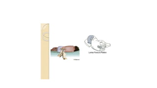
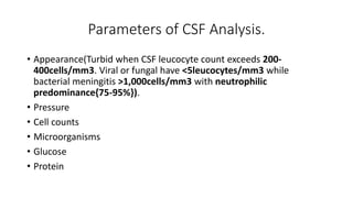
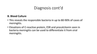
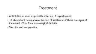
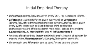
- Symptoms can include fever, headache, nausea, and signs of meningeal irritation like neck stiffness. Diagnosis involves CSF and blood tests. Treatment depends on the suspected cause but may include
Young at heart: Cardiovascular health stations to empower healthy lifestyle b...
Meningitis
1. MENINGITIS
Presented by
DR. AGBOR CHUKWUDI ISRAEL(MBBS, Ebonyi)
Department of Paediatrics
Nigerian Navy Reference Hospital, Ojo, Lagos
17th July 2020
3. Introduction
• Meningitis refers to the inflammation of the meninges.
• Not to be confused with encephalitis which is the inflammation of the
brain parenchyma.
5. Bacterial Causes
• Common cause: Neisseria meningitides (in children 1month to
12month).
• Streptococcus pneumoniae and haemophilus influenza type b
• less common pathogens such as pseudomonas aeruginosa,
Staphylococcus aureus, coagulase negative staphylococci, Salmonella
spp and listeria monocytogenes.
6. Viral Causes
Viral meningitis comprises most aseptic meningitis syndromes. The viral
agents for aseptic meningitis include the following:
• Enteroviruses(Polio virus, Echovirus, Coxsackievirus).
• Herpesvirus (hsv-1,2, VZV, EBV)
• Paramyxovirus(Mumps, Measles)
• Togavirus(Rubella)
• Rhabdovirus(Rabies)
• Retrovirus(HIV)
7. Fungal Causes
• This is rare in healthy individuals but it is a higher risk in those who
have AIDS, other forms of immunodeficiency or immunosuppression.
• The most common agents are Cryptococcus neoformans, Candida spp
and Histoplasma capsulatum.
8. Epidemiology
• As of march 2017, a total of 1407 suspected cases of meningitis and
211 deaths have been reported from 40 local government areas in
five states of Nigeria since December 2016.
• Zamfara, Katsina and Sokoto account for 89% of these cases.
• 26 LGAs from these five states reported 361 cases in one week alone
in march 2017. Three of these LGAs share borders with Niger.
• The most affected age group is 5 to 14 year olds and they are
responsible for half of reported cases.
• Both sexes are almost equally affected.
9. Epidemiology cont’d
• Lack of immunity to specific pathogens associated with young age remains
the major risk factor for meningitis.
• Additional risk factors include close contacts(household, daycare center)
with individuals having invasive disease caused by N. meningitides and H.
influenza type b, crowding and poverty.
• Person to person contact through respiratory tract secretions or droplet is
usually the mode of transmission.
• Defects of the complement system (C5-C8) have been associated with
recurrent meningococcal infection.
• Spenic dysfunction(SCD) or asplenia(trauma, congenital defects) is
associated with an increased risk of pneumococcal, H. influenza type b,
and, rarely, meningococcal sepsis and meningitis.
11. Clinical Manifestations
• More often, meninigitis is preceded by several days of fever
accompanied by upper respiratory tract or gastrointestinal symptoms,
followed by nonspecific signs of CNS infection such as increasing
lethargy and irritability.
• The signs and symptoms are related to the non specific findings
associated with a systemic infection and to manifestations of
meningeal irritation.
• Non specific findings include; anorexia, poor feeding, headache,
myalgia, arthralgia, tachycardia, hypotension.
12. Clinical Manifestations cont’d
• Various cutaneous signs such as petechiae,purpura or an
erythematous macular rash can also be seen.
• Common presentation of bacterial meningitis is; sudden onset,
rapidly progressive manifestations of shock, purpura, DIC, reduced
level of consciousness(leading to coma or death wthin 24hours).
• Meningeal irritation is manifested as; Nuchal rigidity, back pain,
Kernig sign, Brudzinski sign.
• Altered mental status is common and may be the consequence of
increased ICP, cerebritis or hypotension. It manifestations include;
irritability, lethargy, stupor, obtundation and coma.
13.
14. Signs of Meningeal Irritation
• Nuchal Rigidity: Impaired neck flexion resulting from muscle spasm of
the extensor muscles of the neck.
• Kernig Sign: Flexion of the hip 90 degrees with subsequent pain with
extension of the leg.
• Brudzinski Sign: Involuntary flexion of the knees and hips after
passive flexion of the neck while supine.
15.
16.
17. Diagnosis
• A. CerebroSpinal Fluid(CSF) Study
Analysis of the CSF reveals microorganisms on gram stain and culture.
CSF is collected through lumbar puncture.
Immediate Lumbar Puncture is contraindicated in the following;
• Evidence of increased ICP, HTN.
• In patients in whom positioning for the LP would further compromise
cardiopulmonary function.
• Infection of the skin overlying the site of the LP.
18.
19. Parameters of CSF Analysis.
• Appearance(Turbid when CSF leucocyte count exceeds 200-
400cells/mm3. Viral or fungal have <5leucocytes/mm3 while
bacterial meningitis >1,000cells/mm3 with neutrophilic
predominance{75-95%}).
• Pressure
• Cell counts
• Microorganisms
• Glucose
• Protein
20.
21.
22. Diagnosis cont’d
B. Blood Culture
• This reveals the responsible bacteria in up to 80-90% of cases of
meningitis.
• Elevations of C-reactive protein, ESR and procalcitonin seen in
bacteria meningitis can be used to differentiate it from viral
meningitis.
23. Diagnosis cont’d
C. Cranial CT Scan
This is of limited use in acute bacterial meningitis. CT in cerebral
oedema may show slit-like lateral ventricle and areas of low
attenuation.
25. Treatment
• Antibiotics as soon as possible after an LP is performed.
• LP should not delay administration of antibiotics if there are signs of
increased ICP or focal neurological deficits.
• Steroids and antipyretics.
26. Initial Empirical Therapy
• Vancomycin (60mg/kg/24hr, given every 6hr). For <3months infants.
• Cefotaxime (300mg/kg/24hr, given every 6hr) or Ceftriaxone
(100mg/kg/24hr administered once per day or 50mg/kg/dose, given
every 12hr). These can be used because third generation
cephalosporins are efficient against meningitis caused by sensitive
S.pneumoniae, N. meningitidis, and H. influenzae type b
• Patients allergic to beta lactam antibiotics and >1month of age can be
treated with Chloramphenicol 100mg/kg/24hr given every 6hr.
• Vancomycin and Rifampicin can be used for the persons above.
27. Corticosteroids
• Intravenous dexamethasone 0.15mg/kg/dose given every 6hr for 2
days is recommended in children older than 6weeks with acute
bacterial meningitis caused by H. influenzae type b.
• Corticosteroid recipients in the above group have a shorter duration
of fever(inflammation is reduced), lower CSF protein and lactate
levels, and a reduction in sensorineural hearing loss (result of
cochlear infection).
28. Complications
• Acute: Seizures, increased ICP, Cranial nerve palsies and stroke.
• Subdural effusions: common in infants.
• Prolonged fever: >10days fever is seen in 10% of patients. Thisis
usually caused by intercurrent viral infection, nosocomial or
secondary bacterial infection, thrombophlebitis or drug reaction.
• Optic Neuritis
• Cortical Blindness
• Syndrome of Inappropraite Anti-Diuretic Hormone
30. Prevention
• Vaccination according to the NPI schedule.
• Antibiotic prophylaxis of susceptible at-risk contacts. Rifampicin is
used.
• Vaccination- N. meningitidis conjugated quadrivalent( A,C,Y, W135)
vaccines in two forms(Menactra and Menveo).
-H.influenzae type b conjugate vaccine
-S. pneumoniae- Pneumococcal conjugate vaccine(PCV)
31. Prognosis
• Appropriate antibiotic therapy and supportive care have reduced the
mortality of bacterial meningitis after the neonatal period to <10%.
• Highest mortality rates are observed with pneumococcal meningitis.
• Sensorineural hearing loss is the most common sequel of bacterial
meningitis and usually, is already present at the time of initial
presentation. This is as a result of cochlear infection and occurs in as
many as 30% of patients with pneumococcal meningitis, 10% with
meningococcal and 5-20% of those with H. influenzae type b
meningitis.